Nasal Rhinosporidiosis - A Report of Two Cases
- 1. Otorhinolaryngologist at the Bettina Ferro de Souza University Hospital - Hospital Complex, UFPA. Belém / PA. Brazil
- 2. Otorhinolaryngologist at the Bettina Ferro de Souza University Hospital - Hospital Complex, UFPA. Belém / PA. Brazil
- 3. Otorhinolaryngologist at the University Hospital Bettina Ferro de Souza - Hospital Complex, UFPA. Belém / PA. Brazil
- 4. Otorhinolaryngologist at the University Hospital Bettina Ferro de Souza - Hospital Complex, UFPA. Belém / PA. Brazil
- 5. Otorhinolaryngologist at the University Hospital Bettina Ferro de Souza - Hospital Complex, UFPA. Belém / PA. Brazil
- 6. Otorhinolaryngology resident at the University Hospital Bettina Ferro de Souza - Hospital Complex, UFPA. Belém / PA. Brazil
- 7. Otorhinolaryngology resident at the University Hospital Bettina Ferro de Souza - Hospital Complex, UFPA. Belém / PA. Brazil
ABSTRACT
Nasal obstruction is an important entity in childhood due to its several early and late complications. One of its differential diagnoses is rhinosporidiosis, an endemic disease in areas like South America, caused by the
fungus Rhinosporidium seeberiwhich. The disease can affect the nasal mucosa in regions such as the septum, middle nasal turbinate and floor. The main form of therapy is surgical excision of the lesion. The authors of the
paper present two cases of pediatric rhinosporidiosis in the state of Pará/Brazil, an Amazon region in which its population has an important cultural relationship with rivers, streams and seas.
KEYWORDS
Rhinosporidiosis
• Epistaxis
• Neoplasm
• Nasal obstruction
CITATION
de Souza Andrade J, Gondim Barbosa AL, de Sousa FX, dos Santos Brandão A, e Silva LF, et al. (2020) Nasal Rhinosporidiosis - A Report of Two Cases. Ann Otolaryngol Rhinol 7(3): 1244.
INTRODUCTION
Nasal obstruction is a common and non-specific symptom in childhood, which can be related to both benign and malignant diseases [1]. As a cause of obstruction, nasal cavity masses in childhood are rare injuries, however very important because of their morbidity and mortality. They have nonspecific symptoms in this age group, which leads to a diagnosis that is often wrong and late [2]. In addition, nasal tumors have a large number of differential diagnoses, such as granulomas, and their early characterization is important due to their risk of malignancy or of being lymphoproliferative diseases [3].
An important, yet often overlooked, differential diagnosis of nasal granulomatosis is rhinosporidiosis, especially in its endemic areas, such as South America, India and Africa. This disease is caused by the fungus Rhinosporidium seeberi, which mainly affects children, adolescents and young adults [4].
The form of transmission of rhinosporidiosis is through contagion with the pathogen by contaminated water, through transepithelial penetration. The lesions caused by the fungus are presented as polyp or vascularized tumor, mainly in the nasal mucosa of the septum, inferior nasal concha or nasal floor [5].
The present work presents a two cases report of rhinosporidiosis in a child and an adolescent that occurred in the state of Pará, a region of Brazil with a tropical climate and a population related to riverside activities.
CASE PRESENTATION
Case 1
D.C.M/, 8 years old, male, brown, natural and resident in the city of Viseu, northeast of Pará. Inhabitant of an area located on the banks of the Gurupi and Pará rivers, where he practices activities on the beach and in the river, fishing being the main one.He sought medical attention with a case of small volume epistaxis with daily episodes for 7 months, associated with obstruction of the right nasal cavity, where the progressive growth of tumor lesion was seen. On physical examination, a granulomatous exophytic lesion with an irregular, painless and friable surface was observed in the right nasal cavity, with a diameter of 3 cm in its largest visible axis, not completely obstructing the nasal cavity. In nasal endoscopy, the lesion extended from area II to IV of `“Cottle”, mainly close to the floor. Excisional biopsy of the lesion was performed under general anesthesia by direct visualization without complications (Figure 1A),
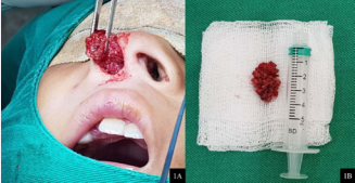
Figure 1: 1A - Exophytic lesion from the right nasal cavity in the immediate moment of its exeresis; 1B - Macroscopic anatomical specimen measuring around 03 cm, with firm-elastic consistency, but friable, reddish and irregular surface.
in which intraoperative insertion in the septal region was observed (Figure 1B). The anatomopathological examination revealed that it was rhinosporidiosis (Figure 2).
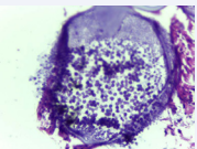
Figure 2: Histopathological study, with hematoxylin-eosin, 400x magnification, showing sporangium bordering the remaining respiratory mucosa and containing numerous spores.
The patient progressed satisfactorily in the immediate and late postoperative period, with no remaining lesions, and no complaints according to the parents.
Case 2
B.R.O., 13 years old, female, from the riverside region of the city of Cametá - Pará. She was admitted in a specialized service with a history of low volume epistaxis for one year. The patient showed a history of exposure and practice of immersion baths in rivers, streams and springs by her house. In the anterior rhinoscopy (Figure 3A),
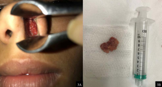
Figure 3: 3A - Anterior rhinoscopy with total obstruction of the left nasal cavity, with an irregular granulomatous aspect, reddish; 3B - Surgical specimen after excision measuring around 2.5 cm, firmelastic, irregular, reddish.
she had a vegetative, reddish lesion, on the floor of the left nasal cavity, friable to touch, painless, totally obstructing the left nasal cavity. In the surgical exeresis, it was observed that the lesion was inserted in the septal region, measuring around 2.5 cm (Figure 3B), and it was removed without complications under direct visualization. The anatomopathological confirmed the diagnosis of rhinosporidiosis (Figure 4A).
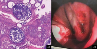
Figure 4: 4A - Histopathological study, with hematoxylin-eosin, 100x magnification, showing sporangium inserted in the respiratory mucosa and containing numerous spores inside. Formation of vacuoles and the presence of germinal centers with monolymphocytic proliferation and non-caseous granuloma; 4B - Immediate postoperative state showing left nasal cavity with signs of surgical manipulation without obstructive elements, bloody appearance and no signs of remaining lesions.
The patient presented a satisfactory postoperative period (Figure 4B), with no complaints or remaining injuries.
DISCUSSION
The two reported cases are in the state of Pará, located in the north of Brazil in the tropical region of the Amazon rainforest. The local population has ingrained in its culture the relationship with rivers, streams and seas, especially in rural regions, which, together with low levels of basic sanitation, exposes the population to contact with the fungus R. seeberi [6].
The interior of Pará still has, unfortunately, a public that is more unassisted and lacks education on health issues [6]. Sociocultural factors, such as immersion bath (in a recreational and personal hygiene way), are important points for research in the anamnesis of this population, as they are risk factors for diseases such as cholesteatoma and rhinosporidiosis, for example [7]. Therefore, the patient’s socioeconomic status is a risk factor for important nosological entities within the field of otolaryngology.
The age groups most affected by rhinosporidiosis are children, adolescents and young adults, with a predominance of males. The rate of nasal involvement reaches 70% of the cases, most commonly in the septal mucosa, inferior nasal concha and nasal floor, however other regions of involvement are reported in the literature, such as conjunctival mucosa, lacrimal sac, lungs, liver, external genitalia and anal region [8]. The lesions appear as a polypoid or vascular mass, with an irregular surface, sometimes pediculated. The main complaints of those affected are nasal obstruction, epistaxis and mucus-purulent rhinorrhea [9].
The treatment of rhinosporidiosis with the best results is surgical excision of the lesion, which also provides its histopathological diagnosis. The literature also mentions drug therapies such as the use of dapsone, but none of them has a satisfactory result as the surgical approach [10].
The reported cases are in agreement with the information available in the literature, in which the two patients of school age are residents of the riverside population. As symptoms, they presented epistaxis and nasal obstruction, with the presence of unilateral exophytic lesion in the nasal cavity. The treatment performed was surgical excision of the lesion, with diagnostic confirmation by histopathological study. Both evolved without complications and remaining injuries.
CONCLUSION
Physicians should be alert to diseases that, despite having a low incidence, present a differential diagnosis with nasal tumors in the pediatric age group.
The two cases described demonstrate the importance of educating and raising awareness among the population most in need of resources and information, in addition to prioritizing the recurrence of entities whose socioeconomic risk factor and cultural practices are important.


