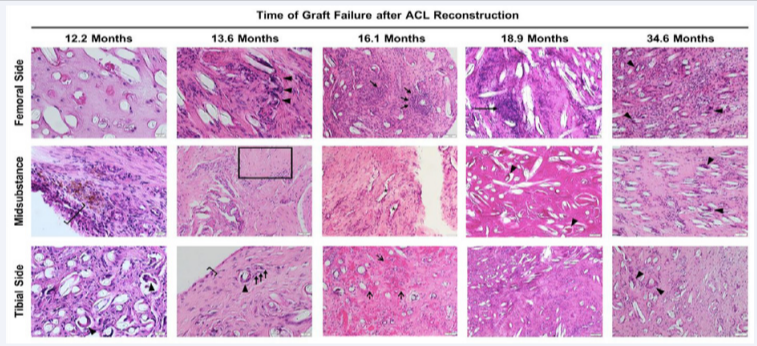
Figure 4 Histology of intra-articular graft biopsies. H&E staining revealed the PLLA scaffold fully infiltrated by cells and connective tissue. In some regions, collagen fiber alignment was observed ( ), as well as a peripheral synovial layer ([ ) and new vasculature (*). A chronic inflammatory response was seen in all biopsies, marked by the presence of foreign body cells ( ), macrophages and lymphocytes ( ), and eosinophils ( ).