Ectopic Eruption of Maxillary Third Molar Tooth in the External Acoustic Meatus: A Case Report
- 1. Department of Otolaryngology Head and Neck Surgery, Sun Yat-Sen Memorial Hospital, China
- 2. Department of Otolaryngology, The University of Hong Kong- Shenzhen Hospital, China
- 3. Department of Otolaryngology Head and Neck Surgery, the Third Affiliated Hospital of Sun Yat-Sen University, China
ABSTRACT
Ectopically erupted tooth is a rare disease, indicating that a tooth erupts into regions other than the oral cavity. We present a case of an ectopic maxillary third molar tooth in the external acoustic meatus, and discuss the diagnosis and treatment of this entity.
KEYWORDS
Schwannoma, Neurilemmoma, Base of tongue
CITATION
Jiang X, Chen Q, Chen Y, Si Y, Liu Y, et al. (2014) Ectopic Eruption of Maxillary Third Molar Tooth in the External Acoustic Meatus: A Case Report. Ann Otolaryngol Rhinol 2(1): 1016.
CASE REPORT
A 16-year old girl was referred to the ENT department for a neoplasm in left external acoustic meatus with a history of 2 months. Physical examination revealed that there was a light red, lobulated, smoothy neoplasm fixed in the upper wall of the meatus. It was solid, 0.5cm x 0.8cm in size. The tympanic membrane was found integrity without congestion or effusion by endoscopy (Figure 1A&B).
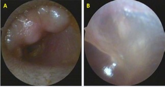
Figure 1: A. Endoscopy revealed a neoplasm in the external acoustic meatus; B. The tympanic membrane was intact.
Audiogram and acoustic immittance was normal. HRCT indicated a lobulated, well-defined, bone density mass in the left external acoustic meatus. It was pedicled to the meatus wall without bone destruction (Figure 2A).
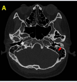
Figure 2: A. HRCT demonstrated the mass;
The patient underwent resection of the mass through the meatus under general anesthesia. After incision of the outer skin layer, a white, firm, irregular mass was exposed. We removed the mass from its pedicle with a osteotome (Figure 2B),
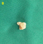
Figure 2:B. The resected ectopic tooth;
finding that the external acoustic meatus was smoothy without bone defect. Then the skin was replaced, and the meatus was packed with Merocel. Given that the lobulated mass looked like a molar, a postoperative orthopanotomography was taken, revealing the absence of her left maxillary third molar (Figure 2C).
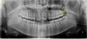
Figure 2:C. Orthopanotomography showed the left maxillary third molar tooth was missing.
The pathology report proved the mass was a molar.
DISCUSSION
Ectopically erupted tooth is rarely seen, with a prevalence varying from 0.1%~1% in the existing literatures, most of which are case reports [1,2]. The etiology is still unclear, while different theories considering trauma, infection, cyst, tumorand developmental abnormalities [3,4]. Because the external auditory canal and tooth structures share a similar embryologic origin, abnormal development of specific first pharyngeal arch derivatives could logically affect the anatomic and physiologic properties of both structures [5]. Cleft lip and cleft palate are common congenital defects associating with ectopically erupted tooth [6].Diverse locations of an ectopically erupted tooth include the nasal cavity, chin, mandibular condyle, coronoid process, palate, orbit, maxillary sinus [3,6]. It could even erupt through the skin. Unilateral occurrence seems to be more frequent than bilateral, and eruption in maxillary is more common than in mandibular [2]. However, an ectopically erupted tooth in the external acoustic meatus has not been described in the literature so far.
Patients with ectopically erupted tooth often present for a variety of symptoms according to the locations of eruption, such as stuffy nose, chronic or recurrent sinusitis, recurrent haemoptysis, epiphora, headaches and facial numbness or pain [6]. The patient in our report felt no discomfort but a neoplasm in her external acoustic meatus accidentally. Sometimes, they can be asymptomatic and got diagnosis by physical examination or CTscan [1,4,6]. Orthopanotomography is useful for the differential diagnosis of ectopic eruption tooth and supernumerary tooth. The former has a total number of normal teeth less than 32, while the latter has a normal number [3].
Generally, surgery is the effective treatment of ectopic eruption tooth without recurrence. For example, the ectopically erupted tooth in maxillary sinus or nasal cavity can be removed by endoscopic sinus surgery or Caldwell-Luc operation [3]. The patient in our report received a resection of the ectopically erupted tooth from its pedicle in left external acoustic meatus. The girl recovered quickly after the surgery with no complication, and had been followed up for 8 months without recurrence.
FINANCIAL DISCLOSURE
This work was supported by the National Natural Science Foundation of China (81371082), China.


