Scaphal Autograft for Secondary Nasal Dorsum Repair
- 1. Aesthetic Plastic Surgery, Paris 75006, France
ABSTRACT
The reconstruction of the nasal dorsum can be difficult in certain secondary cases, when there is a loss of volume; The usual technique consists generally in taking a bone, or a cartilaginous graft, or resorting to a silicone foreign body, among others techniques; Among the cartilaginous grafts available between the costal cartilages or the ear concha, we can add a rather unknown donor site: the scapha, which allows to take a long flat cartilaginous graft 4 to 7 cm long, behind the fold of the antihelix, which structure it should be preserved; around forty patients have been operated on since 1990 by the author, with a favorable and stable result over time.
KEYWORDS
Rhinoplasty; Secondary rhinoplasty; Saddle nose; Scapha graft; Antihelix
CITATION
Mitz V (2020) Scaphal Autograft for Secondary Nasal Dorsum Repair. Ann Otolaryngol Rhinol 7(1): 1235.
INTRODUCTION
The reconstruction of the nasal dorsum can be difficult in certain secondary cases, when there is a loss of volume; The usual technique consists generally in taking a bone, or a cartilaginous graft, or resorting to a silicone foreign body, among others techniques; Among the cartilaginous grafts available between the costal cartilages or the ear concha, we can add a rather unknown donor site: the scapha, which allows to take a long flat cartilaginous graft 4 to 7 cm long, behind the fold of the antihelix, which structure it should be preserved; around forty patients have been operated on since 1980 by the author, with a favorable and stable result over time [1].
MATERIALS AND METHODS
The technique for removing the scapha cartilage by a posterior approach
1. Local anesthesia with dilute adrenaline lidocaine, Including the anterior surface to be able to remove the cartilage more easily
2. posterior ear incision, 4 to 7 mm behind the free edge of the ear
3. passage under posterior perichondral detachment on demand, of the future cartilage graft
4. 2 parallel incisions are realized on the scapha, allowing to define a tongue from 5 to 7 cm long
5. Detachment of the tongue from the anterior skin surface of the scapha, with a soft elevator
6. Scapha removal by cutting the ends of the graft
7. Hemostasis
8. Sutures of the skin incision with absorbable thread
9. Cleaning of the removed cartilage, placed in physiological saline
RESULTS AND DISCUSSION
Results
About 40 cases have been operated on since 1994; although we do not have a very exact count of patients reviewed in the long term, the fact remains that 5 or 6 patients had an insufficient result; the vast majority of patients are extremely satisfied with the result, especially since the operation is simple; We did not observe, in this short series of cases, any infection, or a keloid scar; a photo of a 26-year-old case is shown in this article (Figure 1)
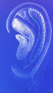
Figure 1: Scaphal groove drawing.
Figure 2
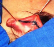
Figure 2: A Posterior Incision is used.
Figure 3
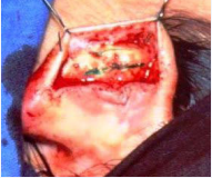
Figure 3: The Subperichondral Dissection.
Figure 4
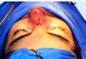
Figure 4: Exemple of an Onlay Scaphal Partial Graft.
Figure 5
Figure 5: Exemple of an Alar Onlay Graft.
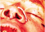
Figure 6
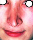
Figure 6: Patient A before Revision Rhinoplasty.
Figure 7
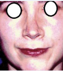
Figure 7: Patient A; one year after Scaphal Autograft to Dorsum.
Figure 8
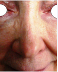
Figure 8: Patient B; good appearance 26 years after a scaphal graft
Figure 9
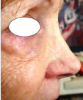
Figure 9: Patient B, the profile after 26 years.
Figure 10
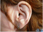
Figure 10: Patient B, Normal Ear.
Figure 11
Figure 11: Ppatient B, the result after graft take, notice the minimal deformity
Figure 12
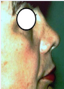
Figure 12: patient C, the saddle nose before.
Figure 13
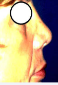
Figure 13: Patient C, result after one year.
Figure 14
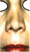
Figure 14: Patient C, face view before.
Figure 15
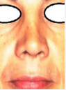
Figure 15: Patient C, face view after.
Discussion
Benefits of scapha cartilage: The intervention is very simple, it removes cartilage in the same operating field; unlike costal cartilage, This thin cartilage will not tend to twist, or, like the iliac bone graft, give a rigid nose with a hard tip; the cartilage, once removed from the ear, makes it possible to obtain a longer, flat, a thin dorsum, in one piece, better than if a conchal graft was is used.
Indications of scapha cartilage: Several deficient structures of the nose can be improved by scapha cartilage:
Firstly, the longitudinal defects of the nasal bridge, because the narrowness of the graft allows the dorsum to be nicely redone; Second, defects in the wing cartilages, whether asymmetry, an iatrogenic rupture, or a projection of the tip Third, scapha cartilage can be used to support an insufficient nasal septum, to fill a hollow between the clean bone and the septum, revealing an unsightly indentation.
Disadvantages of scapha cartilage: The amount of usable cartilage is low, although we can use the two scaphas of the ears to insert a double thickness of cartilage; Once removed the cartilage can curl discreetly on itself; it is possible to crush it in a Jost crusher, or to incise discreetly the cartilaginous graft on the concave side. Our patients who obtained an insufficient result by the scapha method, benefited in 4 cases from a silicone implant of Shirakabe. And another recent case has been corrected for perceived insufficiency of dorsal projection, by injection of a dense hyaluronic acid.
CONCLUSION
Grafting of the cartilage taken behind the ear , the Scapha graft, makes it possible to obtain a narrow and long tongue of thin cartilage; this graft preserves the anatomy of the ear, and keeps the fold of the super antihelix; even, in some cases, it helps to stick a little protruding ear; the cartilage of the scapha has allowed, us in almost 40 cases, to obtain a satisfactory result, in order to correct a saddle nose, an asymmetrical tip due to a defect in an alar cartilage, or to create a columellar strut more flexible than a bone graft; the scapha graft is thus a good alternative to be used instead or with the conch of the ear.


