The
- 1. Department of Otorhinolaryngology, Tan Tock Seng Hospital, National University of Singapore, Singapore
ABSTRACT
Objective: To describe the anatomy of the bony mesentery of the anterior ethmoid artery.
Study Design: Descriptive anatomy based on coronal and sagittal CT sections, endoscopic images of a post-surgical case and a cadaveric dissection specimen.
Methods: A short review of medical literature in textbooks and journals to show past and current assumptions. A descriptive presentation based on images above illustrating an important detail in surgical anatomy.
Results: The presence of the medial aspect of a supraorbital ethmoid cell dictates the length of the bony mesentery and the vertical distance of the anterior ethmoid artery from the skull base. This part of the skull base is not the ethmoid roof as previously presumed in early teachings but refers to the roof of the supraorbital ethmoid cell, which is continuous with the posterior table of the frontal sinus.
Conclusion: The bony mesentery of the anterior ethmoid artery projects horizontally forward from the skull base and may confer more bony protection posteriorly against inadvertent injury during surgery than previously thought. When clearing the posterior outflow tract of the frontal sinus in a posterior to anterior direction, an instrument should not be pushed simultaneously in a superior direction as this could cause anterior ethmoid artery damage.
KEYWORDS
Anterior ethmoid artery, Bony mesentery, Avulsion, Orbital complications, Endoscopic sinus surgery, Frontal mucocele
CITATION
Siow JK (2016) The “Mesentery” of the Anterior Ethmoid Artery. Ann Otolaryngol Rhinol 3(1): 1084.
INTRODUCTION
The surgical significance of the anterior ethmoid artery is well recognized. Damage to this important vessel during endoscopic sinus surgery leading to epistaxis, orbital hematoma and possibly blindness has been described early in the development of endoscopic sinus surgery [1]. It is often presumed that the presence of the bony mesentery to which the anterior ethmoid artery is attached increases the vulnerability of the anterior ethmoid artery to damage during surgery. This presentation aims to show that this increased vulnerability is only so if an instrument is simultaneously pushed upward during clearance of the frontal sinus outflow tract in a posterior to anterior direction.
MATERIALS AND METHODS
Mesentery in its general anatomical sense
Anatomy texts, in general, define mesentery as a double layer of peritoneum attached to the abdominal wall and enclosing in its fold a portion or all of one of the abdominal viscera, conveying to it its vessels and nerves [2]. This term is generally used to describe the peritoneal folds, which support the small intestines, sigmoid colon, appendix, transverse colon, and portions of the ascending and descending colon.
Mesentery in relation to the anterior ethmoid artery
The mesentery of the anterior ethmoid artery often cited in text books [3-9] and in some papers [10-13] is a term, which arose from the observation of the relationship of the anterior ethmoid artery to the skull base in CT scan assessments and in the practice of endoscopic sinus surgery. This anatomic term has not been adequately elaborated in detail and deserves a proper description so that its relevance to surgical anatomy and reference in the medical literature can be better appreciated.
The Mesentery of the Anterior Ethmoid Artery in textbooks
An authoritative text book published in 1991, described the anterior ethmoid artery, surrounded by a thin walled bony channel, as often being connected to the roof of the ethmoid by a bony mesentery with an interspace of as much as 5 mm. An accompanying schematic drawing showed the anterior ethmoid artery in a bony canal traversing the ethmoid below an air space, which was enclosed by a dome described as the frontal bone providing for the roof of the ethmoid. A Blakesley forceps was shown positioned to be able to grasp the anterior ethmoid artery through this “hanging” bony canal [3].These descriptions were based on observations of coronal CT sections with a mention that anatomic preparations in the sagittal plane were of little assistance to surgical anatomy in an endoscopic surgical procedure. The cells in this region were then named as orbitoethmoid cells as the term supraorbital ethmoid cell [4] had not been coined yet. The description of the anterior ethmoid artery in this way influenced generations of authors. A more recent textbook would caution about the vulnerability of the anterior ethmoid artery seemingly suspended on a mesentery from the skull base within the frontal recess as seen on coronal CT section. This was reported to occur in approximately 36% of the time and placed a patient at increased risk for intra-operative bleeding and orbital hematoma [5]. Another author stressed that the location of the anterior ethmoid arteries should be noted as to whether they were positioned in the bony skull base or hanging lower in mesentery, where they might be at risk for injury during surgery when clearing remaining anterior ethmoid cells toward the frontal outflow tract [6]. The anterior ethmoid artery has been described to run in a canal which was oblique and which ran at a variable distance as much as 17 mm below the roof of the ethmoid to which it is attached by a bony mesentery [7]. Thus it was cautioned that if the anterior ethmoid artery was not recognized as being on a mesentery, the artery might be severed as septations on the skull base were removed [8]. A more detailed anatomical description stated that the longer the lateral lamella of the cribriform plate, i.e. the deeper the olfactory fossa, and with that the higher the ethmoidal roof superior to the level of the cribriform plate, the more likely the ethmoid artery is found travelling freely through the ethmoid cavity and penetrating through the lateral lamella of the cribriform plate [9]. These presentations impress upon the reader the very high vulnerability of the anterior ethmoid artery where the tip of an instrument could potentially be inserted superior to this suspended anterior ethmoid artery from posteriorly when clearing the frontal outflow tract and thus lead to transection or avulsion of the anterior ethmoid artery.
The Mesentery of the Anterior Ethmoid Artery in Medical Literature
In a 2006 study on endoscopic anterior ethmoid artery ligation in cadavers, it was concluded that the anterior ethmoid artery should be in a mesentery for an effective clip to be placed [10]. In this study, the presence of the anterior ethmoid canal mesentery was noted on coronal CT and confirmed on endoscopic dissection. This finding it was observed validated the authors’ departmental policy to identify a “hanging” anterior ethmoid canal on coronal CT scans preoperatively to avoid trauma to the anterior ethmoid artery during endoscopic sinus surgery. The sagittal CT section chosen to illustrate the presence of this anterior ethmoid canal mesentery however did not show a supraorbital ethmoid cell and so the bony mesentery of the anterior ethmoid artery was not illustrated. In a 2009 study on the technical feasibility of transnasal endoscopic anterior artery ligation in cadavers, it was concluded that effective clipping could only be performed in 50% of cases with a mesentery and was not technically feasible in cases when the anterior ethmoid artery was without a mesentery [11]. The picture of the procedure showed a clip applied onto a dehiscent anterior ethmoid artery from anteriorly, however the mesentery of the anterior ethmoid artery was not described in detail.
A 2010 study of 141 patients based on cone beam tomography analysis showed the angle of the olfactory fossa and the position of the anterior ethmoid artery to be significantly different when the anterior ethmoid artery was noted to be “hanging” compared to when it was integrated into the skull base [12]. A 2012 similar study by the same authors analyzed 116 patients below 18 years old and 749 adult patients identifying differences in the course of the anterior ethmoid artery whether it was embedded in the skull base or was less or more than 3 mm from the skull base aiming to identify the “dangerous ethmoid” [13].
A Detailed Description of the Bony Mesentery of the Anterior Ethmoid Artery
This detailed description is to complement the work of recent authors by illustrating correlations between CT scan descriptions with endoscopic views post-surgery and in a cadaveric specimen. The digitally reconstructed sagittal CT Section in fine 1mm cut detail has enabled a better appreciation of frontal recess anatomy and of the bony mesentery of the anterior ethmoid artery. The pneumatization superior to the anterior ethmoid artery is due to the presence of the medial aspect supraorbital ethmoid cell. The pneumatization inferior to the anterior ethmoid artery is due to the presence of a suprabullar recess (Figure 1).
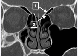
Figure 1: In this coronal CT scan section, the anterior ethmoid artery is identified at the “pinching” of the medial orbital wall, and is seen to traverse across the frontal recess area suspended from the skull base. This conjuresthe possibility of anterior ethmoid artery avulsion during surgery. Arrow 1 shows the medial aspect of the left supraorbital ethmoid cell. Arrow 2 shows the left suprabullar recess.
The bony mesentery of the anterior ethmoid artery is the party wall between these two pneumatized spaces. The anterior ethmoid artery lies along the free anterior edge of the bony mesentery. This bony mesentery extends horizontally forward from the junction of the inferior aspect of the roof of the supraorbital ethmoid cell (continuous with the posterior table of the frontal sinus) and the roof of the suprabullar recess (Figure 2).
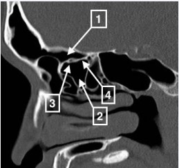
Figure 2: Arrow 1 shows the medial aspect of the supraorbital ethmoid cell. Arrow 2 shows the suprabullar recess. Arrow 3 shows the anterior ethmoid artery. Arrow 4 shows the bony mesentery of the anterior ethmoid artery. A coronal CT section through the anterior ethmoid artery would thus show pneumatized air spaces superior and inferior to the anterior ethmoid artery thus conjuring the “hanging” artery.
The roof of the supraorbital ethmoid cell is in part continuous with the posterior table of the frontal sinus and in part projects anteriorly ending with a free edge forming a party wall between the medial aspect of the supraorbital ethmoid cell and the frontal sinus (Figure 3).
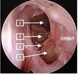
Figure 3: This diagram depicts the left side of the patient. Arrow 1 shows the supraorbital ethmoid cell. Arrow 2 shows the frontal recess. Arrow 3 shows the anterior ethmoid artery. Arrow 4 shows the inferior aspect of the bony mesentery of the anterior ethmoid artery which is the roof of the suprabullar recess.
The more prominent the medial aspect of a supraorbital ethmoid cell, the longer would this bony mesentery of the anterior ethmoid artery be. The superior surface of this bony mesentery is the floor of the medial aspect of the supraorbital ethmoid cell and the inferior surface of this bony mesentery is the roof of the suprabullar recess (Figure 3). Conversely, if the medial aspect of a supraorbital ethmoid cell were small or absent, then the bony mesentery would be shorter or absent. When the bony mesentery is absent, the anterior ethmoid artery is embedded in the skull base. The anterior ethmoid artery has been described frequently but not always to lie posterior to the supraorbital ethmoid cell opening and to travel in a bony mesentery below the skull base [14] (Figure 3). The position of the anterior ethmoid artery has been noted to be variable and when the ethmoid sinuses were more pneumatized and when in particular a supraorbital cell was present, the artery lay below the skull base [15]. The skull base mentioned in this instance is often misunderstood as the roof of the ethmoid sinus [3]. A sagittal CT section shows that the skull base referred to is the part of the roof of the supraorbital ethmoid cell continuous with the posterior table of the frontal sinus separated from the bony mesentery by the pneumatization of the supraorbital ethmoid cell below the posterior table of the frontal sinus (Figure 2).
It has been reported that greater pneumatization of supraorbital ethmoid cells resulted in significantly farther distance from the skull base of the anterior ethmoid artery and this observation was extended to suggest that the supra-orbital ethmoid cell be used as a landmark for the anterior ethmoid artery [16]. This is indeed so as when the anterior ethmoid artery is easily seen on a coronal CT scan, (Figure 1) the pneumatization seen above the anterior ethmoid artery is always the medial aspect of the supraorbital ethmoid cell.
The oblique course of the anterior ethmoid artery is usually from postero-lateral to antero-medial making the medial aspect of the bony mesentery usually a little longer than the lateral aspect.
The anterior ethmoid artery is thus not suspended vertically on a bony mesentery from the skull base as early interpretations of coronal CT sections or less detailed presentations of sagittal CT sections [10] would suggest. Sagittal CT sections of fine 1mm cut detail show clearly that the anterior ethmoid artery is suspended horizontally on a bony mesentery from the skull base (Figure 2).
Surgical Significance
When a narrow tipped instrument is directed in a posterior to anterior direction along the skull base, this bony mesentery, which extends from the skull base as a bony shelf in a horizontal direction toward its free edge containing the anterior ethmoid artery would prevent the tip of this instrument from entering the medial aspect of a supraorbital ethmoid cell from posteriorly (Figure 4).
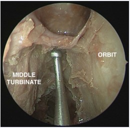
Figure 4: In this cadaveric specimen on the left side, a ball probe is placed at the base of the bony mesentery of the anterior ethmoid artery at the junction of the ethmoid roof and the inferior aspect of the roof of the supraorbital ethmoid cell which is continuous with the posterior table of the frontal sinus. The anterior ethmoid artery at the free edge of this bony mesentery will not be damaged if the tip of the ball probe is moved from posteriorly to anteriorly following the natural lie of the bony mesentery.
Thus this bony mesentery would help to protect the anterior ethmoid artery from inadvertent avulsion. However when removing cells from the skull base in a posterior to anterior direction the surgical instrument should not also be simultaneously pushed in a superior direction as this could cause the tip of the instrument to enter the medial aspect of a supraorbital ethmoid cell posterior to the anterior ethmoid artery through the bony mesentery thus setting the anterior ethmoid artery up for avulsion (Figure 5).
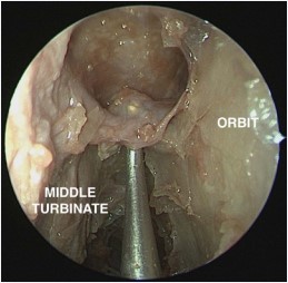
Figure 5: In this same cadaveric specimen on the left side, for the tip of the ball probe to ascend superior to the anterior ethmoid artery from posteriorly to be in a position to avulse the anterior ethmoid artery, the bony mesentery has to be broken through with a deliberate superior thrust of the ball probe.
Caution is very strongly advised in the endoscopic management of a frontal mucocele. The expansion of a frontal mucocele could cause dehiscence of this protective bony mesentery and lead to an unprotected anterior ethmoid artery without a horizontal bony shelf. Ironically this situation would not be readily appreciated on a coronal CT scan on account of the opacification of this region by the presence of the frontal mucocele.
RESULTS AND DISCUSSION
e bony mesentery of the anterior ethmoid artery is a small approximately rhomboid shaped shelf of bone, covered superiorly and inferiorly by sinus mucosa, straddling between medial aspect of a supraorbital ethmoid cell and the suprabullar recess. The anterior ethmoid artery runs in its free edge at its anterior aspect usually in a posterolateral to anteromedial direction in its course from the orbit to the lateral lamella of the cribriform plate. The presence and length of the bony mesentery is dependent upon the amount of pneumatization of the supraorbital ethmoid cell. The greater the pneumatization of the medial aspect of a supraorbital ethmoid cell, the longer is the bony mesentery. The bony mesentery of the anterior ethmoid artery extends horizontally from the junction of the inferior aspect of the roof of the supraorbital ethmoid cell and the roof of the suprabullar recess.
Although this horizontal projection of the bony mesentery of the anterior ethmoid artery provides bony protection to the anterior ethmoid artery posteriorly making it somewhat less vulnerable to damage in surgery, caution is always strongly advised as the consequences of orbital hematoma are most dire. The presence of a frontal mucocele, very likely to cause dehiscence of the bony mesentery of the anterior ethmoid artery sets up the situation of easy inadvertent avulsion during surgery
CONCLUSION
The bony mesentery of the anterior ethmoid artery projects horizontally forward from the skull base and may confer more bony protection posteriorly against inadvertent injury during surgery than previously thought. When clearing the posterior outflow tract of the frontal sinus in a posterior to anterior direction, an instrument should not be pushed simultaneously in a superior direction as this may cause anterior ethmoid artery damage.
REFERENCES
2. Farlex Partner Medical Dictionary © Farlex 2012.
4. Owen RG Jr, Kuhn FA. Supraorbital ethmoid cell. Otolaryngol Head Neck Surg. 1997; 116: 254-261.
8. Wormald PJ, Tewfik MA. Practical Rhinology. CRC Press; 2010; 49.
14.Chua AG, Kennedy DW. The Frontal Sinus. Springer Verlag Berlin Heidelberg; 2005; 195.


