Loading

Annals of Clinical and Experimental Metabolism

Gastric Carcinoid Tumors

Review Article | Open Access | Volume 2 | Issue 2

  • 1. Department of Gastroenterology, Kocaeli University, Turkey
  • 2. Department of Gastroenterology, Mardin State Hospital, Turkey
+ Show More - Show Less
Corresponding Authors
Hülagü S, Department of Gastroenterology, Kocaeli University, Umuttepe, Izmit, Turkey, Tel: 902623031005;
ABSTRACT

With the widespread use of diagnostic endoscopy, gastric carcinoids are increasingly encountered in clinical practice. World Health Organization (WHO) decided to use the term neuroendocrine tumors (NET) instead of carcinoid tumor in 2010. Type I and type II NETs associated with hypergastrinemia are more common, while sporadic type III is rare. Type I NETs constitute 70-80% of gastric nets; type II 5-8%, type III 15-20%. These tumors are usually asymptomatic and have been diagnosed by gastroscopy for other reasons and rarely for bleeding or anemia. Gastric NETs are highly heterogeneous in their clinical behavior. Type I and type II gastric NETs can be frequently followed and treated endoscopically in sizes smaller than 1 cm. sporadic types may require oncologic surgery or systemic treatment.

KEYWORDS

Carcinoid tumors ; Stomach ;Gastric neuroendocrine tumors ; Diagnosis ; Therapy.

CITATION

Hülagü S, Y?lmaz H (2017) Gastric Carcinoid Tumors. Ann Clin Exp Metabol 2(2): 1017.

ABBREVIATIONS

NET: Neuroendocrine Tumors; WHO: World Health Organization; ECL: Enterochromaffin-like Cells; SEER: Surveillance Epidemiology and End Results; IR: Incidence Rate, CCK-2: Cholecystokinin-2 Receptors; MENI: Multiple Endocrine Neoplasia type 1; ZES: Zollinger–Ellison Syndrome; GI: Gastro Intestinal; EUS: Endoscopic Ultrasonography; ESD: Endoscopic Submucosal Dissection; SSAs: Somatostatin Analogs

INTRODUCTION

The term “carcinoid” was first used by German pathologist Siegfried Oberndorfer, and is defined as a small submucosal tumor in the small intestine at a 48-year-old women’s autopsy [1]. Since then, a great deal of knowledge has been gained about the physiology, pathology, and management of these tumors. Italian pathologist Carlo Capella defended not to use the term “carcinoid”, instead he introduced the term “neuroendocrine tumors (NET)” in 1995. Later, “NET” was accepted by the World Health Organization (WHO) for common terminology in 2010 [2]. Recently the term “NET” is accepted worldwide.

NETs are a heterogeneous group of tumors regarding clinical behavior, histology and prognosis, which arise from enterochromaffin-like cells (ECL) spread to the whole gastrointestinal tract. In this review, we will scope to an overview of recent developments in gastric NETs and the latest approach to classification, diagnosis, and treatment.

EPIDEMIOLOGY

The incidence of neuroendocrine tumors seems to have increased globally considering the recent medical literature. Two major reasons of this raising up is firstly; common nomenclature was not used for NETs in the past years, and in many studies the incidence was low because of the absence of tumor anatomical localization, secondly the widespread use of imaging methods and surveillance endoscopy have increased the awareness of these tumors even if they are asymptomatic. Gastric NETs are representing 8% of the neuroendocrine tumors of the gastrointestinal tract [3]. In the USA, according to Surveillance Epidemiology and End Results (SEER) database, the gastric NETs incidence rate (IR) was 0.03 per 100000 populations per year between 1973-1977 and it increased to 0.33 between 2003-2007 [4]. IR of gastric NETs was 0.16 and 0.15 for male and female, respectively, in England between 2000-2006 and it was 0.08 at Austria between 2004-2005 [5,6]. There is also a remarkable increase in incidence from 39% to 88% in gastric NET in Norwegian population over a decade between the 1990s and 2000s [7].

PATHOPHYSIOLOGY AND CLASSIFICATION

Following food intake, gastrin is released physiologically from the G cells in the antrum and binds to the cholecystokinin-2 receptors (CCK-2) of ECL cells localized in the gastric corpus and fundus, leading to histamine release from these cells. Histamine leads to acid production from parietal cells. Gastrin is suppressed by somatostatin release from D cells in the antrum. If gastric acid secretion decreases (achlorhydria), hypergastrinemia will occur and lead to trophic effects on ECL cells which cause hyperplasia. Hypergastrinemia is inadequate for arising NET from ECL cells alone, whereby the long-term PPI use or vagotomy, does not seem to be associated with gastric NETs. Genetic (loss of heterozygosity of the MEN-I gene locus on 11q13 was identified of 17-73 %of type I NETs), epigenetic (LINE1 hypomethylation was identified 50% gastric NETs), bacterial (in an animal model it has shown H. pylori lipopolysaccharides and peptidoglycans has an influence on the proliferation of ECL cells) and dietary (smoking status) factors can contribute to tumor development [8-10].

Gastric NETs are solid lesions which are usually yellowish white color and could be present endoscopically as mucosal nodular, polypoid, ulcerous lesions or submucosal lesions, as well. Randi et al. described three subtypes of gastric NETs; type I and type II are associated with hypergastrinemia and type III are formed sporadically [11].

Type I gastric NETs constitute 70-80% of all gastric NETs. They are multiple carcinoid tumors that are smaller than 1 cm in diameter and tend to be localized in the corpus and antrum [12]. It is the type associated with atrophic gastric [13]. The metastasis rate is <5% [14]. Gastric acidity is low and serum gastrin level is high in type I patients.

Type II gastric NETs constitute 5-8 %of the gastric NETs. They are also multiple, smaller than 1cm and localized at the fundus, corpus, and antrum of the stomach. Gastric acidity and serum gastrin levels of type II patients are both high. Type II lesions are associated with Multiple Endocrine Neoplasia type 1(MENI) and Zollinger–Ellison Syndrome (ZES). These lesions are moderately malignant and metastasis rate is below 10% [15].

Type III Gastric NETs are about 15-20 %of the gastric NETs. This type is the most aggressive type of tumors and metastasis rate is over 50% [16]. This lesion is solitary, bigger than 2 cm and generally localized to antrum of the stomach. Patients are mostly male and it occurs sporadically.

WHO classified NETs into three groups based on cell proliferations, which is defined by the number of mitoses per 10 high-power microscopic fields (HPF), and on the percentage of tumor cells positively immune-labeled for Ki-67 antigen(Labeling Index, LI): grade 1; mitoses ≤ 2/10 HPF and Ki-67 LI ≤ 2% and grade 2 mitoses 2–20/10 HPF and Ki-67 LI 3–20% and grade 3 mitoses >20 HPF orKi-67 LI >20% [17].

Grade 1 and grade 2 tumors are well differentiated and show organoid architecture with trabecular, gyriform pattern, neoplastic cells are uniform and a plenty content of secretory granules responsible for intense and diffuse staining for general neuroendocrine markers (synaptophysin and chromogranins). The nuclear chromatin is regular and mitosis is not common. Grade 3 neoplasms are poorly differentiated and have highly pleomorphic, atypical nuclei, a solid growth pattern, and nonischemic necrosis areas, mitosis are always rich and often atypical [18].

Currently Chromogranin A (CgA) is the most widely used biomarker for the diagnosis of NETs. Chromogranin A is a watersoluble acidic glycoprotein of 68 kDa located in the matrix of large secretory granules of neural and endocrine cells. These protein levels, which can be measured in serum and plasma, correlate with the NET mass [19]. CgA may be found in healthy tissues, and serum levels may also increase in other diseases such as renal failure, cardiac diseases, prostate cancer and small cell lung cancer. Measurable values are affected by proton pump inhibitors and food consumption [20]. Sensitivity is 60-90% but its specificity is below 50% due to reasons listed above. There is also no standard in existing CgA kits and there is wide variability between laboratory measurements [21].

Pankreostatin is a chromogranin derivative and could be used as a tumor marker for carcinoids. Its advantage over he chromogranin A is not being affected by the proton pump inhibitor usage [20]. 

NSE) is a protein that occupies the cytosol of the neuron and neuroendocrine cells and enzymatically acts on the glycolytic cycle and can indicate the NET. It is not a better indicator than CgA and at the present time clinical use is not common [22].

Synaptophysinis a transmembrane glycoprotein of the presynaptic vesicles. Commonly expressed by the neuroendocrine cells, which reveals a good biomarker potential. However, it is not specific because it is also found in adrenal cortical cells and adenomas.

CD56 or neural cell adhesion molecule is a sialoglyco protein which interacts adhesion between two cells. CD56 is widely expressed at the cell membrane of neuroendocrine cells but also at the tumors of kidney, endomterium and, ovary that causes reduction of its specificity as a carcinoid biomarker [23,24].

From the aspect of immunohistochemical staining, good and poorly differentiated gastric neuroendocrine tumors are stained diffusely and strongly with chromogranin, synaptophysin, and NSE [25]. Chromogranin A and synaptophysin are potent markers of neuroendocrine differentiation and may show immunohistochemical staining up to 90% of gastric NET’s [26].

DIAGNOSIS

Type I NETs are non-functioning tumors so symptoms due to the tumors themselves are rare unless developing an ulcer or bleeding. B12 and iron deficiency symptoms can occur because type I gastric NETs are associated with atrophic gastritis and pernicious anemia. This lesion is generally detected by upper GI endoscopy performed for another reason. Gastroscopy is usually revealed multiple polypoid lesions with a background of the atrophic gastric mucosa. In case of gastric NETs, three biopsies from the mid part of the gastric-body mucosa border along the great curve and three biopsies from the antralmucosa should be obtained for the diagnosis.

It has been described earlier with more extensive biopsy sampling of 2 from antrum 4 from the body and 4 from fundus mucosa correlates with the rate of the neuroendocrine tumors diagnosis in patients with chronic atrophic gastritis [27,28]. The possibility of carcinoma should be considered and biopsies should be taken from the greatest polyp, for assessing ECL cell hyperplasia and atrophic gastritis biopsies also should be taken surrounding mucosa [29].

Most type II patients who have peptic ulcer complaints are unresponsive to treatment due to increased gastric acid secretion [30]. A large number of polypoid lesions are detected by endoscopy, while gastritis and ulcer are observed in the surrounding mucosa.

Type III gastric NET patients may present with abdominal pain, weight loss, anemia of iron deficiency, upper gastrointestinal bleeding. Symptoms of itching and bronchospasm secondary to excessive histamine release may be seen [31]. Chromogranin A- a glycoprotein which is released from the secretory granules of ECL cells- and serum gastrin levels can be used as biological markers for diagnostic purpose. Immunohistochemical staining with chromogranin A and synaptophysin is also used for diagnosis of the NETs (Figure 1).

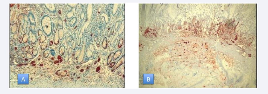
Figure 1 A: Gastric NET limitedtomucosa, 40x magnification B: Gastric NET limitedtomucosa, 10x magnification.

Gastroscopy commonly detects a single ulcerovegetant lesion with a diameter greater than 2 cm. Narrow band imaging or magnified endoscopy may provide additional benefit in detecting type I lesions smaller than 1 cm which may have a micronodular appearance. Lesions larger than 1 cm should be evaluated by endoscopic ultrasonography (EUS) for submucosal invasion or lymph node metastasis [32]. Since type II lesions tend to be locally advanced, they must be visualized by EUS, scintigraphy, CT or MRI. If these diagnostic modalities are insufficient for diagnosis of NETs and gastrinomas, PET-CT with 68 Ga-DOTANOC may have an additional contribution and can help localize gastrinomas [33]. Type III lesions often require cross-sectional imaging like in adenocarcinomas which frequently undergo distant metastases.

Conventional imaging methods such as CT, MRI, and PET / CT have been frequently used for diagnosis of primary and metastatic NETs. FDG-PET / CT may have a limited power to identify these tumors because the rate of glucose degradation of well differentiated NETs is close to normal [34].

Positron emitting radionuclide gallium 68 added to the chelator DOTA and somatostatin analogs to obtain radio labeled tracer DOTA- NOC (68Ga-DOTA,1-Nal3]-octreotide) or DOTATATE (DOTA-DPhe,1Tyr3-octreotate) for better detection of somatostatin receptor expressing tumors. Kayani et al., found 68Ga-DOTATATE PET/CT sensitivity 82% where the 18F-FDG PET/CT sensitivity 66% in a cohort of 38 patients and detection of the low-grade NETs was significantly higher than FDG PET/CT with the 68Ga-DOTATATE PET/CT [35].

68Ga-DOTA-NOC PET/CT is significantly superior identifying both primer and metastatic NETs over contrast enhanced CT. In a study where 109 patients were screened, six patients (5.5%) had chance of surgical treatment for lesions which were not exposed by conventional imaging modalities, DOTA NOC revealed additional resectable lesions therefore, 8(6.4%) patients surgical planning changed, 4 (3.6%) patients had spared from unnecessary surgery with detection of advanced disease stage [36].

68 Ga-DOTATATE has an affinity for somatostatin receptor subtype 2 whereas 68 Ga-DOTA-NOC has a broader receptor affinity for subtype 2, 3, 5and is therefore superior to the 68GaDOTATATE aspect of imaging. In a single center study from London, the lesion based sensitivity of 68Ga-DOTANOC PET was 93.5%, compared with 85.5% for 68Ga-DOTATATE PET for detecting gastroenteropancreatic NETs. (P < 0.005). DOTA peptide imaging methods also give the information about the somatostatin receptor condition of the tumors, and patients may be candidates for peptide-based radionuclide therapy or the somatostatin analogs treatments [37].

MANAGEMENT

Clinical management and treatment for gastric NETs rely upon metastasis risk which is indicated by tumor size, the presence of muscular wall infiltration, increased tumor cell proliferation. The treatment of type I and type II Gastric NETs is quite similar and in this respect, they differ from type III. Sato at al., pointed that throughout 7 years of endoscopic surveillance tumors ≤ 10 mm in diameter did not display advancement.8 tumors smaller than 10 mm among 82 was capillary invasive but none of them revealed recurrence or distant metastasis after endoscopic treatment during follow-up.(38) Uygun et al., also reported a median 7 years of follow up of type I NETs after endoscopic treatment and denoted 100% rate of overall survival rate and disease?free survival [39]. Because of slow progression and low risk of metastasis type I and type II lesions, which are smaller than 1 cm, can be taken under endoscopic surveillance annually [40]. Follow up of the small type I and type II tumors should include gastroscopy and EUS where further modalities such as octreoscan and radionuclides imaging did not reveal additional pathology over conventional imaging [41]. Lesions that are 1-2cm size should be checked with EUS primarily for the invasion of muscularispropria or local lymph node metastasis (Figure 2).

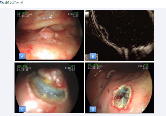
Figure 2 A: Gastric NET B: NET confinedtosubmucosadetectedby EUS C: Sub mucosal view of NET D: Lesion basis after removal of lesion

Endoscopic treatments should be considered for lesion which is limited to the mucosa and submucosa. Endoscopic mucosal resection, endoscopic mucosal resection with a ligation device or endoscopic submucosal dissection (ESD) may be selected as an endoscopic procedure, but any of them should be performed with experienced endoscopists. ESD is based on the stepwise dissection of the lesion with special endoscopic knives following injection of fluids that will form a submucosal cushion below the lesion (Figure 2). Among our ESD cases, four gastric NETs were observed and total en bloc resection was achieved in all of the cases. Li and colleagues performed 100% en bloc and complete resection in a wider series of 23 patients. The endoscopic appearance of NETs is often followed by increased capillary on the lesions. These lesions tend to bleed during ESD because of their hypervascularity and even late bleeding can be observed after 1 week [42,43].

Medical treatment options include Somatostatin analogs and the newly developed oral CCK-2 receptor antagonist Netazepide, which are reported to prevent progression of type I and type II lesions by reducing the size and number [44,45]. In a study of 5 patients who were treated with 20 mg octreotide LAR every 28 daya for gastric NETs for a year had a reduction of tumor size and the number of visible tumors was decreased by more than 50 % [46]. Significant gastric carcinoid recurrence was not observed in 12 months after patients treated with octreotide LAR [47].

Treatment with SSAs (somatostatin analogs) in type I NETs leads to an actual tumor load reduction and the decrease of serum gastrin levels suggesting anti-proliferative effect of SSA on ECL cells. Complete disappearance of the tumors at 1 year of treatment was achieved 73% of the patients [48,49].

Surgical treatment is indicated in the presence of six or more lesions where three of them are larger than 1 cm or a single lesion larger than 2 cm when muscularispropria invasion or lymph node metastasis are present [50,51]. In cases of type II gastric NETs if gastrinoma can be located, it should be removed and proton pump inhibitors should be used to prevent peptic complications. Due to the high risk of metastasis, all type III lesions that are not at an advanced disease stage should be treated according to surgical oncologic principles which surgical intervention should aim complete resection of all detectable tumor manifestations [52]. Surgery should provide partial or total gastrectomy with lymph node dissection that surgical treatment not differs from gastric adenocarcinomas. The applicability of resection with no residual tumor depends on the localization of the primary lesion, the proportions of local metastasis, the number and localization of liver metastasis. In case of liver metastases, tumor localized to one lobe or located separately in two lobes but fewer than five metastases could be fully resectable [53,54].

Metastatic lesion of neuroendocrine tumor in the liver could be curable when they are operated, and patients who had the chance of metastasectomy have 5-year survival rates of 71–85% [55,56].

Tumor burden should be reduced by metastasectomy and cytoreductive surgical methods in appropriate patients with advanced disease [57]. Ablation treatments can be used alone or in conjunction with surgery [58]. Hepatic artery embolization techniques are useful in the widespread disease where surgery cannot be performed [59]. SSAs, oral target of rapamycin (mTOR) inhibitor Everolimus, Peptide Receptor Radionuclide Therapy are the treatment modalities for systemic treatment. Mammalian target of rapamycin (mTOR) is a serine-threonine kinase that regulates cell growth, proliferation, and survival signaling reaction to metabolic stress factors and inhibition of the mTOR pathway has in vitro antiproliferative effects on NET cells [60]. Everolimus is an oral inhibitor of mTOR which had assessed in phase III trial of RADIANT-4. 302 patients with advanced, progressive, well differentiated NETs of the lung or gastrointestinal tract received either everolimus 10 mg or placebo. Median progression free survival was 11 months in the Everolimus group while it was 3.9 months patients who received placebo. Reduction in the estimated risk of progression was 52% with Everolimus [61].

Peptide receptor radionuclide therapy (PRRT) targets somatostatin receptor expressing well differentiated NETs by radio labeled somatostatin analogs with yttrium-90 or lutetium-177. DOTA used as a chelator for somatostatin and yttrium to obtain radionuclide 177Lu-DOTA0-Tyr3-octreotate. Recently a phase III trial of NETTER-1 mention that PRRT has promising results of progression free survival in patients with neuroendocrine tumors with an overall response rate of 18% [62]. In conclusion, overall 5-year survival rate of the gastric NETs is 98% for type I, 100% for type II, 60% for type III and 39% for the poorly differentiated gastric NETs [63].

ACKNOWLEDGEMENTS

Sadettin Hülagü researched and wrote the review as the first author. Hasan Y?lmaz contributed and reviewed the article.

REFERENCES

1. Oberndorfer S. Carcinoid tumors of the dunndarms. Frankfurt Z Pathol. 1907; 1: 426-432.

2. Solcia E, Kloppel G, Sobin LHX. Histological typing of endocrine tumours. Springer Science & Business Media. 2012.

3. Modlin IM, Lye KD, Kidd M. A 5-decade analysis of 13,715 carcinoid tumors. Cancer. 2003; 97: 934-959.

4. Lawrence B, Gustafsson BI, Chan A, Svejda B, Kidd M, Modlin IM. The epidemiology of gastroenteropancreatic neuroendocrine tumors. Endocrinol Metab Clin North Am. 2011; 40: 1-18.

5. Ellis L, Shale MJ, Coleman MP. Carcinoid tumors of the gastrointestinal tract: trends in incidence in England since 1971. Am J Gastroenterol. 2010; 105: 2563-2569.

6. Niederle MB, Hackl M, Kaserer K, Niederle B. Gastroenteropancreatic neuroendocrine tumours: the current incidence and staging based on the WHO and European Neuroendocrine Tumour Society classification: an analysis based on prospectively collected parameters. Endocr Relat cancer. 2010; 17: 909-918.

7. Hauso O, Gustafsson BI, Kidd M, Waldum HL, Drozdov I, Chan AK, et al. Neuroendocrine tumor epidemiology: contrasting Norway and North America. Cancer. 2008; 113: 2655-2664.

8. Kidd M, Miu K, Tang LH, Perez-Perez GI, Blaser MJ, Sandor A, et al. Helicobacter pylori lipopolysaccharide stimulates histamine release and DNA synthesis in rat enterochromaffin-like cells. Gastroenterology. 1997; 113: 1110-1117.

9. Stricker I, Tzivras D, Nambiar S, Wulf J, Liffers S-T, Vogt M, et al. Site-and grade-specific diversity of LINE1 methylation pattern in gastroenteropancreatic neuroendocrine tumours. Anticancer Res. 2012; 32: 3699-3706.

10.D’Adda T, Keller G, Bordi C, Höfler H. Loss of heterozygosity in 11q13- 14 regions in gastric neuroendocrine tumors not associated with multiple endocrine neoplasia type 1 syndrome. Lab Invest. 1999; 79: 671-677.

11.Rindi G, Luinetti O, Cornaggia M, Capella C, Solcia E. Three subtypes of gastric argyrophil carcinoid and the gastric neuroendocrine carcinoma: a clinicopathologic study. Gastroenterology. 1993; 104: 994-1006.

12.Kidd M, Gustafsson B, Modlin IM. Gastric carcinoids (neuroendocrine neoplasms). Gastroenterology Clinics of North America. 2013; 42: 381-397.

13.Bordi C. Neuroendocrine pathology of the stomach: the Parma contribution. Endocr Pathol. 2014; 25: 171-180.

14.Crosby DA, Donohoe CL, Fitzgerald L, Muldoon C, Hayes B, O’Toole D, et al. Gastric neuroendocrine tumours. Dig Surg. 2012; 29: 331-348.

15.Norton JA, Melcher ML, Gibril F, Jensen RT. Gastric carcinoid tumors in multiple endocrine neoplasia-1 patients with Zollinger-Ellison syndrome can be symptomatic, demonstrate aggressive growth, and require surgical treatment. Surgery. 2004; 136: 1267-1274.

16.Rindi G, Azzoni C, La Rosa S, Klersy C, Paolotti D, Rappel S, et al. ECL cell tumor and poorly differentiated endocrine carcinoma of the stomach: prognostic evaluation by pathological analysis. Gastroenterology. 1999; 116: 532-542.

17.Bosman FT, Carneiro F, Hruban RH, Theise ND. WHO classification of tumours of the digestive system. WHO. 2010.

18.Klimstra DS, Modlin IR, Coppola D, Lloyd RV, Suster S. The pathologic classification of neuroendocrine tumors: a review of nomenclature, grading, and staging systems. Pancreas. 2010; 39: 707-712.

19.Modlin IM, Gustafsson BI, Moss SF, Pavel M, Tsolakis AV, Kidd M. Chromogranin A--biological function and clinical utility in neuro endocrine tumor disease. Ann Surg Oncol. 2010; 17: 2427-2443.

20.Raines D, Chester M, Diebold AE, Mamikunian P, Anthony CT, Mamikunian G, et al. A prospective evaluation of the effect of chronic proton pump inhibitor use on plasma biomarker levels in humans. Pancreas. 2012; 41: 508-511.

21.Lawrence B, Gustafsson BI, Kidd M, Pavel M, Svejda B, Modlin IM. The clinical relevance of chromogranin A as a biomarker for gastroenteropancreatic neuroendocrine tumors. Endocrinol Metab Clin North Am. 2011; 40: 111-134.

22.Baudin E, Gigliotti A, Ducreux M, Ropers J, Comoy E, Sabourin JC, et al. Neuron-specific enolase and chromogranin A as markers of neuroendocrine tumours. British journal of cancer. 1998; 78: 1102- 1107.

23.Zeromski J, Nyczak E, Dyszkiewicz W. Significance of cell adhesion molecules, CD56/NCAM in particular, in human tumor growth and spreading. Folia Histochem Cytobiol. 2000; 39: 36-37.

24.Rayhan N, Sano T, Qian ZR, Obari AK, Hirokawa M. Histological and immunohistochemical study of composite neuroendocrine-exocrine carcinomas of the stomach. J Med Invest. 2005; 52: 191-202.

25.Lloyd RV. Endocrine Pathology: Differential Diagnosis and Molecular Advances. Springer Science & Business Media. 2010.

26.Ishida M, Sekine S, Fukagawa T, Ohashi M, Morita S, Taniguchi H, et al. Neuroendocrine carcinoma of the stomach: morphologic and immunohistochemical characteristics and prognosis. Ame J Surg Pathol. 2013; 37: 949-959.

27.Annibale B, Azzoni C, Corleto VD, Di Giulio E, Caruana P, D’Ambra G, et al. Atrophic body gastritis patients with enterochromaffin-like cell dysplasia are at increased risk for the development of type I gastric carcinoid. Europ J Gastroenterol Hepatol. 2001; 13: 1449-1456.

28.Ravizza D, Fiori G, Trovato C, Fazio N, Bonomo G, Luca F, et al. Longterm endoscopic and clinical follow-up of untreated type 1 gastric neuroendocrine tumours. Dig Liver Dis. 2007; 39: 537-543.

29.Delle Fave G, Kwekkeboom DJ, Van Cutsem E, Rindi G, Kos-Kudla B, Knigge U, et al. ENETS consensus guidelines for the management of patients with gastroduodenal neoplasms. Neuroendocrinology. 2012; 95: 74-87.

30.Nikoul GC, Toubanakis C, NikolaouI P, GiannatouI E, MarinouI K, Safoleasz M, et al. Gastrinomas Associated with MEN-1 syndrome: New insights for the Diagnosis and Management na Series at it Patients. Hepatogastroenterology. 2005: 52: 1668-1676.

31.Rappel S, Altendorf-Hofmann A, Stolte M. Prognosis of gastric carcinoid tumours. Digestion. 1995; 56: 455-462.

32.Singh R, Yao K, Anagnostopoulos G, Kaye P, Ragunath K. Microcarcinoid tumor diagnosed with high-resolution magnification endoscopy and narrow band imaging. Endoscopy. 2008; 40: 12.

33.Naswa N, Sharma P, Soundararajan R, Karunanithi S, Nazar AH, Kumar R, et al. Diagnostic performance of somatostatin receptor PET/ CT using 68Ga-DOTANOC in gastrinoma patients with negative or equivocal CT findings. Abdom Imaging. 2013; 38: 552-560.

34. Adams S, Baum R, Rink T, Schumm-Dräger PM, Usadel KH, Hör G. Limited value of fluorine-18 fluorodeoxyglucose positron emission tomography for the imaging of neuroendocrine tumours. Eur J Nucl Med. 1998; 25: 79-83.

35.Kayani I, Bomanji JB, Groves A, Conway G, Gacinovic S, Win T, et al. Functional imaging of neuroendocrine tumors with combined PET/ CT using 68Ga-DOTATATE (DOTA-DPhe1, Tyr3-octreotate) and 18F-FDG. Cancer. 2008; 112: 2447-2455.

36.Naswa N, Sharma P, Kumar A, Nazar AH, Kumar R, Chumber S, et al. Gallium-68-DOTA-NOC PET/CT of patients with gastroenteropancreatic neuroendocrine tumors: a prospective singlecenter study. AJR Am J Roentgenol. 2011; 197: 1221-1228.

37.Wild D, Bomanji JB, Benkert P, Maecke H, Ell PJ, Reubi JC, et al. Comparison of 68Ga-DOTANOC and 68Ga-DOTATATE PET/CT within patients with gastroenteropancreatic neuroendocrine tumors. J Nucl Med. 2013; 54: 364-372.

38.Sato Y, Imamura H, Kaizaki Y, Koizumi W, Ishido K, Kurahara K, et al. Management and clinical outcomes of type I gastric carcinoid patients: retrospective, multicenter study in Japan. Dig Endosc. 2014; 26: 377- 384.

39.Uygun A, Kadayifci A, Polat Z, Yilmaz K, Gunal A, Demir H, et al. Longterm results of endoscopic resection for type I gastric neuroendocrine tumors. J Surg Oncol. 2014; 109: 71-74.

40.Merola E, Sbrozzi-Vanni A, Panzuto F, Dambra G, Di Giulio E, Pilozzi E, et al. Type I gastric carcinoids: a prospective study on endoscopic management and recurrence rate. Neuroendocrinology. 2012; 95: 207-213.

41.Thomas D, Tsolakis AV, Grozinsky-Glasberg S, Fraenkel M, Alexandraki K, Sougioultzis S, et al. Long-term follow-up of a large series of patients with type 1 gastric carcinoid tumors: data from a multicenter study. Eur J Endocrinol. 2013; 168: 185-193.

42.Li QL, Zhang YQ, Chen WF, Xu MD, Zhong YS, Ma LL, et al. Endoscopic submucosal dissection for foregut neuroendocrine tumors: an initial study. World J Gastroenterol. 2012; 18: 5799-5806.

43.Hulagu S, Senturk O, Aygun C, Kocaman O, Celebi A, Konduk T, et al. Endoscopic submucosal dissection for premalignant lesions and noninvasive early gastrointestinal cancers. World J Gastroenterol. 2011; 17: 1701-1709.

44.Manfredi S, Pagenault M, de Lajarte-Thirouard A-S, Bretagne J-F. Type 1 and 2 gastric carcinoid tumors: long-term follow-up of the efficacy of treatment with a slow-release somatostatin analogue. Eur J Gastroenterol Hepatol. 2007; 19: 1021-1025.

45.Fossmark R, Sordal O, Jianu CS, Qvigstad G, Nordrum IS, Boyce M, et al. Treatment of gastric carcinoids type 1 with the gastrin receptor antagonist netazepide (YF476) results in regression of tumours and normalisation of serum chromogranin A. Aliment Pharmacol Ther. 2012; 36: 1067-1075.

46.Fykse V, Sandvik AK, Qvigstad G, Falkmer SE, Syversen U, Waldum HL. Treatment of ECL cell carcinoids with octreotide LAR. Scand J Gastroenterol. 2004; 39: 621-628.

47.Fykse V, Kristian Sandvik A, Lyder Waldum H. One-year follow-up study of patients with enterochromaffin-like cell carcinoids after treatment with octreotide long-acting release. Scand J Gastroenterol. 2005; 40: 1269-1274.

48.Grozinsky-Glasberg S, Kaltsas G, Gur C, Gal E, Thomas D, Fichman S, et al. Long-acting somatostatin analogues are an effective treatment for type 1 gastric carcinoid tumours. Eur J Endocrinol. 2008; 159: 475- 482.

49.Campana D, Nori F, Pezzilli R, Piscitelli L, Santini D, Brocchi E, et al. Gastric endocrine tumors type I: treatment with long-acting somatostatin analogs. Endocr Relat Cancer. 2008; 15: 337-342.

50.Basuroy R, Srirajaskanthan R, Prachalias A, Quaglia A, Ramage JK. Review article: the investigation and management of gastric neuroendocrine tumours. Aliment Pharmacol Ther. 2014; 39: 1071- 1084.

51.Borch K, Ahrén B, Ahlman H, Falkmer S, Granérus G, Grimelius L. Gastric carcinoids: biologic behavior and prognosis after differentiated treatment in relation to type. Ann Surg. 2005; 242: 64-73.

52.Lawrence B, Kidd M, Svejda B, Modlin I. A clinical perspective on gastric neuroendocrine neoplasia. Curr Gastroenterol Rep. 2011; 13: 101-109.

53.Norton JA, Alexander HR, Fraker DL, Venzon DJ, Gibril F, Jensen RT. Comparison of surgical results in patients with advanced and limited disease with multiple endocrine neoplasia type 1 and Zollinger-Ellison syndrome. Ann Surg. 2001; 234: 495-506.

54.Norton JA, Sugarbaker PH, Doppman JL, Wesley RA, Maton PN, Gardner JD, et al. Aggressive resection of metastatic disease in selected patients with malignant gastrinoma. Ann Surg. 1986; 203: 352-359.

55.Elias D, Lasser P, Ducreux M, Duvillard P, Ouellet J-F, Dromain C, et al. Liver resection (and associated extrahepatic resections) for metastatic well-differentiated endocrine tumors: a 15-year single center prospective study. Surgery. 2003; 133: 375-382.

56.Chen H, Hardacre JM, Uzar A, Cameron JL, Choti MA. Isolated liver metastases from neuroendocrine tumors: does resection prolong survival? J Am Coll Surg. 1998; 187: 88-92.

57.Mayo SC, De Jong MC, Pulitano C, Clary BM, Reddy SK, Gamblin TC, et al. Surgical management of hepatic neuroendocrine tumor metastasis: results from an international multi-institutional analysis. Ann Surg Oncol. 2010; 17: 3129-3136.

58.Mayo SC, Herman JM, Cosgrove D, Bhagat N, Kamel I, Geschwind J-FH, et al. Emerging approaches in the management of patients with neuroendocrine liver metastasis: role of liver-directed and systemic therapies. J Am Coll Surg. 2013; 216: 123-134.

59.Ruutiainen AT, Soulen MC, Tuite CM, Clark TWI, Mondschein JI, Stavropoulos SW, et al. Chemoembolization and bland embolization of neuroendocrine tumor metastases to the liver. J Vasc Interv Radiol. 2007; 18: 847-855.

60.Missiaglia E, Dalai I, Barbi S, Beghelli S, Falconi M, Della Peruta M, et al. Pancreatic endocrine tumors: expression profiling evidences a role for AKT-mTOR pathway. J Clin Oncol. 2009; 28: 245-255.

61.Yao JC, Fazio N, Singh S, Buzzoni R, Carnaghi C, Wolin E, et al. Everolimus for the treatment of advanced, non-functional neuroendocrine tumours of the lung or gastrointestinal tract (RADIANT-4): a randomised, placebo-controlled, phase 3 study. Lancet. 2016; 387: 968-977.

62.Strosberg JR, Wolin EM, Chasen B, Kulke MH, Bushnell DL, Caplin ME, et al. NETTER-1 phase III: Progression-free survival, radiographic response, and preliminary overall survival results in patients with midgut neuroendocrine tumors treated with 177-Lu-Dotatate. 2016.

63.Manfredi S, Leconte B, Uguen T, Le Sourd S, Cernon C, Morcet J, et al. Study of the therapeutic management of gastric carcinoid tumors from data of the French national cohort of neuroendocrine tumors: CARGAS study. Am Soci Clin Oncol. 2015.

Hülagü S, Y?lmaz H (2017) Gastric Carcinoid Tumors. Ann Clin Exp Metabol 2(2): 1017

Received : 25 Apr 2017
Accepted : 12 Jun 2017
Published : 14 Jun 2017
Journals
Annals of Otolaryngology and Rhinology
ISSN : 2379-948X
Launched : 2014
JSM Schizophrenia
Launched : 2016
Journal of Nausea
Launched : 2020
JSM Internal Medicine
Launched : 2016
JSM Hepatitis
Launched : 2016
JSM Oro Facial Surgeries
ISSN : 2578-3211
Launched : 2016
Journal of Human Nutrition and Food Science
ISSN : 2333-6706
Launched : 2013
JSM Regenerative Medicine and Bioengineering
ISSN : 2379-0490
Launched : 2013
JSM Spine
ISSN : 2578-3181
Launched : 2016
Archives of Palliative Care
ISSN : 2573-1165
Launched : 2016
JSM Nutritional Disorders
ISSN : 2578-3203
Launched : 2017
Annals of Neurodegenerative Disorders
ISSN : 2476-2032
Launched : 2016
Journal of Fever
ISSN : 2641-7782
Launched : 2017
JSM Bone Marrow Research
ISSN : 2578-3351
Launched : 2016
JSM Mathematics and Statistics
ISSN : 2578-3173
Launched : 2014
Journal of Autoimmunity and Research
ISSN : 2573-1173
Launched : 2014
JSM Arthritis
ISSN : 2475-9155
Launched : 2016
JSM Head and Neck Cancer-Cases and Reviews
ISSN : 2573-1610
Launched : 2016
JSM General Surgery Cases and Images
ISSN : 2573-1564
Launched : 2016
JSM Anatomy and Physiology
ISSN : 2573-1262
Launched : 2016
JSM Dental Surgery
ISSN : 2573-1548
Launched : 2016
Annals of Emergency Surgery
ISSN : 2573-1017
Launched : 2016
Annals of Mens Health and Wellness
ISSN : 2641-7707
Launched : 2017
Journal of Preventive Medicine and Health Care
ISSN : 2576-0084
Launched : 2018
Journal of Chronic Diseases and Management
ISSN : 2573-1300
Launched : 2016
Annals of Vaccines and Immunization
ISSN : 2378-9379
Launched : 2014
JSM Heart Surgery Cases and Images
ISSN : 2578-3157
Launched : 2016
Annals of Reproductive Medicine and Treatment
ISSN : 2573-1092
Launched : 2016
JSM Brain Science
ISSN : 2573-1289
Launched : 2016
JSM Biomarkers
ISSN : 2578-3815
Launched : 2014
JSM Biology
ISSN : 2475-9392
Launched : 2016
Archives of Stem Cell and Research
ISSN : 2578-3580
Launched : 2014
Annals of Clinical and Medical Microbiology
ISSN : 2578-3629
Launched : 2014
JSM Pediatric Surgery
ISSN : 2578-3149
Launched : 2017
Journal of Memory Disorder and Rehabilitation
ISSN : 2578-319X
Launched : 2016
JSM Tropical Medicine and Research
ISSN : 2578-3165
Launched : 2016
JSM Head and Face Medicine
ISSN : 2578-3793
Launched : 2016
JSM Cardiothoracic Surgery
ISSN : 2573-1297
Launched : 2016
JSM Bone and Joint Diseases
ISSN : 2578-3351
Launched : 2017
JSM Bioavailability and Bioequivalence
ISSN : 2641-7812
Launched : 2017
JSM Atherosclerosis
ISSN : 2573-1270
Launched : 2016
Journal of Genitourinary Disorders
ISSN : 2641-7790
Launched : 2017
Journal of Fractures and Sprains
ISSN : 2578-3831
Launched : 2016
Journal of Autism and Epilepsy
ISSN : 2641-7774
Launched : 2016
Annals of Marine Biology and Research
ISSN : 2573-105X
Launched : 2014
JSM Health Education & Primary Health Care
ISSN : 2578-3777
Launched : 2016
JSM Communication Disorders
ISSN : 2578-3807
Launched : 2016
Annals of Musculoskeletal Disorders
ISSN : 2578-3599
Launched : 2016
Annals of Virology and Research
ISSN : 2573-1122
Launched : 2014
JSM Renal Medicine
ISSN : 2573-1637
Launched : 2016
Journal of Muscle Health
ISSN : 2578-3823
Launched : 2016
JSM Genetics and Genomics
ISSN : 2334-1823
Launched : 2013
JSM Anxiety and Depression
ISSN : 2475-9139
Launched : 2016
Clinical Journal of Heart Diseases
ISSN : 2641-7766
Launched : 2016
Annals of Medicinal Chemistry and Research
ISSN : 2378-9336
Launched : 2014
JSM Pain and Management
ISSN : 2578-3378
Launched : 2016
JSM Women's Health
ISSN : 2578-3696
Launched : 2016
Clinical Research in HIV or AIDS
ISSN : 2374-0094
Launched : 2013
Journal of Endocrinology, Diabetes and Obesity
ISSN : 2333-6692
Launched : 2013
Journal of Substance Abuse and Alcoholism
ISSN : 2373-9363
Launched : 2013
JSM Neurosurgery and Spine
ISSN : 2373-9479
Launched : 2013
Journal of Liver and Clinical Research
ISSN : 2379-0830
Launched : 2014
Journal of Drug Design and Research
ISSN : 2379-089X
Launched : 2014
JSM Clinical Oncology and Research
ISSN : 2373-938X
Launched : 2013
JSM Bioinformatics, Genomics and Proteomics
ISSN : 2576-1102
Launched : 2014
JSM Chemistry
ISSN : 2334-1831
Launched : 2013
Journal of Trauma and Care
ISSN : 2573-1246
Launched : 2014
JSM Surgical Oncology and Research
ISSN : 2578-3688
Launched : 2016
Annals of Food Processing and Preservation
ISSN : 2573-1033
Launched : 2016
Journal of Radiology and Radiation Therapy
ISSN : 2333-7095
Launched : 2013
JSM Physical Medicine and Rehabilitation
ISSN : 2578-3572
Launched : 2016
Annals of Clinical Pathology
ISSN : 2373-9282
Launched : 2013
Annals of Cardiovascular Diseases
ISSN : 2641-7731
Launched : 2016
Journal of Behavior
ISSN : 2576-0076
Launched : 2016
Clinical Research in Infectious Diseases
ISSN : 2379-0636
Launched : 2013
JSM Microbiology
ISSN : 2333-6455
Launched : 2013
Journal of Urology and Research
ISSN : 2379-951X
Launched : 2014
Journal of Family Medicine and Community Health
ISSN : 2379-0547
Launched : 2013
Annals of Pregnancy and Care
ISSN : 2578-336X
Launched : 2017
JSM Cell and Developmental Biology
ISSN : 2379-061X
Launched : 2013
Annals of Aquaculture and Research
ISSN : 2379-0881
Launched : 2014
Clinical Research in Pulmonology
ISSN : 2333-6625
Launched : 2013
Journal of Immunology and Clinical Research
ISSN : 2333-6714
Launched : 2013
Annals of Forensic Research and Analysis
ISSN : 2378-9476
Launched : 2014
JSM Biochemistry and Molecular Biology
ISSN : 2333-7109
Launched : 2013
Annals of Breast Cancer Research
ISSN : 2641-7685
Launched : 2016
Annals of Gerontology and Geriatric Research
ISSN : 2378-9409
Launched : 2014
Journal of Sleep Medicine and Disorders
ISSN : 2379-0822
Launched : 2014
JSM Burns and Trauma
ISSN : 2475-9406
Launched : 2016
Chemical Engineering and Process Techniques
ISSN : 2333-6633
Launched : 2013
Annals of Clinical Cytology and Pathology
ISSN : 2475-9430
Launched : 2014
JSM Allergy and Asthma
ISSN : 2573-1254
Launched : 2016
Journal of Neurological Disorders and Stroke
ISSN : 2334-2307
Launched : 2013
Annals of Sports Medicine and Research
ISSN : 2379-0571
Launched : 2014
JSM Sexual Medicine
ISSN : 2578-3718
Launched : 2016
Annals of Vascular Medicine and Research
ISSN : 2378-9344
Launched : 2014
JSM Biotechnology and Biomedical Engineering
ISSN : 2333-7117
Launched : 2013
Journal of Hematology and Transfusion
ISSN : 2333-6684
Launched : 2013
JSM Environmental Science and Ecology
ISSN : 2333-7141
Launched : 2013
Journal of Cardiology and Clinical Research
ISSN : 2333-6676
Launched : 2013
JSM Nanotechnology and Nanomedicine
ISSN : 2334-1815
Launched : 2013
Journal of Ear, Nose and Throat Disorders
ISSN : 2475-9473
Launched : 2016
JSM Ophthalmology
ISSN : 2333-6447
Launched : 2013
Journal of Pharmacology and Clinical Toxicology
ISSN : 2333-7079
Launched : 2013
Annals of Psychiatry and Mental Health
ISSN : 2374-0124
Launched : 2013
Medical Journal of Obstetrics and Gynecology
ISSN : 2333-6439
Launched : 2013
Annals of Pediatrics and Child Health
ISSN : 2373-9312
Launched : 2013
JSM Clinical Pharmaceutics
ISSN : 2379-9498
Launched : 2014
JSM Foot and Ankle
ISSN : 2475-9112
Launched : 2016
JSM Alzheimer's Disease and Related Dementia
ISSN : 2378-9565
Launched : 2014
Journal of Addiction Medicine and Therapy
ISSN : 2333-665X
Launched : 2013
Journal of Veterinary Medicine and Research
ISSN : 2378-931X
Launched : 2013
Annals of Public Health and Research
ISSN : 2378-9328
Launched : 2014
Annals of Orthopedics and Rheumatology
ISSN : 2373-9290
Launched : 2013
Journal of Clinical Nephrology and Research
ISSN : 2379-0652
Launched : 2014
Annals of Community Medicine and Practice
ISSN : 2475-9465
Launched : 2014
Annals of Biometrics and Biostatistics
ISSN : 2374-0116
Launched : 2013
JSM Clinical Case Reports
ISSN : 2373-9819
Launched : 2013
Journal of Cancer Biology and Research
ISSN : 2373-9436
Launched : 2013
Journal of Surgery and Transplantation Science
ISSN : 2379-0911
Launched : 2013
Journal of Dermatology and Clinical Research
ISSN : 2373-9371
Launched : 2013
JSM Gastroenterology and Hepatology
ISSN : 2373-9487
Launched : 2013
Annals of Nursing and Practice
ISSN : 2379-9501
Launched : 2014
JSM Dentistry
ISSN : 2333-7133
Launched : 2013
Author Information X