MDMA and Rhabdomyolysis in Rats
- 1. Military Medical Academy, Institute for Pathology and Forensic Medicine, Serbia
Abstract
3,4-Methylenedioxymethamphetamine (MDMA) or Ecstasy is a psychoactive drug illegally produced and distributed throughout the world. MDMA leads to an increase in physical strength and stamina, but causes rhabdomyolysis in the skeletal musculature, with the release of intracellular content of skeletal muscles. Rhabdomyolysis is mainly linked with intensive physical labour and ischemia, but may also be caused by genetic, but also direct toxic or mechanical damage. The laboratory findings are dominated by the increase of values for creatine kinase, the increase of values for potassium and hypo and hypercalcemia.
Methods: The experiment was carried out on Wistar rats at average room temperature of 22 ± 2°C. The control group of animals received 1 ml of distilled water, whereas the experimental group received MDMA dissolved in 1 ml of distilled water, in doses of 20 mg/kg and 40 mg/kg. At the beginning of the experiment, prior to administering water and MDMA and every hour after that, the animals core temperature was measured rectally, and blood was taken for biochemical analyses in the eighth hour of the exrpeiment: creatine kinase (CK), Calcium (Ca) and Potassium (K). After eight hours, the animals were sacrificed, a sample of the skeletal muscle was taken and pathohistological material was dyed using the following methods:
Hematoxylin-eosin (HE), Phosphotungstic acid haematoxylin (PTAH) and desmin.
Results: There is a statistically significant difference in the increase of body temperature in animals that received MDMA compared to the control groupbut only in the group that received the dose of 40 mg/kg of MDMA. There is a drop in Ca level in both experimental groups compared to the control group, and the increase of K level in the group that received 40 mg of MDMA. Creatine kinase also shows a statistically significant increase only in the group that received a higher dose of MDMA. The pathohistological examination has shown individual myolysis of myofibrils with occasional fresh bleeding in all the animals that received MDMA.
Conclusion: After the application of MDMA in the 8-hour experiment, all experimental animals are dominated by myofibrils oedema, occasional striated muscle and nuclei loss, with occasional fresh bleeding, while the biochemical indicators of rhabdomyolysis, hyperkalemia and increased values of creatine kinase show statistically significant increase only in the experimental group that received the dose of 40 mg/kg of MDMA.
CITATION
Marinkovi? N, Aleksi? I (2018) MDMA and Rhabdomyolysis in Rats. Ann Forensic Res Anal 5(1): 1053.
KEYWORDS
MDMA; Rats; Rhabdomyolysis; Temperature; Creatine kinase; Calcium; Potassium.
INTRODUCTION
3,4-Methylenedioxymethamphetamine (MDMA) or Ecstasy is a psychoactive drug illegally produced and distributed throughout the world. One of the effects caused by the MDMA is the increase of physical strength and stamina, so after taking Ecstasy - a person may be alert and physically active over a longer period of time [1-4]. MDMA, however, in certain cases may cause rhabdomyolysis, with the release of intracellular content of the skeletal muscles. Due to intracellular changes in the content of muscle cells, rhabdomyolysis is characterised by oedema, grainy structure, partial loss of transverse striation and myofibril destruction [5]. The laboratory findings are dominated by the increase of value of creatine kinase, CK which is a sensitive biochemical rhabdomyolysis indicator [4]. Apart from CK, hyperkalemia and hypercalcemia also occur [4].
METHODS
The experiment was carried out on Wistar rats, male, 6 weeks old, with average weight of 258 g, with free access to food and water, at average room temperature of 22 ± 2°C. Ecstasy tablets were obtained under the authority of the Serbian Ministry of Interior and qualitatively and quantitatively analysed by infrared spectrophotometry, thin layer chromatography and gas chromatography (FTIR- 8300 i GC-17A-Shimadzu), gas chromatography with FID detector, and it has been established that the tablets contain lactose and MDMA in concentration of 15.68%. The tablets were dissolved in distilled water and the animals were divided into three groups of eight members, and two hours before the experiment were placed in the space in which they resided during the experiment. The control group of animals received 1 ml of distilled water, whereas the experimental groups received MDMA dissolved in 1ml of distilled water, in doses of 20 mg/kg and 40 mg/kg. The solution was administered perorally by a probe to all the animals. At the beginning of the experiment, prior to administering water and MDMA and every hour after that, the animals’ core temperature was measured rectally, and blood was taken for biochemical analyses in the eighth hour of the experiment. After this, ether was used on animals for anesthesia and they were sacrificed by decapitation. After the sacrification, one sample of skeletal muscle was taken from the hind right leg, fixated in 5% neutral buffered formalin, paraffin-embedded, serially cut by microtome and dyed using the following methods: Hematoxylin-eosin (HE), Phosphotungstic acid haematoxylin (PTAH) and desmin. The following biochemical analyses were done: creatine kinase, calcium and potassium. The results were processed using the descriptive statistics method.
RESULTS
The animals that received MDMA started to spike their hair and become hyperactive only after 15 minutes after administration, while the control group is sleeping. One and a half hours after the beginning of the experiment, the control group started sniffing around for food, while the experimental group, regardless of the dose of MDMA, does not sniff for food; they only move around the cage, lift themselves on their hind legs and sniff around the cage roof. After two hours, the animals that received 40 mg/kg of MDMA show that they have sweated more than the animals that received 20 mg/kg of MDMA. Two and a half hours after the beginning of the experiment, both experimental groups were sweaty, hyperkinetic, they are sniffing around the cage, while the animals that received 40 mg/kg of MDMA are moving around in circles. After four hours, the animals move less and sweat less. The control group maintains the body temperature within one degree. The temperature at the beginning of the experiment has an average value of 36.83°C, during the first hour it increased by around one degree (37.93°C) and was maintained at the approximate value for eight hours. The experimental group that received 20 mg/kg of MDMA has the body temperature of 36.78°C at the beginning of the experiment. The temperature in the very first hour reaches the value of 39.44°C, in the second hour the increase continues to 40.00°C when it becomes constant at such a high value, which has an average value of 38.43°C in the eighth hour. The experimental group that received 40 mg/ kg of MDMA shows the increase of body temperature in the first hour to 39.60°C and the temperature becomes constant at the approximate value until the seventh hour when a slow decrease in temperature starts. The maximal individual temperature in the group that received 40 mg/kg is 41.30°C measured in the second hour of the experiment, whereas in the group that received 20 mg/ kg of MDMA the maximal temperature was 41.80°C, measured in the sixth hour of the experiment. The maximal average value of temperature in the control group is 37.93°C, in the group that received 20 mg/kg of MDMA is 40.00°C, and in the group that received 40 mg/kg of MDMA is 39.95°C, where all these values are measured during the second hour of the experiment. (Table 1) The values of K show a decrease in the group that received 20 mg/ kg of MDMA, and a statistically significant increase in the group that received 40 mg/kg of MDMA.
|
Table 1: Average value of temperature and statistical significance. |
|||
|
|
0h |
2h |
SV ± SD |
|
Control SV ± SD |
36.83 ± 0.3 |
*37.93 ± 0.4 |
37.285 ± 0.455 |
|
20mg/kg SV ± SD |
36.78 ± 0.4 |
*40.00 ± 0.4 |
39.605 ± 0.825 p<0.196
|
|
40mg/kg SV ± SD |
36.50 ± 0.2
|
*39.95 ± 1.1 |
38.896 ± 0.815 p<0.00059 |
|
•the maximal average value of temperature |
|||
The values of Ca were lower in both experimental groups compared to the control groups, with statistically significant drop in the group that received 40 mg/kg of MDMA. Creatine kinase shows a drop of value in the animals that received 20 mg/kg of MDMA compared to the control group, but without statistical significance. In the animals that received 40 mg/kg of MDMA, a statistically significant increase of creatine kinase value has been found (Table 2).
|
Table 2: Average value of biochemical analyses in the blood and statistical significance. |
|||
|
Analyses
|
Potassium (K) mmol/Ll |
Calcium (Ca) mmol/L |
Creatinin kinase (CK) U/L |
|
control |
4.10 ± 0.368 |
2.968 ± 0.133 |
613.71 ± 43.12 |
|
20 mg/kg |
3.76 ± 1.1 p<0.493 |
2.68 ± 0.06 p<0.65
|
525.00 ± 34.76 p<0.637 |
|
40 mg/kg |
4.36 ± 0.35 p<0.092 |
2.635 ± 0.075 p<0.034
|
1196.00 ± 47.89 p<0.039 |
Pathohistological examination of skeletal muscle in animals from the control group has shown no pathological changes, whereas the animals from the experimental groups show non-specific changes in the form of mild oedema of myofibril, occasional striated muscle and nuclei loss, with occasional fresh bleeding between myofibril (Figure 1). Desmin dyeing shows a partial loss of desmin (Figure 1).
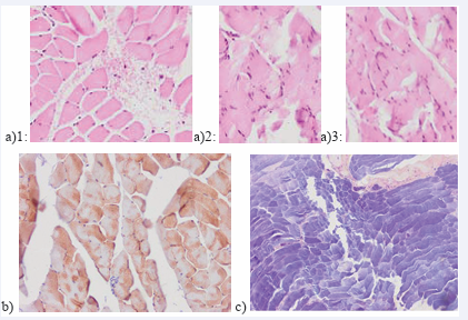
Figure 1: a)1,3: (40 mg/kg MDMA) mild oedema of myofibril, with occasional fresh bleeding between myofibril; a)2: (20 mg/kg MDMA)occasional striated muscle and nuclei loss, with occasional fresh bleeding between myofibril(HE) b) Desmin dyeing shows a partial loss of desmin (40 mg/kg MDMA) c) Oedema of myofibril and nuclei loss (PTAH- 40 mg/kg MDMA)
DISCUSSION
Rhabdomyolysis is a skeletal muscle disintegration syndrome and the presence of disintegrated muscle components in the plasma, while the mechanism of occurrence of rhabdomyolysis is multifactorial. Rhabdomyolysis is primarily linked to intensive physical work and ischemia, but may also be caused by genetic, direct toxic or mechanical damage. 3,4-Methylenedioxymethamphetamine is only one of a number (around 150) substances that may lead to rhabdomyolysis [1]. Described cases of rhabdomyolysis after the use of MDMA are rare, but there are presentations of 18 paediatric patients, fulminant rhabdomyolysis in 16-year old girl after taking 30 tablets, a woman age 37 and alike [6-9]. Miotoxic effect may be primary, such as direct effect of the toxic substance on the muscle function and structure, whereas the secondary is the consequence of pressure or trauma in the muscle due the loss of conscience, coma, etc. 1 It is believed that rhabdomyolysis after the use of MDMA occurs due to the release of catecholamine, increased body temperature and increased muscle activity [1]. The significance of hyperpyrexia is confirmed by the experiment that showed that the use of MDMA with α1 antagonist prazosin and β3 AR antagonist results in a significant reduction of body temperature growth and the increase of creatine kinase level in the blood, which may have therapeutic significance [10]. A sensitive biochemical indicator of rhabdomyolysis is the increase of creatine kinase value in the serum, and that is why some authors recommend routine determination of CK in all forms of poisoning to ensure timely therapy to prevent acute renal insufficiency and extensive rhabdomyolysis [11]. Increase of CK value occurs after 2-12 hours after muscle damage, and the peak value is achieved within three to five days [4]. Early rhabdomyolysis caused by MDMA may be a consequence of hyperthermia, increased energy for excessive physical activity and crush injuries, when the patient is unconscientious for a longer period of time [3]. The cases of rhabdomyolysis have been described 55 hours after taking MDMA in the form of increased CK values [3]. A small amount of MDMA may lead to hyperthermia and rhabdomyolysis, which is in certain cases explained by serotonin syndrome and individual sensitivity [11]. During rhabdomyolysis, intracellular K is released, which leads to hyperkalemia, which in turn may cause heart arrhythmia [1,4]. In our experiment, the hyperkalemia is evident, together with the increased CK value in animals that received a higher dosage of MDMA, which indicated that, regardless of the fact that smaller doses lead to toxic effects, higher doses of MDMA speed up the muscle cell degradation process. In the initial stage of rhabdomyolysis, Ca is accumulated in the muscles, which results in hypocalcemia [1,4]. Later, Ca is mobilized from the necrotic muscle, which leads to hypercalcemia, which in turn may explain the hypocalcemia in our experiment that lasted 8 hours [1,4].
CONCLUSION
3,4-Methylenedioxymethamphetamine leads to an increase of body temperature and to hyperactivity in rats. These two factors, combined with the effects of MDMA, lead to non-specific pathohistological changes in the skeletal muscles during the 8-hour experiment, but also to a statistically significant increase of rhabdomyolysis parameter values, creatine kinase and potassium in the blood. Increased values have been found only in animals that received a higher dose of 3,4-Methylenedioxymethamphetamine (40 mg/kg). Further research is required on rhabdomyolysis after the use of MDMA and the effect of MDMA dosage on the degree of rhabdomyolysis.


