Clinical Presentation of Intralabyrinthine Hemorrhage: A Description Based on 8 Cases
- 1. Department of Otorhinolaryngology, CHRU Hautepierre, France
- 2. Department of Otorhinolaryngology, Ain Shams University, Egypt
- 3. Department of Radiology I, CHRU Hautepierre, France
Abstract
Objectives: Identification of clinical criteria for a fast diagnosis of intralabyrinthine hemorrhage and based on an observation and analysis of clinical findings.
Material and methods: Retrospective study reporting cases of clinically presumed intralabyrinthine hemorrhage identified between 2010 and 2014. Cases were confirmed by magnetic resonance imaging (MRI) after clinical and videonystagmoscopy examination.
Results: Eight cases of intralabyrinthine hemorrhage were collected, as an acute and severe cochleovestibular deficit. A risk factor was noted in six cases. History and physical examination eliminated the main differential diagnoses, except for the internal auditory artery ischemia. 75% of patients had a complete deafness and a rotatory vertigo. Early spontaneous nystagmus was horizontorotatory, beating towards the unaffected side. Videonystagmoscopy reported 75% of atypical positional nystagmus, which may change over time. Three patients presented an initial areflexia of the lateral semicircular canal (positive Head Impulse Test), two of which associated with a silent videonystagmoscopy and poor cochleovestibular recovery. Half suffered secondarily a typical and ipsilateral benign paroxystic positional vertigo in the weeks following the incident, isolated or repetitive. Hearing improvement was rare.
Conclusion: The diagnosis of intralabyrinthine hemorrhage is predominantly suspected on clinical history and association of sudden sensorineural hearing loss and vertigo. Simple clinical findings based on videonystagmoscopy may orient the differential diagnosis between hemorrhage and ischemia. A suspected vascular damage of the inner ear with normal videonystagmoscopy and positive Head Impulse Test seems associated with poor functional outcome.
Keywords
Intralabyrinthine hemorrhage , Ischemia , Videonystagmoscopy ,MRI , Clinical criteria
Citation
Schmaltz H, Vuong H, Rohmer D, Milad P, Molière S, et al. (2016) Clinical Presentation of Intralabyrinthine Hemorrhage: A Description Based on 8 Cases. Ann Gerontol Geriatric Res 3(2): 1040.
ABBREVIATIONS
MRI: Magnetic Resonance Imagery; ILH: Intralabyrinthine Hemorrhage; ACVD: Acute Vestibulocochlear Deficit; IAA: Internal Auditory Artery; VNS: Videonystagmoscopy; VNG: Videonystagmography; VHIT: Video Head Impulse Test; BPPV: Benign Paroxystic Positional Vertigo; AICA: Anterio-Inferior Cerebellar Artery
INTRODUCTION
Intralabyrinthine hemorrhage (ILH) is a rare cause of acute vestibulocochlear deficit (AVCD). Data on this condition are scarce and controversial. ILH presents as severe sudden sensorineural hearing loss combined with a rotatory vertigo, rarely simple balance disorder. The functional prognosis is poor given cochlear symptoms, better given vestibular symptoms, due to central compensation or peripheral recovery. Hearing recovery is rarely reported [1,2]. The incidence of ILH is very rare, but uneasy to estimate. For example, the incidence of sudden sensorineural hearing loss is about 5-20 per 100,000, including all degrees of severity [3]. Association of dizziness varies from 29 to 56%. Xuan Wu and al, on a large series of MRI scans, concluded that an ILH was present in 6.25% of patients diagnosed as severe sudden sensorineural hearing loss [4]. The CT scan of the petrous bone is unable to identify ILH. Contributing factors to ILH are variable and not always identified. The use of anticoagulants is considered a classic cause [1,2,5]. Other underlying coagulopathies may include severe anemia, anti-phospholipid antibodies of lupus [6], hyper viscosity of myeloma or leukemia, acute vascular occlusion in sickle cell disease [7], and fragility of the vessels as in post-irradiation of the head and neck [8]. Factors that have been anecdotally related to ILH include endolymphatic sac tumors [9], mutation of the MethyleneTetraHydroFolate Reductase (MTHFR) gene [10], and abuse of cocaine [11]. Finally, trauma (head trauma, barotrauma, or local surgery) may precipitate ILH.
The diagnosis of ILH is difficult and involves ruling out many differential diagnoses: cochleovestibular neuritis, labyrinthine hydrops or Meniere’s disease, labyrinthitis, vestibulocochlear schwannoma and ischemia of the internal auditory artery (IAA). Basing on clinical examination, ischemia of the IAA is very difficult to distinguish from ILH, and MRI seems to be the standard for differential diagnosis [1,2,6,7,10,12]. Access to emergency MRI is limited to referral centers. The ability to recognize an ILH based on clinical observations could be important in terms of course to follow. The objective of this study is to present clinical findings and analysis from 8 cases of clinically presumed ILH, confirmed by 3T MRI, and try to identify some clinical criteria which may orient the clinician to positive and differential diagnosis.
MATERIALS AND METHODS
This retrospective study included patients with a clinically suspected ILH, confirmed by 3.0 Tesla MRI in the ENT department of the University Hospital between 2010 and 2014. Patients, who did not receive MRI and early infrared videonystagmoscopy (VNS) evaluation, were excluded. MRIs were examined by specialized radiologists in our center. The MRI diagnostic criteria for ILH were spontaneous hyper intense signal of vestibulocochlear structures in T1 and FLAIR sequences, without enhancement after contrast agent injection.
VNS was systematically performed in the first two days, positioning the patient in the plane of the 6 semicircular canals, with a mask allowing a total darkness for the patient and with a screen for the practitioner.VNS evaluation is looking for spontaneous and induced nystagmus (Figure 1).
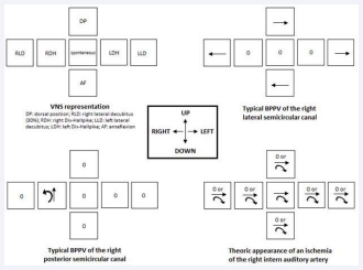
Figure 1 Presentation of VNS results. By convention, the central box of each cross represents spontaneous nystagmus, the patient sitting in front of the clinician. The left boxes represent positional nystagmus in left Dix Hallpike and left lateral decubitus positions. The right boxes represent positional nystagmus in right positions. The upper box represents the 30° dorsal decubitus, and the bottom box represents the anteflexion position. The scheme finally represents a facing patient: a left arrow on the scheme represents a right horizontal nystagmus by definition, and vice versa. A clockwise direction arrow on the schema represents a left rotatory nystagmus, and vice versa. The three last schemes represent the typical positional nystagmus in case of typical BPPV of the right lateral and posterior semicircular canals, and the theoretic appearance of an ischemia of the right intern auditory artery. Abbreviations: VNS: Videonystagmoscopy; BPPV: Benign Paroxystic Positional Vertigo
Data were collected in medical reports. They included past medical history, affected side, initial symptoms, the delay period before specialized consultation, clinical findings as the presence of an initial spontaneous nystagmus, neurological examination, the results of the horizontal Head Impulse Test (testing middle and high frequencies), results of VNS, and the initial pure tone audiometry and videonystagmography (VNG) which was performed within days or weeks after the incident (testing low frequencies). VHIT (Video Head Impulse Test), testing the 6 semicircular canals, was not performed because unavailable in our center at this moment.
RESULTS
8 patients (6 females and 2 men) presented an ILH, in approximately 55 admitted for severe sudden hearing loss, vertigo and MRI. The mean age was 57.9 years (range: 27-80 years).
History examination
The average time before specialized consultation was 6.3 days (0.5-15).Three patients had a diagnosis of hypertension. Four patients were on antiplatelet or anticoagulant therapy, only one was overdosed. In two cases, no known risk factor was identified.
No patient suffered from an autoimmune disease. No patient had medical history of sudden sensorineural hearing loss and vertigo. All patients reported an episode of sudden unilateral hearing loss, associated with ipsilateral tinnitus in five cases and an ipsilateral clogged ear sensation in four cases (Table 1). A true rotatory vertigo was reported in six cases. Two patients reported simple balance disorders. Symptoms shifted in three patients (hearing loss 24-48 hours before vertigo).
Table 1: Initial clinical reports of the 8 patients.
| Patient | Risk Factor | Sudden unilateral Hearing loss | Initial audiometric loss | Rotatory Vertigo | Tinnitus | Clogged ear | Initial spontaneous nystagmus (first consultation) | Initial h-HIT |
| 1 | Hypertension | + | Complete | + | + | 0 | 0 | Unknown |
| 2 | ACT | + | Mild | + | + | + | 0 | Unknown |
| 3 | Hypertension, ACT | + | Complete | + | 0 | 0 | + | + |
| 4 | Hypertension | + | Complete | + | + | + | + | 0 |
| 5 | ACT | + | Complete | Simple balance disorders | 0 | + | 0 | + |
| 6 | 0 | + | Complete | + | + | 0 | + | + |
| 7 | 0 | + | Complete | Simple balance disorders | + | 0 | 0 | 0 |
| 8 | Antiplatelettherapy | + | Severe | + | 0 | + | 0 | 0 |
Physical examination
Body temperature, tympanic membrane and neurologic examination were normal in every case. A fistula test was negative. Only three patients, whose consultation period was less than 5 days, presented with initial nystagmus (horizontal or horizontorotatory) beating towards the healthy side. The condition resolved within days, except in one case. Significant segmental deviation was present in 50% of cases. Horizontal Head Impulse Test (h-HIT or Halmagyi maneuver) was positive on the affected side in three patients out of six tested. VNS evaluation discovered atypical positional nystagmus were discovered in 6 patients (75%), combined with dizziness (Figure 2).
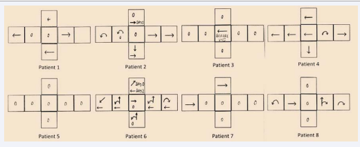
Figure 2 VNS results of the eight patients. Convention of the cross scheme is presented on Figure (1). Diagonal arrows represent oblique nystagmus. In cases 2 and 6, positional nystagmus changed according to the day of examination (day 0, 1 or 2).
Further evaluation
The reported symptoms were consistent with audiometry: six patients had complete deafness, one a severe sudden sensorineural hearing loss and one mild a hearing loss (30 dB, 50 dB scotoma at 500 Hz). When VNG was performed, two patients had areflexia in caloric tests, i.e. no response after 30°C and 15°C irrigation. Four patients showed vestibular unilateral weakness (mean 70%; 60-80), half of which were compensated (VNG was done on the 10th and 30th day from the incident, respectively). The other two patients who were not centrally compensated had spontaneous nystagmus and their VNG was done early, on the 5th and 6th day from the incident, respectively.
3.0 Tesla MRI was performed for all patients, 1 to 16 days after initial symptoms (mean 4 days) (Figure 3).
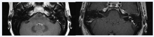
Figure 3 MRI of a left ILH: spontaneous hyperintense signal in T1 (left) and FLAIR (right) sequences.
Unilateral ILH was found on MRI in 8 cases, always ipsilateral to cochlear symptoms. No case of multiple etiologies is reported. All patients were treated by 48 hours of intravenous corticosteroids and oral relay, for a total period of 7-10 days.
Clinical evolution
The mean clinical follow-up in this case series was 8.1 months (range: 1-36 months). Over the course of follow-up, hearing improved in both patients who didn’t have initial complete hearing loss, though recovery was minimal and limited to low frequencies. Vertigo always improved, despite the persistence of minimal balance disorders in seven subjects. One patient still has recurrent episodes of vertigo poorly explained, presented by crises. Half of the patients suffered typical and ipsilateral benign paroxystic positional vertigo (BPPV) in the months following the incident, isolated or repetitive. Two of them were BPPV of the lateral semicircular canal, one of the posterior semicircular canal, and one of both. A recovery in the HIT was evident in two cases out of the three that were initially pathological. The clinical course of the VNG among these patients cannot be described due to inconsistent measurement during the follow-up period.
DISCUSSION
ILH is a rare but severe AVCD, combining deafness (75% of the series) to a vestibular deficit usually responsible for a severe initial rotatory vertigo. Vestibular symptoms vary from 21 to 92% in literature [4,13]. Hearing recovery is rare and limited to mild impairment and low frequencies [2,13]. In 75% of our patients, VNS objectified atypical positional nystagmus. Hemorrhagic risk factor was found in most cases, but no obvious etiology. To our knowledge, this retrospective study is the first trying to identify some original clinical criteria which may orient to positive and differential diagnosis of ILH, before 3T MRI; positional nystagmus in VNS as well as h-HIT are very uncommonly reported [2].
The diagnosis of ILH involves ruling out some differential diagnoses. Emergencies are vascular diseases (AICA or IAA ischemia), labyrinthitis and fistula to a functional degree. Clinical background is essential (Table 2).
Table 2: Main otologicdifferential diagnoses of ILH.
| Context | Evolution | Cochlear symptoms | Emergency | Imaging | |
| Ischemia | Thrombo-embolism risk factors | Unfavorable | (+) | (+++) | CT, MRI |
| Meniere’s disease | 0 | Recurrences | Low frequencies, initial recovery | (-) | +/- MRI |
| BPPV | 0 | Favorable | (-) | (-) | 0 |
| Labyrinthitis | Fever, otitis, autoimmune disease | (-) | (+) | (++) | MRI +/- CT |
| Schwannoma | 0 | Mostly progressive, fluctuating | (+/-) | (+/-) | MRI |
| Fistula | Head, surgical or baro-trauma | Fluctuating | (+) | (++) | CT |
| Abbreviations: BPPV: Benign Paroxystic Positional Vertigo; MRI: Magnetic Resonance Imagering; CT: CT Scan | |||||
Labyrinthitis (viral, bacterial, postoperative or autoimmune) needs to be identified with a clear cause (fever, acute otitis). Autoimmune labyrinthitis is a complex differential diagnosis, but hearing loss is bilateral,
Fluctuating and progressive, and often occurs in patients with other systemic involvement or biological autoimmunity. It often responds to a corticosteroid or immunosuppressive therapy [14].
Labyrinthine fistula often presents a traumatic context and a fluctuating deafness. Facing a combination of vertigo and severe deafness, vestibular neuritis and BPPV can be eliminated a priori. Ménière’s disease mostly affects low frequencies first, with a high rate of auditory recovery and recurrences.
Vestibular or vestibulocochlear schwannoma can present with many clinical aspects of ILH [15]. Symptoms are generally dissociated and progressive. Some other central causes have to be eliminated facing a sudden sensorineural hearing loss and vertigo, as arachnoid cysts, multiple sclerosis, hyperleukocytosis with abnormal microvascular perfusion for example. MRI is essential for this diagnosis.
Ischemia of the IAA remains a complex but urgent diagnosis to eliminate, suffering from the lack of comparative study.
When a patient presents with an AVCD, fast detection of vascular cause is imperative, whether central or peripheral, ischemic or hemorrhagic. The priority is to distinguish peripheral from central causes. The distinction between hemorrhagic and ischemic damage is certainly important, but prognosis and management of both entities are not strictly codified so far. In our opinion, a cardiologic consultation should be imperative, since a stroke of the inner ear (ischemic or hemorrhagic), like any other organ, reflects cardiovascular morbidity.
Specific clinical and radiological data about strict inner ear ischemia are rare. Occlusion of the IAA is not evident in MRI, unlike the ILH. Clinical orientation may be contributive, but we did not gather enough ischemia cases to compare clinical findings of both entities. The IAA, also called labyrinthine artery, is usually a branch of the antero-inferior cerebellar artery (AICA). Infarction of the AICA is usually associated when a labyrinthine infarction occurs [16], and Lee and al reported that 92% of patients with AICA infarction show labyrinthine infarction [17]. It means that IAA ischemia could be associated with AICA symptoms. AICA occlusion is exceptional and associates inconstant symptoms as deafness, vertigo, dysarthria, hemiataxia Isolated cochleovestibular symptoms are scarce. A systematic neurological examination is essential to eliminate central etiologies of cochleovestibular deficit.
The cochlea is an organ with no collaterals, supplied by a branch of the AICA. A complete and definitive occlusion of the IAA leads to total deafness and vestibular areflexia, but consequences of segmental ischemia are not well known. More proximal occlusions may result in more extensive ischemic lesions and consequent symptoms. A permanent ischemia causes necrosis of the membranous labyrinth of the supplied territory, followed by fibrosis and ossification in the following weeks. The possibility of reversal of the vascular lumen is questionable: an occlusion of the IAA for 60 seconds results in a drop of cochlear potentials, which recover completely if the occlusion is lower than 8 minutes, but disappear irretrievably if the occlusion lasts beyond 60 minutes [19]. We can hypothesize that small emboli in the inner ear vascularity could cause a transient cochlear ischemia, with a possible recovering in case of reperfusion. Association of a typical BPPV and a sudden sensorineural hearing loss is described in recent studies [20]. It may reflect an inner ear suffering from viral or ischemic cause. For example, a transient ischemia of the IAA may be insufficient for a total and definitive areflexia. Transient occlusion of the cochleovestibular artery may explain the cochlear dysfunction; occlusion of the anterior vestibular artery may be responsible of utricular damage and BPPV.
Common clinical tools are used in our department
Horizontal Head Impulse Test or Halmagyi test: tested in the plane of the lateral semicircular canal in 6 patients (h-HIT), it was initially positive in 3 patients. 2 patients recovered a normal h-HIT a few weeks later, which could be explained by an initial functional stunning linked to the hemorrhagic stroke. This maneuver does not evaluate complete vestibular function i.e. only high frequencies, as opposed to the caloric test, which assesses low frequencies. One patient in our series had both positional nystagmus and positive h-HIT, meaning that at least one part of the vestibule was functional. The positivity of this sign denotes areflexia of the lateral canal at high frequencies, but does not sign a total or definitive vestibular areflexia. The test is very sensitive to the presence of a severe paresis, but doesn’t detect moderate vestibular weakness [21]. h -HIT is rarely positive in case of stroke [22]. Anyway, it is a very useful bedside test. VNS: this simple test is looking for induced nystagmus in the positional maneuvers of every semi-circular canal. 75% of our patients had atypical positional nystagmus, which implies residual vestibular activity. In 1973, Fluur proved the need of residual canalicular function to produce nystagmus by showing that its experimental destruction leads to the disappearance of nystagmus [23]. In both cases of silent VNS (assumption loss of vestibular function), the horizontal HIT was positive: this combination may reflect major vestibular damage. Besides, those two patients sustained their initial hearing losses and a greater than 80% vestibular unilateral weakness. A suspected ILH with normal VNS and positive HIT/VHIT could be associated with poor functional outcome: no positional nystagmus in VNS (destruction of the labyrinthine function according to Fluur) and areflexia of the lateral canal in high frequencies (HIT). Associated with a profound initial hearing loss, would it be a poor prognosis factor? Lee and al. recently reported 12 cases of ILH; they found out that the abrupt onset of hearing loss is a poor prognostic factor, and that ILH is general has definitely a poor prognosis [13].
VHIT: it usually completes the HIT and analyses the 6 canals. Our experience of this test is very recent in our center. Pezier and al reported a case of left ILH [2]. The VHIT showed damage of the left lateral canal only, associated with obvious unilateral weakness in caloric testing. VEMP (Vestibular Evoked Myogenic Potential), testing the otolith organs, showed utriculo-saccular functional impairment. Combinations of vestibular tests are necessary to specify the vestibular dysfunction. Half of our series suffered some weeks later from BPPV on the affected side, isolated or repetitive. One patient still has recurrent episodes of vertigo poorly explained, presented by crises, which may correspond to a labyrinthine hydrops. The ILH could exacerbate these episodes by clotting and by a modification of fluid density and pressure in the inner ear, or changes in the ionic distribution. Kim and al created an experimental endolymph model, added to a blood sample [24]. Density of blood is greater than density of endolymph, so blood products were clotted at the bottom of the tube. On the fifth day, the red blood cells were hemolyzed. However, our radiologists noticed MRI signal abnormalities for up to 7 months in some of our patients. Similarly, Schuknecht noted a prolonged lifespan of red blood cells in fluids of the inner ear after cadaveric dissection [25]. Could these distant BPPV be related to persistent endolymphatic blood clots or debris? BPPV could also be generated by the release of otoconia from the suffering macula utriculi. Dissections on cadavers having suffered a hemorrhagic diathesis revealed perilymphatic more than endolymphatic compartment involvement [26]. Hemorrhage would occur initially in the perilymph space, only reaching the endolymph in case of rupture of the membranous labyrinth. A small ILH of the perilymph space would be better tolerated than a massive ILH with endolymphatic effusion [26,27]. Some hearing recoveries could match this situation, as we noticed in two cases. According to Fluur, canal pathway is spontaneously inhibited by otolith pathway [23]. In case of perilymphatic hemorrhage, nerve endings in this space can be damaged, which may explain disinhibition and positional nystagmus. Macular damage is also possible. 3T MRI is a sensitive and specific tool. In healthy subjects, fluids of the inner ear display an isointense signal in T1 weighted and FLAIR sequences, variable signals in T2 weighted images and are not enhanced in the T1 sequence after gadolinium injection. In patients with ILH, there is a spontaneous hyperintense signal in T1 sequence after 48 hours, that is not enhanced by contrast injection, a hyperintense signal on FLAIR and a variable signal in T2 [1,2,6,7,10]. This investigation eliminates the main differential diagnoses for ILH; only lipoma, infection, and labyrinthitis show spontaneous hyperintense signal in T1. It is therefore essential to achieve a T1 sequence before contrast injection [10,12]. Although MRI is the gold standard to detect ILH, the clinician can meet obstacles. 3T MRI access is often limited to referral centers, especially in emergency; however, ILH diagnosis should be made quickly. MRI is noisy and could theoretically worsen the auditory condition. False negative can occur. Indeed, the hyperintense signal in T1 appears only after 48 hours because of the production of met-hemoglobin [4,12], thus early MRI may give false negative results [24]. Later, the FLAIR sequence becomes indispensable, as the hyperintense T1 signal decreases after a few weeks due to the resorption of met-hemoglobin [4,5]. From a clinical viewpoint, an early clinical orientation could orient MRI interpretation and even make the diagnosis before MRI in some cases, to adapt medical care. Data from our series are insufficient to do without MRI for the moment, but physician may already move towards it.
Our findings lead to some hypotheses. In case of sudden unilateral loss of cochleovestibular functions, priority is to eliminate central etiologies. Then, interpreting clinical signs could maybe help distinguishing between cochleovestibular ischemia and ILH, whose management may be different (Table 3). Ischemia of the IAA may reflect a more severe ischemic stroke.
Table 3: Hypothesis for vascular damage.
| Clinicalfindings | Probable diagnosis |
|
Sudden hearing loss + spontaneous initial nystagmus + atypical positional nystagmus |
ILH |
|
Sudden hearing loss + spontaneous initial nystagmus + silent VNS ? |
Severe ILH |
|
Sudden hearing loss +/- cerebellar or neurologic symptoms + spontaneous initial nystagmus + silent VNS |
Fixed IAA Ischemia |
|
Sudden hearing loss + BPPV? |
Transient / Focal IAA Ischemia |
| Abbreviations: ILH: Intralabyrinthine Hemorrhage; VNS: Videonystagmoscopy; BPPV: Benign Paroxystic Positional Vertigo; IAA: Internal Auditory Artery | |
Our series failed to find strong clinical criteria of ILH, due to a small size and the lack of comparison with an IAA ischemia series. Converging arguments may guide the diagnosis. First, risk factors are different. Hemorrhagic damage seems rather unsystematic (VNS with atypical positional nystagmus, variable h-HIT and caloric responses) and unstable (possible partial recovery). We could hypothesize that a fixed ischemic injury could be more systematic (function of the affected branch; spontaneous initial nystagmus without positional nystagmus) and/or fixed (irreversible cell necrosis in case of fixed occlusion). Due to serious lack of data, these hypotheses must be supported by new prospective and comparative studies (ischemia versus hemorrhage).
CONCLUSION
ILH is a rare condition. 3 Tesla MRI of the ear and/or brain is the gold standard to rule out the main differential diagnoses and confirms ILH after 48-72 hours. It provides an increase in sensitivity for identification of the structural integrity of the inner ear- both cochlea and vestibular labyrinth. However, ILH may be suspected clinically before MRI. The clinical diagnosis of ILH is based on clinical history and correlation with association of sensorineural hearing loss and vertigo. Clogged ear and tinnitus are recommended to be included in reports of all patients with sensorineural hearing loss. Clinical differential diagnosis with IAA ischemia is still tough, but some arguments may orient the clinician towards ILH: risk factors for hemorrhage, atypical positional nystagmus in VNS (nonsystematic, nor fixed), better recovering in case of normal h-HIT. The small size of our series prevents strong conclusions, but our patients with an AVCD, vascular risk factors, positive HIT and normal VNS seem to have a poor hearing prognosis. Such a scenario may be indicative of a severe ILH or vestibulocochlear ischemia. It seems essential to detect vascular damages of the inner ear, as it may reflect a general risk of ischemia and even announce a central ischemic stroke. Largest prospective studies should be led to specify these findings.
REFERENCES
1. Salomone R, Abu TA, Chaves AG, Bocalini MCC, Vicente A de O, Riskalla PE. Sudden hearing loss caused by labyrinthine hemorrhage. Braz J Otorhinolaryngol. 2008; 74: 776-779. 2. Pézier T, Baráth K, Hegemann S. Partial Recovery of Audiological, Vestibular, and Radiological Findings following Spontaneous Intralabyrinthine Haemorrhage. Case Rep Otolaryngol. 2013; 2013: 941530. 3. Kuhn M, Heman-Ackah SE, Shaikh JA, Roehm PC. Sudden sensorineural hearing loss: a review of diagnosis, treatment, and prognosis. Trends Amplif. 2011; 15: 91-105. 4. Wu X, Chen K, Sun L, Yang Z, Zhu Y, Jiang H. Magnetic resonance imaging-detected inner ear hemorrhage as a potential cause of sudden sensorineural hearing loss. Am J Otolaryngol. 2014; 35: 318-323. 5. Naganawa S, Ishihara S, Iwano S, Sone M, Nakashima T. Detection of presumed hemorrhage in the ampullar endolymph of the semicircular canal: a case report. Magn Reson Med Sci. 2009; 8: 187-191. 6. Sugiura M, Naganawa S, Teranishi M, Sato E, Kojima S, Nakashima T. Inner ear hemorrhage in systemic lupus erythematosus. Laryngoscope. 2006; 116: 826-828. 7. Whitehead RE, MacDonald CB, Melhem ER, McMahon L. Spontaneous labyrinthine hemorrhage in sickle cell disease. AJNR Am J Neuroradiol. 1998; 19: 1437-1440. 8. Poh AC, Tan TY. Sudden deafness due to intralabyrinthine haemorrhage: a possible rare late complication of head and neck irradiation. Ann Acad Med Singapore. 2007; 36: 78-82. 9. Jagannathan J, Butman JA, Lonser RR, Vortmeyer AO, Zalewski CK, Brewer C, et al. Endolymphatic sac tumor demonstrated by intralabyrinthine hemorrhage. Case report. J Neurosurg. 2007; 107: 421-425.
10.Braverman I, Ben David J, Shupak A. MTHFR polymorphism: associated intralabyrinthine hemorrhage. Otolaryngol Head Neck Surg. 2009; 141: 541-542. 11.Nicoucar K, Sakbani K, Vukanovic S, Guyot JP. Intralabyrinthine haemorrhage following cocaine consumption. Acta Otolaryngol. 2005; 125: 899-901. 12.Vellin JF, Bozorg Grayeli A, Cyna-Gorse F, Refass A, Bouccara D, Sterkers O. Labyrinthine hemorrhage caused by anticoagulant therapy. Ann Otolaryngol Chir Cervicofac. 2005; 122:194-197. 13.Lee JW, Park YA, Park SM, Kong TH, Park SY, Bong JP, et al. Clinical Features and Prognosis of Sudden Sensorineural Hearing Loss Secondary to Intralabyrinthine Hemorrhage. J Audiol Otol. 2016; 20: 31-35. 14.McCabe BF. Autoimmune sensorineural hearing loss. Ann Otol Rhinol Laryngol. 2007; 116: 875-879. 15.Lee SH, Choi SK, Lim YJ, Chung HY, Yeo JH, Na SY, et al. Otologic manifestations of acoustic neuroma. Acta Otolaryngol. 2015; 135: 140-146. 16.Amarenco P, Rosengart A, DeWitt LD, Pessin MS, Caplan LR. Anterior inferior cerebellar artery territory infarcts. Mechanisms and clinical features. Arch Neurol. 1993; 50: 154-161. 17.Lee H, Ahn B-H, Baloh RW. Sudden deafness with vertigo as a sole manifestation of anterior inferior cerebellar artery infarction. J Neurol Sci. 2004; 222: 105–107. 18.Pardal Refoyo JL, Pérez Plasencia D, Beltrán Mateos LD. Ischemia of the anterior vestibular artery (Lindsay-Hemenway syndrome). Review and comments. Acta Otorrinolaringológica Esp. 1998; 49: 599-602. 19.Tabuchi K, Kusakari J, Ito Z, Takahashi K, Wada T, Hara A. Effect of nitric oxide synthase inhibitor on cochlear dysfunction induced by transient local anoxia. Acta Otolaryngol. 1999; 119: 179-184. 20.El-Saied S, Joshua B-Z, Segal N, Kraus M, Kaplan DM. Sudden hearing loss with simultaneous posterior semicircular canal BPPV: possible etiology and clinical implications. Am J Otolaryngol. 2014; 35: 180- 185. 21.Beynon GJ, Jani P, Baguley DM. A clinical evaluation of head impulse testing. Clin Otolaryngol Allied Sci. 1998; 23: 117-122. 22.Newman-Toker DE, Kattah JC, Alvernia JE, Wang DZ. Normal head impulse test differentiates acute cerebellar strokes from vestibular neuritis. Neurology. 2008; 70: 2378-2385. 23.Fluur E, Siegborn J. Interaction between the utricles and the vertical semicircular canals. Bilateral selective sectioning of anterior or posterior ampullar nerves or unilateral selective sectioning of the two vertical ampullar nerves, followed by tilting around their bitemporal or longitudinal axis. Acta Otolaryngol. 1974; 77: 80-85. 24.Kim CH, Shin JE, Park HJ, Koo JW, Lee JH. Concurrent posterior semicircular canal benign paroxysmal positional vertigo in patients with ipsilateral sudden sensorineural hearing loss: is it caused by otolith particles. Med Hypotheses. 2014; 82: 424-427. 25.Schuknecht HF. Deafness following blows to the head: a clinical and experimental study. Am Acad Ophthalmol Otolaryngol. 1951; 55: 407- 417. 26.Schuknecht HF, Igarashi M, Chasin WD. Inner ear hemorrhage in leukemia. A case report. Laryngoscope. 1965; 75: 662-668. 27.Lee HY, Jung SY, Park MS, Yeo SG, Lee SY, Lee SK. Feasibility of threedimensional fluid-attenuated inversion recovery magnetic resonance imaging as a prognostic factor in patients with sudden hearing loss. Eur Arch Otorhinolaryngol. 2012; 269: 1885-1891.


