Histopathological Disease Activity Predicts Early Consolidation after Arthrodesis in Charcot Neuroosteoarthropathy: A Retrospective Cohort Study
- 1. Sektion für Diabetische und Neuropathische Fußerkrankungen, Zentrum für Fuß- und Sprunggelenkschirurgie, Schönklinik München, Deutschland, Germany
- 2. Klinik für Orthopädie und Unfallchirurgie, Albert-Ludwigs-Universität Freiburg, Germany
- 3. MVZ for Histology, Cytology and molecular Diagnostic Trier GmbH, Germany
- 4. Privat Orthopedic Doctor´s office Seintsch Illgner, Koblenz, Germany
- 5. MUM – Muskuloskelettales Universitätszentrum München, Ludwig-Maximilians Universität München, München, Deutschland, Germany
- 6. Orthopädische Klinik König-Ludwig-Haus, Justus-Maximilians-Universität Würzburg, Würzburg, Deutschland, Germany
- *. These two authors are contributed equally
abstract
Charcot neuroosteoarthropathy (CN) is a progressive destructive disorder of the foot and ankle most commonly associated with diabetic neuropathy [1-28]. Advanced disease stages may lead to severe deformity, joint instability, ulceration, and an increased risk of lower limb amputation [1-29].
Keywords
• Charcot Neuroosteoarthropathy
• Diabetic Foot
• Arthrodesis
• Histopathology
• Bone Fusion
• Prognostic Factors
Citation
Mehlhorn AT, Huber V, Krenn V, Illgner U, Walther M. (2026) Histopathological Disease Activity Predicts Early Consolidation after Arthrod esis in Charcot Neuroosteoarthropathy: A Retrospective Cohort Study. Ann Sports Med Res 13(1): 1239.
INTRODUCTION
Charcot neuroosteoarthropathy (CN) is a progressive destructive disorder of the foot and ankle most commonly associated with diabetic neuropathy [1-28]. Advanced disease stages may lead to severe deformity, joint instability, ulceration, and an increased risk of lower limb amputation [1-29].
Charcot neuroosteoarthropathy (CN) is a progressive destructive disorder of the foot and ankle most commonly associated with diabetic neuropathy [1-28]. Advanced disease stages may lead to severe deformity, joint instability, ulceration, and an increased risk of lower limb amputation [1-29].
However, clinical overlap between stages is frequently observed, and radiographic findings do not always reflect the underlying biological activity of the disease process [29-31]. Recent reviews emphasize the multifactorial pathophysiology of Charcot neuroosteoarthropathy and highlight the limited availability of objective biomarkers to guide surgical timing [30,31].
The histopathological Charcot Score (HCS) was introduced to quantify inflammatory, degenerative, and reparative tissue changes in intraoperative specimens [7]. The score evaluates histological features of bone, cartilage, synovial tissue, and inflammatory infiltration and provides a structured assessment of local tissue-level activity [7,8].
Although the HCS provides face validity as a histological activity marker, its potential prognostic value for postoperative outcomes has not yet been investigated.
The aim of the present study was therefore to determine whether histopathological activity quantified by the HCS correlates with early clinical and radiographic outcomes following hindfoot and ankle arthrodesis in patients with CN.
We hypothesized that lower histopathological activity would be associated with improved osseous consolidation and better functional outcome.
BACKGROUND
The optimal timing of reconstructive surgery in Charcot neuroosteoarthropathy (CN) remains controversial,particularly when clinical signs of disease activity overlap with later stages of consolidation. The histopathological Charcot Score (HCS) quantifies inflammatory and reparative tissue changes in intraoperative specimens; however, its prognostic relevance remains unestablished. This study investigated whether histopathological activity correlates with early clinical and radiographic outcomes following hindfoot and ankle arthrodesis for CN.
METHODS
A retrospective cohort study including 62 patients undergoing corrective internal arthrodesis for CN between 2017 and 2023 was performed. Intraoperative tissue samples were analyzed using the HCS (range 0–21). Clinical outcome was assessed six months postoperatively using a modified Reinker–Carpenter grading system. Early osseous consolidation was evaluated by computed tomography at three months using a standardized fusion score (0–6 points). Associations between HCS and outcome parameters were analyzed using Spearman’s correlation. Multivariable regression analysis assessed potential confounding effects of age and sex.
MATERIALS AND METHODS
Study design
A retrospective cohort study was conducted including consecutive patients who underwent corrective internal arthrodesis for Charcot neuroosteoarthropathy between 2017 and 2023. Institutional review board approval was obtained prior to data collection.
Patients
Inclusion criteria were:
• Confirmed diagnosis of Charcot neuroosteoarthropathy
• Eichenholtz stage II or III clinically and radiographically
• Corrective hindfoot or ankle arthrodesis
• Availability of intraoperative tissue specimen
• Minimum follow-up of six months
A total of 62 patients met the inclusion criteria. Mean patient age was 63.5 ± 11.6 years. Twenty-four patients were female and thirty-eight were male.
Because of the retrospective study design, detailed metabolic parameters such as HbA1c levels, smoking status, renal function, and vascular status were not consistently available for all patients and could therefore not be incorporated into multivariable adjustment.
Histopathological Charcot Score (HCS)
Intraoperative tissue samples were processed according to standardized histopathological protocols.
The histopathological Charcot Score (HCS) evaluates inflammatory, degenerative, and reparative tissue changes in intraoperative specimens and has previously been described by Illgner et al. [7]. The conceptual framework of semi-quantitative histopathological scoring systems has previously been applied in musculoskeletal pathology and osteomyelitis diagnostics [8,9].
The HCS evaluates seven histological parameters representing inflammatory, degenerative, and reparative tissue changes. Each parameter is scored from 0 to 3, resulting in a total score ranging from 0 to 21.
Activity levels were categorized as:
• 0–6 points: low activity
• 7–13 points: moderate activity
• 14–21 points: high activity
No patients in the present cohort demonstrated high activity scores.
It should be noted that the original HCS publication did not correlate histopathological findings with Eichenholtz stage. Therefore, HCS should be interpreted as a measure of tissue-level biological activity rather than a radiographic staging surrogate.
Clinical Outcome
Clinical outcome was assessed six months postoperatively using a modified Reinker–Carpenter grading system ranging from 0 to 3 points. This score evaluates pain, deformity correction, and functional capacity.
Radiographic Outcome
Computed tomography (CT) has been shown to provide a more reliable assessment of hindfoot fusion than conventional radiographs and is widely used for evaluating arthrodesis consolidation [11-13].
Computed tomography (CT) imaging was performed three months postoperatively according to institutional protocol.
Fusion was assessed at three joints:
• First tarsometatarsal joint
• Talonavicular joint
• Calcaneocuboid joint
Each joint was scored from 0 to 2 points depending on the degree of osseous bridging (>50%, 25–50%, <25%), resulting in a maximum fusion score of 6 points.
The three-month CT evaluation reflects early consolidation rather than definitive fusion.
Statistical Analysis
Clinical outcome scores were ordinal in nature; therefore, non-parametric statistics were applied. Spearman correlation coefficients were calculated to assess monotonic associations between HCS and outcome parameters. To contextualize effect size, the proportion of explained variance (r²) was calculated.
Multivariable linear regression model were constructed to evaluate whether HCS remained associated with outcome measures after adjustment for age and sex. Statistical significance was defined as p < 0.05.
RESULTS
Patient Characteristics
Median HCS was 7 (range 4–13). Twenty-nine patients demonstrated low histopathological activity (HCS ≤6), while thirty-three patients demonstrated moderate activity (7–13). Median clinical score at six months was 1 (range 0–3). Median CT consolidation score at three months was 4 (range 0–6).
Correlation Analysis
HCS demonstrated a significant negative correlation with clinical outcome (r = −0.33, p = 0.01) (Figure 1A-1B). A similar negative correlation was observed between HCS and CT consolidation score (r = −0.34, p = 0.008) (Figure 2A-2B).
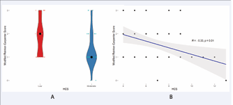
Figure 1 1A-1B. Lower Histopathological Charcot Scores were associated with better clinical outcomes as measured using the modified Reinker-Carpenter score in patients with Charcot Neuroosteoarthropathy treated with arthrodesis.
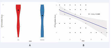
Figure 2 2A-2B. Lower Histopathological Charcot Scores were associated with better bony consolidation as measured on CT scans 3 months postoperatively in patients with Charcot Neuroosteoarthropathy treated with arthrodesis.
The corresponding explained variance (r² ≈ 0.11–0.12) indicates that histopathological activity represents a measurable contributor to variability in early postoperative outcomes.
Subgroup Analysis
Patients with low histopathological activity demonstrated significantly higher median clinical scores (p = 0.007) and CT consolidation scores (p = 0.044) compared with patients with moderate activity.
Regression Analysis
Age and sex were not significant predictors of clinical or radiographic outcome (p > 0.05).
DISCUSSION
The present study evaluated whether histopathological disease activity quantified by the Histopathological Charcot Score is associated with early clinical and radiographic outcomes following hindfoot and ankle arthrodesis for Charcot neuroosteoarthropathy.
Charcot neuroosteoarthropathy represents a complex complication of diabetic neuropathy characterized by progressive joint destruction, deformity, and a high risk of ulceration and amputation [1-29]. Despite advances in diabetic foot care, surgical decision-making in Charcot reconstruction remains challenging, particularly regarding the optimal timing of surgery.
Current treatment algorithms are primarily based on the Eichenholtz staging system, which relies on clinical and radiographic findings [2]. However, several recent studies and reviews have highlighted the limitations of purely radiographic staging and emphasized the importance of biological disease activity [30,31].
The present findings suggest that histopathological activity may represent such a biological parameter. Higher HCS values were associated with inferior clinical outcome and reduced early osseous consolidation.
The observed correlation coefficients (r ≈ −0.33 to −0.34) correspond to moderate associations according to conventional interpretation standards. Bone healing in Charcot neuroosteoarthropathy is influenced by multiple interacting factors, including metabolic control, vascular status, neuropathy severity, and mechanical stability [30,31]. In this multifactorial context, the observed explained variance of approximately 11–12% suggests that histopathological activity represents a measurable and clinically relevant contributor to early postoperative outcome variability.
Subgroup analysis further demonstrated superior clinical and radiographic outcomes in patients with lower histopathological activity. This observation supports the hypothesis that ongoing inflammatory activity at the tissue level may impair early consolidation.
Radiographic assessment in the present study was performed using CT imaging at three months postoperatively. CT has previously been shown to provide superior reliability in assessing hindfoot arthrodesis compared with conventional radiographs [11-13]. Although this time point reflects early consolidation rather than definitive fusion, early osseous bridging is clinically relevant in this high-risk patient population.
Several limitations must be acknowledged. First, the retrospective study design introduces potential selection bias. Second, important systemic factors influencing bone healing in Charcot neuroosteoarthropathy - such as glycemic control, smoking status, renal function, and vascular status - were not consistently available. These factors have previously been identified as relevant determinants of surgical outcome in Charcot reconstruction [30,31].
Despite these limitations, the present study provides preliminary evidence that histopathological disease activity correlates with early postoperative healing dynamics following arthrodesis for Charcot neuroosteoarthropathy.
From a clinical perspective, histopathological evaluation of intraoperative tissue samples may provide additional biological information when determining surgical timing. Future prospective studies integrating standardized metabolic assessment, validated patient reported outcome measures, and longer-term imaging follow-up are required to further clarify the prognostic value of histopathological disease activity.
CONCLUSION
Higher histopathological disease activity was associated with inferior early consolidation and functional outcome following arthrodesis for Charcot neuroosteoarthropathy. Although the effect size was moderate, histopathological assessment may provide additional information regarding biological disease activity at the time of surgery. Further prospective studies are required to confirm the prognostic role of histopathological activity in surgical decision making for Charcot neuroosteoarthropathy.
ACKNOWLEDGEMENTS
The authors thank the histopathology laboratory staff for their technical assistance in specimen processing.
REFERENCES
- Rogers LC, Frykberg RG. The Charcot foot. Med Clin North Am. 2013; 97: 847-856.
- Eichenholtz S. Charcot joints. Springfield (IL): Charles C Thomas; 1966.
- Illgner U, Podella M, Rümmler M, Wühr J, Büsch HG, Wetz HH. Rekonstruktive Chirurgie beim Charcot-Fuss: 5-Jahres-Langzeitoutcome [Reconstructive surgery for Charcot foot. Long-term 5-year outcome]. Orthopade. 2009; 38: 1180-1186.
- Koller A, Hafkemeyer U, Fiedler R, Wetz HH. Rekonstruktive Fusschirurgie bei diabetisch-neuropathischer Osteoarthropathie [Reconstructive foot surgery in cases of diabetic-neuropathic osteoarthropathy]. Orthopade. 2004; 33: 983-991.
- Pinzur MS, Sostak J. Surgical stabilization of nonplantigrade Charcot arthropathy of the midfoot. Am J Orthop (Belle Mead NJ). 2007; 36: 361-365.
- Pinzur MS. Neutral ring fixation for high-risk nonplantigrade Charcot midfoot deformity. Foot Ankle Int. 2007; 28: 961-966.
- Illgner U, Mehlhorn AT, Osada N, Krenn V. Histopathologischer Charcot-Score an intraoperativen Gewebeproben vom Fuß : Prospektive Untersuchung [Histopathological Charcot score on intraoperative tissue samples from the foot : A prospective investigation]. Orthopade. 2019; 48: 693-703.
- Krenn V, Waldstein W, Najm A, Perino G, Gaulke R. Histopathologische Klassifikationsprinzipien rheumatischer Gelenkerkrankungen : Beitrag der Pathologie zur Diagnose [Histopathological classification principles of rheumatic joint diseases : Contribution of pathology to the diagnosis]. Orthopade. 2018; 47: 941-948.
- Reinker KA, Carpenter CT. Ilizarov applications in the pediatric foot. J Pediatr Orthop. 1997; 17: 796-802.
- Cerrato RA, Aiyer AA, Campbell J, Jeng CL, Myerson MS. Reproducibility of computed tomography to evaluate ankle and hindfoot fusions. Foot Ankle Int. 2014; 35: 1176-1180.
- Coughlin MJ, Grimes JS, Traughber PD, Jones CP. Comparison of radiographs and CT scans in the prospective evaluation of the fusion of hindfoot arthrodesis. Foot Ankle Int. 2006; 27: 780-787.
- Jones CP, Coughlin MJ, Shurnas PS. Prospective CT scan evaluation of hindfoot nonunions treated with revision surgery and low-intensity ultrasound stimulation. Foot Ankle Int. 2006; 27: 229-235.
- Raspovic KM, Wukich DK. Self-reported quality of life in patients with diabetes: a comparison of patients with and without Charcot neuroarthropathy. Foot Ankle Int. 2014; 35: 195-200.
- Robinson AH, Pasapula C, Brodsky JW. Surgical aspects of the diabetic foot. J Bone Joint Surg Br. 2009; 91: 1-7.
- Rettedal D, Parker A, Popchak A, Burns PR. Prognostic Scoring System for Patients Undergoing Reconstructive Foot and Ankle Surgery for Charcot Neuroarthropathy: The Charcot Reconstruction Preoperative Prognostic Score. J Foot Ankle Surg. 2018; 57: 451-455.
- Mittlmeier T, Klaue K, Haar P, Beck M. Should one consider primary surgical reconstruction in charcot arthropathy of the feet? Clin Orthop Relat Res. 2010; 468: 1002-1011.
- Simon SR, Tejwani SG, Wilson DL, Santner TJ, Denniston NL. Arthrodesis as an early alternative to nonoperative management of charcot arthropathy of the diabetic foot. J Bone Joint Surg Am. 2000; 82: 939-950.
- Easley ME, Trnka HJ, Schon LC, Myerson MS. Isolated subtalar arthrodesis. J Bone Joint Surg Am. 2000; 82: 613-624.
- Haskell A, Pfeiff C, Mann R. Subtalar joint arthrodesis using a single lag screw. Foot Ankle Int. 2004; 25: 774-777.
- Pell RF 4th, Myerson MS, Schon LC. Clinical outcome after primary triple arthrodesis. J Bone Joint Surg Am. 2000; 82: 47-57.
- De Heus JA, Marti RK, Besselaar PP, Albers GH. The influence of subtalar and triple arthrodesis on the tibiotalar joint. A long-term follow-up study. J Bone Joint Surg Br. 1997; 79: 644-647.
- Mann RA, Beaman DN, Horton GA. Isolated subtalar arthrodesis. Foot Ankle Int. 1998; 19: 511-519.
- Donley BG, Ward DM. Implantable electrical stimulation in high-risk hindfoot fusions. Foot Ankle Int. 2002; 23: 13-18
- Flemister AS Jr, Infante AF, Sanders RW, Walling AK. Subtalar arthrodesis for complications of intra-articular calcaneal fractures. Foot Ankle Int. 2000; 21: 392-399
- Ljung P, Kaij J, Knutson K, Pettersson H, Rydholm U. Talonavicular arthrodesis in the rheumatoid foot. Foot Ankle. 1992; 13: 313-316.
- Hastings MK, Johnson JE, Strube MJ, Hildebolt CF, Bohnert KL, Prior FW, et al. Progression of foot deformity in Charcot neuropathic osteoarthropathy. J Bone Joint Surg Am. 2013; 95: 1206-1213.
- Pinzur MS. Surgical treatment of the Charcot foot. Diabetes Metab Res Rev. 2016; 32: 287-291.
- Wukich DK, Schaper NC, Gooday C, Bal A, Bem R, Chhabra A, et al. Guidelines on the diagnosis and treatment of active Charcot neuro-osteoarthropathy in persons with diabetes mellitus (IWGDF 2023). Diabetes Metab Res Rev. 2024; 40: e3646.
- Argyropoulos M, Wynell-Mayow W, Johnson O, Faroug R, Johal KS, Deol RS, Hakmi A, Mordecai S. Charcot neuro-osteoarthropathy: a review of key concepts and an evidence-based surgical management algorithm. Front Clin Diabetes Healthc. 2024; 5: 1344359.
- Khan O, Kavarthapu M, Edmonds M, Kavarthapu V. Surgical management of Charcot foot - The advancements over the past decade. J Clin Orthop Trauma. 2023; 47: 102317.
- Dubský M, Bém R, Sojáková D, Fejfarová V, Hughes M, Jude EB. Charcot foot disease: A new approach. Drugs Aging. 2025; 42: 837-851.