The Calpain Homeostasis Model: A Physiological Framework Explaining the Sequential Collapse of Neuronal Homeostasis after Concussion
- 1. College of Dental Medicine, Western University of Health Sciences, USA
- 2. College of Osteopathic Medicine of the Pacific, Western University of Health Sciences, USA
Abstract
Concussion presents a fundamental biological paradox. Although the initiating mechanical injury lasts only milliseconds, neurological dysfunction may evolve over months or years, leading to persistent cognitive, emotional, and behavioral impairment. Repetitive concussions further increase the risk of chronic traumatic encephalopathy (CTE) and other neurodegenerative disorders, yet the mechanisms linking a transient biomechanical insult to progressive neurological disease remain poorly understood.
Here we introduce the Calpain Homeostasis Model (CHM), a unifying conceptual framework proposing that persistent disruption of calpain homeostasis provides the mechanistic link between the initial injury and delayed neurological dysfunction. Under physiological conditions, calpain-1 and calpain-2 coordinate adaptive proteolytic remodeling while preserving neuronal homeostasis. Following concussion, sustained calcium dysregulation and injury-induced signaling shift the physiological balance toward persistent calpain-2 activation, converting adaptive remodeling into maladaptive proteolysis and initiating self-amplifying cycles of cellular dysfunction.
Within this framework, excitotoxicity, mitochondrial dysfunction, oxidative stress, proteostatic failure, neuroinflammation, and synaptic degeneration are viewed as interconnected manifestations of the sequential collapse of neuronal homeostasis rather than independent pathological processes. The CHM provides a mechanistic explanation for the temporal dissociation between the brief mechanical injury and the prolonged evolution of neurological dysfunction. It also establishes a translational framework in which calpain-derived proteolytic fragments serve as biomarkers of pathological calpain activation, while restoration of calpain homeostasis—particularly through selective calpain-2-derived inhibition—represents a promising disease-modifying therapeutic calpain-2 strategy.
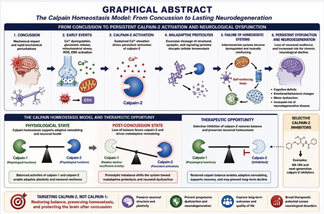
Graphical Abstract The Calpain Homeostasis Model: from concussion to persistent neurological dysfunction.
A transient mechanical insult initiates calcium dysregulation, glutamate signaling, mitochondrial stress, oxidative stress, and injury-associated signaling that favor persistent calpain-2 activation. Sustained calpain-2 activity shifts the calpain system from physiological, adaptive proteolytic remodeling toward maladaptive proteolysis and progressive disruption of interconnected structural, metabolic, proteostatic, neuroimmune, and network homeostatic systems. Self-reinforcing interactions among these systems progressively reduce neuronal resilience and can sustain neurological dysfunction after the initiating mechanical insult has resolved. Under physiological conditions, regulated calpain-1 and calpain-2 activities maintain calpain homeostasis; following concussion, the balance shifts toward persistent calpain-2 activity. Selective calpain-2 inhibition is proposed to restore this balance while preserving beneficial calpain-1-dependent functions, thereby interrupting downstream homeostatic failure and promoting recovery.
Keywords
• Concussion
• Mild traumatic brain injury
• Calpain-2
• Neuronal homeostasis
• Neurodegeneration
• Biomarkers
Citation
Baudry M, Bi X (2026) The Calpain Homeostasis Model: A Physiological Framework Explaining the Sequential Collapse of Neuronal Homeo stasis after Concussion. Ann Sports Med Res 13(1): 1240.
ABBREVIATION
AD: Alzheimer’s Disease; AMPA: α-Amino-3 hydroxy-5-methyl-4-isoxazolepropionic acid; ATP: Adenosine Triphosphate; CHM: Calpain Homeostasis Model; CNS: Central Nervous System; CRMP: Collapsin Response Mediator Protein; CTE: Chronic Traumatic Encephalopathy; DCX: Doublecortin; ER: Endoplasmic Reticulum; ERK: Extracellular Signal-Regulated Kinase; GFAP: Glial Fibrillary Acidic Protein; LMP: Lysosomal Membrane Permeabilization; LTP: Long-Term Potentiation; mTBI: Mild Traumatic Brain Injury; NMDA: N-Methyl-D-Aspartate; NfL: Neurofilament Light Chain; P13BP: PTPN13 Breakdown Product; PSD: Postsynaptic Density: PSD-95: Postsynaptic Density Protein-95; PTEN: Phosphatase and Tensin Homolog; ROS: Reactive Oxygen Species; SAP97: Synapse-Associated Protein 97; STEP: Striatal-Enriched Protein Tyrosine Phosphatase; SNTF: αII-Spectrin N-Terminal Fragment; SUMO: Small Ubiquitin Like Modifier; TBI: Traumatic Brain Injury; UPS: Ubiquitin Proteasome System
Concussion: An Unresolved Clinical Challenge
Concussion presents one of the central biological paradoxes in clinical neuroscience. Although the initiating mechanical injury lasts only milliseconds, its biological consequences often continue to evolve for months or even years, ultimately producing persistent cognitive impairment and increasing the risk of chronic neurodegenerative disease [1,2]. Millions of individuals sustain concussions each year as a consequence of sports participation, military service, motor vehicle accidents, falls, and recreational activities (https://www. barrowneuro.org/condition/concussion/). Although traditionally considered a transient injury from which most patients recover completely, increasing clinical and experimental evidence indicates that concussion may initiate complex biological processes that persist long after the initial mechanical insult has resolved [3]. While many individuals recover within days or weeks, a substantial proportion develop persistent post-concussion symptoms characterized by cognitive impairment, memory deficits, emotional disturbances, sleep disorders, headaches, and reduced quality of life. Repetitive concussions further increase the risk of chronic traumatic encephalopathy (CTE), Alzheimer’s disease, and other neurodegenerative disorders, emphasizing that even mild brain injuries may have long-lasting neurological consequences [4].
One of the defining characteristics of concussion is the striking temporal dissociation between the initiating injury and the subsequent evolution of neurological dysfunction. The biomechanical forces that initiate concussion act over milliseconds, yet molecular, cellular, and network abnormalities continue to evolve over hours, days, months, and in some cases years. This temporal dissociation strongly suggests that the primary injury serves as a trigger rather than the principal driver of disease progression. Understanding the mechanisms that transform an acute biomechanical event into chronic neurological disease remains one of the central challenges in concussion research. The central question is therefore not whether concussion activates multiple pathological pathways—that is now well established—but rather how these diverse processes become integrated into a progressive biological program that continues to evolve long after the initiating mechanical insult has ended.
Over the past several decades, considerable progress has been made in identifying the molecular events associated with concussion. Mechanical deformation of neuronal membranes initiates the well-characterized neurometabolic cascade, characterized by ionic imbalance, excessive glutamate release, calcium influx, mitochondrial dysfunction, oxidative stress, impaired axonal transport, cytoskeletal disruption, neuroinflammation, altered protein homeostasis, and progressive synaptic dysfunction [5,6]. Each of these mechanisms has been extensively investigated and clearly contributes to the pathophysiology of concussion. Despite remarkable progress in defining these mechanisms individually, considerably less is understood about how they become organized into a progressive and self-reinforcing biological process. This conceptual limitation has also influenced therapeutic development. Numerous experimental interventions targeting excitotoxicity, oxidative stress, mitochondrial dysfunction, inflammation, apoptosis, or protein aggregation have demonstrated encouraging results in preclinical studies, yet few have translated into effective clinical therapies [3-9]. One possible explanation is that these approaches target individual downstream manifestations of injury rather than the upstream regulatory mechanisms that coordinate the broader pathological response. If concussion represents the progressive failure of an interconnected biological system rather than the sum of independent pathological pathways, successful therapeutic intervention may require restoration of the regulatory mechanisms that normally maintain neuronal homeostasis.
Increasing evidence suggests that this may indeed be the case. This perspective shifts attention from individual molecular pathways toward the mechanisms that preserve—or fail to preserve—neuronal homeostasis following injury. Rather than progressing through a series of isolated pathological events, concussion appears to involve progressive disruption of multiple interconnected homeostatic systems responsible for maintaining neuronal structure, metabolism, intracellular protein quality control, neuroimmune regulation, and network function. The remarkable overlap between these abnormalities and those observed in chronic neurodegenerative disorders further suggests that concussion and neurodegeneration may share common biological mechanisms despite their distinct initiating insults [10,11].
In this review, we propose the Calpain Homeostasis Model (CHM), a unifying and experimentally testable framework in which persistent disruption of calpain homeostasis provides the mechanistic transition linking transient mechanical injury to the sequential collapse of neuronal homeostasis. Building upon recent advances demonstrating fundamentally distinct physiological roles for calpain-1 and calpain-2 [12], we propose that concussion initiates a progressive shift from adaptive proteolytic remodeling toward persistent calpain-2 mediated pathological proteolysis. Within this framework, sustained disruption of calpain homeostasis initiates a sequential collapse of structural, metabolic, proteostatic, inflammatory, and network homeostasis that ultimately culminates in persistent neurological dysfunction. This model not only provides a mechanistic explanation for the delayed evolution of concussion pathology but also identifies preservation of calpain homeostasis as a potential therapeutic strategy capable of restoring multiple homeostatic systems simultaneously (Box 1).
Box 1 summarizes the central concepts of the Calpain Homeostasis Model and serves as a roadmap for the mechanistic framework developed throughout this review.
Calpains: From Calcium-Activated Proteases to Guardians of Neuronal Homeostasis
Box 1. The Calpain Homeostasis Model (CHM): Principles and Testable Predictions
Definition
The Calpain Homeostasis Model proposes that neuronal function depends on the dynamic regulation of calpain-mediated proteolysis. Under physiological conditions, coordinated activation of calpain-1 and calpain-2 permits adaptive structural and molecular remodeling while preserving neuronal integrity. Following concussion, persistent disruption of this regulatory balance shifts calpain activity toward sustained calpain-2-dependent proteolysis, initiating self-reinforcing disturbances across multiple interconnected homeostatic systems that progressively drive the transition from adaptive remodeling to maladaptive remodeling and ultimately to neuronal dysfunction.
Central Principles
Principle 1 – Physiological proteolysis is adaptive.
Calpains are not primarily degradative enzymes, but highly regulated mediators of proteolytic remodeling required for normal neuronal function.
Principle 2 – Calpain homeostasis depends on balanced calpain signaling.
Neuronal function depends not simply on the amount of calpain activity, but on the precise temporal, spatial, and quantitative coordination of calpain-1 and calpain-2. This dynamic balance enables physiological proteolytic remodeling while preventing excessive or sustained proteolysis that compromises neuronal function.
Principle 3 – Persistent disruption of calpain homeostasis converts adaptive remodeling into maladaptive remodeling.
Following concussion activity, sustained calcium dysregulation and injury-induced signaling shift the physiological balance of calpain signaling toward persistent calpain-2 predominance, producing pathological proteolysis and progressive cellular and CNS dysfunction.
Principle 4 – Progressive neurological dysfunction reflects systems failure.
Excitotoxicity, mitochondrial dysfunction, impaired proteostasis, neuroinflammation, axonal degeneration, and synaptic dysfunction are interconnected and mutually reinforcing manifestations of progressive disruption of nervous system homeostasis rather than isolated pathological pathways.
Principle 5 – Restoration of calpain homeostasis is a therapeutic objective.
Disease-modifying therapies should aim not simply to inhibit proteolysis but to re-establish physiological regulation of calpain activity and thereby restore neuronal and glial homeostasis. Accordingly, the therapeutic objective is not merely to suppress proteolysis, but to recover the physiological balance that supports adaptive remodeling, preserves neuronal homeostasis, and promotes long-term neurological resilience.
Testable Predictions of the Calpain Homeostasis Model
• Restoration of calpain homeostasis should normalize multiple downstream pathological pathways simultaneously.
• Biomarkers reflecting pathological calpain activity should predict disease progression more accurately than biomarkers reflecting generalized tissue injury.
• Selective calpain-2 inhibition should preserve physiological calpain-1-dependent plasticity while preventing maladaptive remodeling.
Central Concept
Persistent disruption of calpain homeostasis transforms adaptive neuronal remodeling into sequential collapse of interconnected homeostatic systems.
Calpains comprise a highly conserved family of intracellular calcium-dependent cysteine proteases that participate in numerous physiological and pathological processes throughout the body. Among the fifteen calpain family members identified in mammals, the ubiquitous isoforms calpain-1 (μ-calpain) and calpain-2 (m-calpain) are particularly abundant in the central nervous system, where they regulate diverse aspects of neuronal structure and function [13]. Since their discovery nearly five decades ago [14,15], calpains have been recognized as uniquely positioned to translate intracellular calcium signals into rapid proteolytic remodeling of neuronal proteins. For many years, calpains were viewed primarily as destructive enzymes whose activation contributed to neuronal injury following ischemia, traumatic brain injury, epilepsy, and neurodegenerative diseases [16-21]. This interpretation was supported by extensive evidence demonstrating calpain-mediated proteolysis of cytoskeletal proteins, ion channels, receptors, kinases, phosphatases, and numerous additional substrates following pathological elevations in intracellular calcium [21-28]. Consequently, calpain activation became widely recognized as a hallmark of neuronal injury, and pharmacological inhibition of calpains emerged as an attractive therapeutic strategy for limiting secondary neuronal damage [29-33].
However, this interpretation created a fundamental biological paradox. Pharmacological inhibition of calpains emerged as an attractive therapeutic strategy—one that later proved more complex than initially anticipated. Evolution has conserved calpains throughout vertebrate evolution, and their expression is remarkably high in neurons under physiological conditions. It seemed unlikely that enzymes maintained with such precision across species would serve primarily pathological functions. This paradox stimulated a reassessment of calpain biology that fundamentally altered our understanding of their physiological roles.
Accumulating evidence now demonstrates that calpains function primarily as regulatory proteases rather than degradative enzymes [34]. This shift in perspective has profound implications for understanding neuronal physiology. If calpains coordinate adaptive proteolytic remodeling, their physiological role extends far beyond protein cleavage itself. Rather, they become integral components of the homeostatic machinery that enables neurons to continuously adapt while preserving structural and functional stability [35]. In contrast to lysosomal proteases or the proteasome, which generally mediate complete protein degradation, calpains typically perform limited proteolysis that modifies protein activity, localization, stability, or interactions with other proteins. Through selective cleavage of key regulatory molecules, calpains rapidly remodel signaling pathways, cytoskeletal organization, membrane trafficking, and transcriptional responses without requiring de novo protein synthesis [36,37]. In this respect, calpain-mediated proteolysis represents a form of post-translational regulation analogous to phosphorylation, ubiquitination, or SUMOylation, providing neurons with an efficient mechanism for rapidly adapting to changing physiological demands. Unlike protein degradation, limited proteolysis generates functional diversity by modifying existing proteins rather than eliminating them.
The recognition that calpain-1 and calpain-2 perform distinct—and often opposing—functions represented a second major conceptual advance [38]. Although the two enzymes share extensive structural homology and many substrates, genetic, biochemical, and pharmacological studies have demonstrated that they participate in different signaling pathways and produce markedly different biological outcomes. Calpain-1 is preferentially activated during physiological synaptic activity and contributes to long-term potentiation, learning and memory, neuronal survival, and adult neurogenesis. In contrast, calpain-2 contributes to physiological synaptic remodeling under tightly regulated conditions but becomes persistently activated following CNS injury.
These discoveries fundamentally changed the interpretation of calpain activation in the nervous system. This evolution in thinking represents a paradigm shift from viewing calpains as mediators of pathological protein degradation to recognizing them as fundamental regulators of neuronal homeostasis whose dysregulation contributes to disease. Rather than determining whether calpains are simply “active” or “inactive,” neuronal function depends on the precise temporal, spatial, and quantitative regulation of individual calpain isoforms. Physiological proteolysis must be sufficiently dynamic to permit continuous synaptic remodeling while remaining tightly constrained to preserve neuronal integrity. Loss of this regulatory balance has consequences that extend far beyond excessive protein cleavage, disrupting the coordinated biological processes that maintain neuronal structure, metabolism, intracellular quality control, and network function [12]. Thus, neuronal dysfunction reflects not excessive proteolysis alone, but disruption of the regulatory system that normally governs adaptive proteolytic remodeling.
This emerging perspective provides the foundation for the conceptual framework developed throughout this review. The CHM proposes that the principal function of the calpain system is not simply to regulate proteolysis but to preserve neuronal homeostasis by coordinating adaptive structural remodeling with long-term cellular stability. Under physiological conditions, this dynamic balance is maintained through tightly regulated interactions among intracellular calcium signaling, calpastatin, phosphorylation, subcellular compartmentalization, and the complementary activities of calpain-1 and calpain-2. Following concussion, persistent disturbances in calcium homeostasis and injury-induced signaling shift this balance toward sustained calpain-2 activation, initiating a cascade of maladaptive proteolysis that extends well beyond individual protein substrates. The following section develops this concept in detail and introduces the Calpain Homeostasis Model (CHM), which serves as the central framework for understanding how disruption of physiological proteolytic regulation can drive the progressive failure of neuronal homeostasis following concussion.
THE CALPAIN HOMEOSTASIS MODEL
Physiological Calpain Homeostasis: Regulated Proteolysis as a Mechanism for Neuronal Remodeling
Physiological calpain homeostasis is founded on the concept that calpains function as highly regulated proteolytic signaling enzymes rather than degradative proteases. Through spatially and temporally restricted limited proteolysis, calpains rapidly remodel protein function, localization, and interactions, thereby enabling adaptive structural and functional plasticity without requiring complete protein turnover. This concept is particularly relevant in the nervous system, where neurons continuously remodel synaptic connections in response to experience. Synaptic plasticity, dendritic spine remodeling, axonal growth, adult neurogenesis, and adaptive responses to environmental stimuli all require rapid and spatially restricted reorganization of complex protein assemblies [39]. Limited proteolysis provides an efficient mechanism for this remodeling because it permits rapid modification of existing proteins without requiring their complete turnover or new protein synthesis. Within this context, calpains emerge not as destructive enzymes but as essential regulators of neuronal remodeling.
The physiological actions of calpains are tightly regulated at multiple levels. Transient increases in intracellular calcium initiate activation, whereas phosphorylation, phospholipid interactions, subcellular localization, membrane association, and inhibition by calpastatin determine both the magnitude and duration of proteolytic activity [15-42]. These complementary mechanisms restrict calpain activation to specific subcellular compartments and narrow temporal windows, thereby allowing precise remodeling of selected protein complexes while preserving overall neuronal integrity. This is further accomplished by the association of calpain-1 and calpain-2 to different PDZ binding proteins due to the existence of different PDZ binding motifs in their C-terminal domains [38]. Accordingly, neuronal function depends not simply on whether calpains are activated, but on where, when, for how long, and which calpain isoform is activated.
Functional Specialization of Calpain-1 and Calpain-2
One of the most significant advances in calpain biology has been the recognition that the two ubiquitous isoforms, calpain-1 and calpain-2, are not functionally redundant. Although they share substantial structural similarity and many substrates, accumulating genetic, pharmacological, and biochemical evidence demonstrates that they participate in distinct signaling pathways and often exert opposing biological effects (Figure 1).
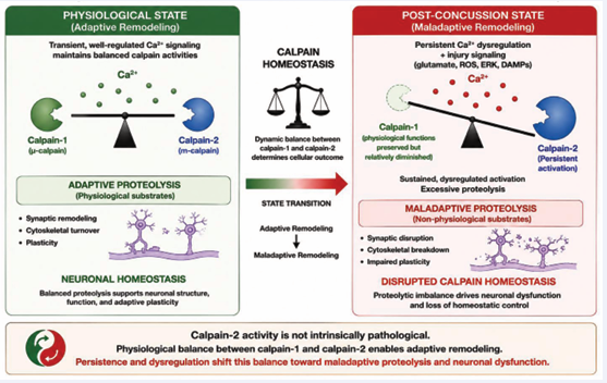
Figure 1: Physiological calpain homeostasis and its disruption following concussion.
Under physiological conditions, the complementary and tightly regulated activities of calpain-1 and calpain-2 maintain calpain homeostasis and support adaptive neuronal remodeling. Calpain-1 predominantly promotes synaptic plasticity, neuronal survival, and other adaptive responses, whereas calpain-2 contributes to physiological synaptic and homeostatic remodeling when its activation is appropriately restricted. Following concussion, persistent calcium dysregulation and injury associated signaling shift this balance toward sustained calpain-2 activation. The resulting transition from adaptive to maladaptive proteolytic remodeling represents a central feature of disrupted calpain homeostasis and promotes progressive neuronal dysfunction.
Calpain-1 is preferentially activated by physiological patterns of synaptic activity and contributes to mechanisms underlying long-term potentiation, learning and memory, neuronal survival, and adult neurogenesis [12]. Activation of calpain-1 promotes signaling pathways that facilitate adaptive synaptic remodeling and structural plasticity while supporting neuronal resilience following physiological stimulation.
In contrast, calpain-2 exhibits a distinct activation profile and signaling repertoire. Under physiological conditions, calpain-2 participates in the regulation of synaptic remodeling and homeostatic plasticity, thereby preventing excessive synaptic strengthening and maintaining network stability [43]. Persistent disruption of calpain homeostasis shifts proteolytic remodeling from adaptive regulation toward pathological remodeling, thereby disrupting multiple homeostatic systems discussed in subsequent sections [12]. These observations indicate that the biological consequences of calpain activation cannot be predicted solely from total enzymatic activity. Instead, they depend critically on the relative activation of calpain-1 and calpain-2, their temporal dynamics, and the signaling context in which activation occurs.
The Calpain Homeostasis Model
The accumulating evidence summarized above suggests a broader conceptual framework for understanding calpain function in both health and disease. We propose that normal neuronal function is maintained through physiological calpain homeostasis, a dynamic regulatory state that coordinates the activation of calpain-1 and calpain-2 in time, space, and magnitude to permit continuous neuronal remodeling while preserving neuronal integrity. Within the CHM, pathophysiology is viewed not simply as excessive calpain activity but as a state transition in which persistent disruption of calpain homeostasis converts adaptive remodeling into maladaptive remodeling. Importantly, calpain homeostasis should not be viewed as a static equilibrium. Neurons continuously experience physiological fluctuations in intracellular calcium associated with synaptic transmission, learning, metabolic activity, and environmental adaptation [44-46]. Accordingly, calpain activity is likewise dynamic, constantly adjusting to changing physiological demands. Homeostasis therefore reflects the capacity of the regulatory system to maintain appropriate proteolytic remodeling despite continual perturbation.
Within this framework, calpain-1 and calpain-2 fulfill complementary rather than redundant functions. Calpain-1 predominantly supports adaptive remodeling associated with synaptic plasticity, neuronal survival, and repair. Calpain-2 contributes to physiological remodeling under tightly controlled conditions but also serves as an important constraint that limits excessive or inappropriate structural plasticity. Under normal circumstances, multiple regulatory mechanisms—including transient calcium signaling, calpastatin inhibition, phosphorylation, and spatial compartmentalization—ensure that calpain-2 activation remains appropriately restricted.
Following injury, persistent disturbances in calcium and intracellular signaling overwhelm these regulatory mechanisms, shifting proteolytic remodeling from adaptive to maladaptive. Accordingly, we propose that the transition from adaptive to maladaptive neuronal remodeling represents the defining feature of disrupted calpain homeostasis.
This framework reconciles numerous observations that have previously appeared unrelated. It explains how identical enzymes can mediate both physiological plasticity and pathological degeneration, why pan-calpain inhibition has generally produced disappointing therapeutic results, and why selective inhibition of calpain-2 can preserve beneficial calpain-1-dependent functions while preventing progressive neurodegeneration. Viewed from this perspective, restoration of calpain homeostasis becomes the therapeutic objective, whereas selective calpain-2 inhibition represents one potential strategy for achieving it.
Implications for Concussion
Concussion provides an ideal biological context in which to examine the consequences of disrupted calpain homeostasis because the transient mechanical insult is followed by a prolonged period of progressive biological remodeling. We propose that persistent disruption of calpain homeostasis provides one such mechanism. Rather than functioning simply as another downstream consequence of calcium dysregulation, persistent calpain-2 activation may represent a critical molecular transition linking the initial injury to delayed synaptic dysfunction, axonal degeneration, neuroinflammation, and chronic neurodegeneration. The following sections examine how disruption of calpain homeostasis propagates across multiple interconnected homeostatic systems, ultimately leading to neuronal dysfunction after concussion.
FROM MECHANICAL IMPACT TO SUSTAINED CALPAIN-2 ACTIVATION: A MOLECULAR CASCADE
One of the central unresolved questions in concussion research is how a mechanical insult lasting only milliseconds initiates molecular processes that continue for days, weeks, or even years. Although numerous mechanisms have been implicated—including excitotoxicity, mitochondrial dysfunction, oxidative stress, neuroinflammation, impaired axonal transport, and protein aggregation [5,6] —these processes are often presented as parallel pathological events with unclear mechanistic relationships. Such a view provides only limited insight into the sequence of events that transforms an acute biomechanical injury into chronic neurodegeneration.
We propose that these seemingly disparate mechanisms can be integrated into a single molecular cascade centered on disruption of calpain homeostasis (Figure 2). In this model, the primary biomechanical insult initiates a series of tightly linked events that progressively favor sustained activation of calpain-2 while overwhelming the regulatory mechanisms that normally maintain physiological proteolytic homeostasis. Once activated, calpain-2 reinforces multiple downstream homeostatic disturbances through selective proteolysis of key regulatory proteins. This framework provides a mechanistic explanation for the delayed evolution of concussion pathology and suggests that calpain-2 occupies a central position linking the initial mechanical insult to progressive neuronal dysfunction. The cascade of events leading to sustained calpain-2 activation is summarized below.
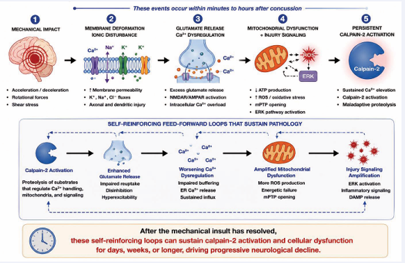
Figure 2: From mechanical impact to persistent calpain-2 activation.
Concussion produces rapid mechanical deformation of neuronal membranes and axons, initiating ionic disturbances, glutamate release, and intracellular Ca²? accumulation. Persistent calcium dysregulation, together with mitochondrial dysfunction, oxidative stress, and injury-associated signaling pathways including ERK activation, favors sustained calpain-2 activation. Calpain-2-mediated proteolysis further compromises mitochondrial function, calcium regulation, and cellular integrity, establishing self-reinforcing feed-forward mechanisms that can maintain pathological signaling after the initiating mechanical insult has resolved. This transition provides a mechanistic link between the acute biomechanical event and delayed neuronal dysfunction.
Mechanical Injury and the Neurometabolic Cascade
The immediate consequence of concussion is the rapid distortion of neuronal membranes, axons, glial cells, and cerebral microvasculature produced by rotational and translational acceleration forces. Although gross structural disruption is generally absent, these mechanical forces produce widespread transient membrane perturbations, axonal stretch, and disruption of mechanosensitive ion channels. The resulting ionic imbalance initiates the well characterized neurometabolic cascade that has become a defining feature of concussion pathology [5-47].
Mechanical deformation rapidly disrupts ionic homeostasis, producing potassium efflux, sodium and calcium influx, and excessive glutamate release. Restoration of ionic gradients markedly increases ATP demand at a time when cerebral blood flow is transiently reduced, creating the characteristic neurometabolic crisis. Although many neurons gradually recover from this initial disturbance, intracellular calcium concentrations often remain elevated for prolonged periods, particularly within injured axons and dendritic compartments. Persistent calcium dysregulation represents one of the earliest molecular abnormalities linking the primary mechanical injury to delayed secondary injury mechanisms.Importantly, calcium does not simply activate multiple independent pathological pathways; rather, it serves as the common initiating signal that drives the progressive disruption of calpain homeostasis.
Persistent Calcium Dysregulation and Selective Calpain-2 Activation
A defining feature of concussion is that intracellular calcium homeostasis often remains disturbed long after restoration of membrane integrity and resolution of the initial ionic imbalance. Calcium may remain elevated within injured axons because of persistent membrane permeability, impaired mitochondrial calcium buffering, dysfunction of endoplasmic reticulum calcium stores, and sustained activation of glutamatergic signaling pathways [5-48]. This prolonged disturbance distinguishes concussion from many physiological forms of neuronal activation and provides the molecular environment necessary for activation of pathological signaling cascades.
Although both calpain-1 and calpain-2 are calcium dependent proteases, their activation is not determined solely by intracellular calcium concentration [49,50]. Instead, each isoform is regulated by distinct molecular mechanisms that confer markedly different physiological functions (Table 1).
Table 1: Distinct regulation and functions of calpain-1 and calpain-2.
|
Property |
Calpain-1 |
Calpain-2 |
|
Activation |
Transient |
Sustained after injury |
|
Calcium signal |
Physiological/localized |
Persistent + ERK-dependent |
|
Physiological role |
Synaptic plasticity |
Homeostatic remodeling |
|
Biological outcome |
Adaptive remodeling |
Maladaptive remodeling when dysregulated |
|
Predominant biological consequence |
Survival and plasticity |
Degeneration and dysfunction |
The distinction between the two isoforms has important implications for concussion. During physiological neuronal activity, transient calcium signals predominantly recruit calpain-1, promoting adaptive structural remodeling that underlies synaptic plasticity and learning. Following concussion, however, prolonged calcium dysregulation, together with sustained ERK activation and additional injury-induced signaling pathways, shifts increasingly the balance toward sustained calpain-2 signaling [12,38]. Concussion therefore produces not simply more calpain activity, but a qualitative shift in calpain signaling from adaptive to maladaptive proteolytic remodeling. Once this transition occurs, calpain-2 initiates proteolytic cleavage of numerous proteins, which further exacerbate calcium dysregulation and cellular stress, creating self-reinforcing feed-forward mechanisms that reinforce continued calpain-2 activation [51,52]. The result is a self-amplifying cycle in which disruption of calpain homeostasis drives neuronal dysfunction despite the absence of ongoing mechanical injury. Importantly, this transition is not likely to occur as a discrete event but rather as a progressive shift in the balance of calpain signaling, consistent with the gradual evolution of post-concussive pathology observed clinically.
This model explains how a transient biomechanical event can initiate a chronic molecular process that evolves over days, weeks, and potentially years. Rather than acting as one component of the secondary injury cascade, disruption of calpain homeostasis may constitute the mechanistic convergence point through which multiple pathological pathways become integrated and mutually reinforce one another.
COLLAPSE OF STRUCTURAL HOMEOSTASIS
Synaptic Dysfunction: The Earliest Manifestation of Disrupted Calpain Homeostasis
The earliest functional consequences of concussion emerge at the synapse, long before widespread neuronal death becomes evident. Although the mechanical insult is transient, alterations in synaptic transmission, dendritic spine morphology, and network connectivity develop rapidly and correlate closely with the onset of cognitive deficits observed following mild traumatic brain injury [53]. These observations indicate that impaired neuronal communication, rather than irreversible cell loss, represents the initial pathological consequence of concussion. Within the framework of the CHM, this early synaptic dysfunction reflects the transition from adaptive to maladaptive proteolytic activation. Under physiological conditions, transient activation of calpain-1 contributes to activity-dependent remodeling of synaptic protein complexes that underlies learning and memory [43]. Limited proteolysis of selected substrates permits rapid modification of receptor trafficking, cytoskeletal organization, and intracellular signaling while preserving overall synaptic architecture. These tightly regulated events allow neurons to continuously adapt to changing patterns of activity without compromising structural integrity.
Concussion fundamentally alters this physiological balance. Persistent calcium dysregulation and sustained ERK activation progressively shift proteolytic activity toward calpain-2, transforming a normally adaptive remodeling process into one characterized by dismantling of synaptic organization. Rather than selectively modifying individual proteins, maladaptive proteolytic remodeling disrupts the coordinated molecular architecture that maintains synaptic function. Consequently, deficits in neurotransmission arise not because a single protein is lost, but because the highly organized network of structural and signaling proteins required for efficient synaptic communication disintegrates (Figure 3).

Figure 3: Progressive failure of interconnected homeostatic systems following disruption of calpain homeostasis.
Persistent disruption of calpain homeostasis promotes interacting failures across multiple levels of neuronal organization. Structural homeostasis is compromised through synaptic dysfunction, dendritic spine loss, postsynaptic density disorganization, cytoskeletal destabilization, and impaired axonal transport. Metabolic homeostasis subsequently deteriorates through mitochondrial dysfunction, loss of energetic reserve, oxidative stress, and impaired calcium buffering. Failure of lysosomal, autophagic, and ubiquitin-proteasome pathways disrupts proteostatic homeostasis, leading to accumulation of damaged proteins and organelles and release of danger-associated molecular patterns (DAMPs). Persistent neuronal distress then promotes failure of neuroimmune resolution and chronic glial activation, ultimately contributing to disruption of neural network organization and function. These levels of homeostatic failure are neither discrete nor strictly sequential but overlap and mutually reinforce one another, progressively reducing neuronal resilience and promoting persistent neurological dysfunction.
Experimental studies have identified numerous synaptic proteins as calpain substrates, including spectrin, PSD-95, SAP97, NMDA receptors and associated scaffolding proteins, AMPA receptors and regulatory proteins, CRMP family members, and several cytoskeletal adaptors [23 57]. Although cleavage of each substrate contributes to synaptic dysfunction, their collective proteolysis produces a far greater biological consequence: destabilization of the postsynaptic density, disruption of receptor anchoring, impairment of intracellular signaling, and loss of dendritic spine stability. Thus, persistent calpain-2 activation targets the organizational framework of the synapse rather than isolated molecular components.
Importantly, these early synaptic alterations provide a plausible explanation for one of the defining clinical characteristics of concussion. Many patients exhibit persistent impairments in attention, working memory, processing speed, and executive function despite the absence of detectable neuronal loss on conventional neuroimaging [58]. Such findings strongly suggest that dysfunction of surviving neuronal circuits, rather than extensive cell death, underlies the earliest neurological manifestations of concussion. Within this context, disruption of calpain homeostasis offers a mechanistic explanation linking transient biomechanical injury to prolonged cognitive dysfunction through progressive impairment of synaptic connectivity. Accordingly, synaptic dysfunction should be viewed not simply as one manifestation of secondary injury but as the earliest stage of a progressive calpain-2-driven neurodegenerative cascade. If sustained proteolytic dysregulation persists, the structural destabilization initiated at synapses progressively extends to axons, mitochondria, lysosomes, and ultimately the entire neuron. Thus, structural homeostatic failure evolves hierarchically, beginning at individual synapses before progressively involving larger levels of neuronal organization.
Dendritic Spine Remodeling: From Physiological Plasticity to Pathological Spine Loss
Dendritic spines are the principal sites of excitatory synaptic transmission and undergo continuous activity-dependent remodeling that underlies learning, memory, and adaptive behavior [59]. Accumulating evidence indicates that calpains play central roles in this physiological remodeling process. Transient activation of calpain-1 following synaptic activity contributes to the structural and molecular reorganization required for long-term potentiation (LTP). Through limited proteolysis of selected cytoskeletal and signaling proteins, calpain-1 facilitates actin remodeling, receptor trafficking, and stabilization of newly potentiated synapses [38]. Importantly, these proteolytic events are highly localized and tightly regulated, permitting structural plasticity without compromising synaptic integrity.
In contrast, persistent activation of calpain-2 fundamentally alters the nature of dendritic spine remodeling. Rather than promoting adaptive structural modification, calpain-2 activation drives progressive spine shrinkage and elimination through excessive proteolysis of proteins responsible for maintaining the actin cytoskeleton, postsynaptic scaffolds, and adhesion complexes [12].
Experimental models of traumatic brain injury consistently demonstrate rapid reductions in dendritic spine density within hippocampal and cortical circuits, changes that closely parallel the development of cognitive deficits [53]. More broadly, genetic deletion or selective inhibition of calpain-2 attenuates neuropathological changes and improves functional recovery after TBI and repeated concussion, supporting a role for persistent calpain-2 activation in maladaptive neuronal remodeling [60,61]. Additional studies indicate that calpain-1 and calpain-2 also play opposite roles in the regulation of spine morphology and neurogenesis [62]. These observations further indicate that dendritic spine loss is not an inevitable consequence of mechanical injury itself but rather reflects activation of a downstream proteolytic program that remains amenable to therapeutic intervention.
Within the CHM, dendritic spine remodeling represents the first structural manifestation of the transition from physiological to pathological proteolysis. Under normal conditions, calpain-1-dependent remodeling continuously reshapes synaptic connections while maintaining network stability. Following concussion, disruption of calpain homeostasis shifts this balance toward calpain-2 dependent spine elimination, thereby reducing synaptic connectivity and progressively impairing information processing across neuronal networks.
This distinction has important implications for understanding persistent post-concussion symptoms. Cognitive deficits are often detectable despite minimal neuronal loss because neuronal computation depends not only on the survival of individual neurons but also on the integrity of the synaptic networks that connect them. Progressive loss of dendritic spines therefore provides a plausible structural substrate for the memory impairment, decreased cognitive flexibility, and reduced processing speed that characterize many patients following concussion. Once synaptic architecture begins to collapse, disruption extends beyond individual spines to involve the postsynaptic density, dendritic cytoskeleton, axonal transport systems, and intracellular organelles. Progressive spine loss extends this failure from individual synapses to neuronal circuits.
Disassembly of the Postsynaptic Density: Collapse of the Synaptic Signaling Platform
The postsynaptic density (PSD) is a highly organized signaling platform that coordinates neurotransmitter receptor function with intracellular signaling, cytoskeletal organization, and synaptic plasticity. Physiological remodeling of the PSD is a continuous process that enables synapses to modify their strength in response to neuronal activity. During learning, transient activation of calpain-1 contributes to this remodeling by selectively modifying protein interactions within the PSD, thereby facilitating receptor trafficking, structural reorganization, and stabilization of potentiated synapses.
Persistent calpain activation following concussion is expected to destabilize this process, with calpain-2 activity providing a potential driver of maladaptive PSD remodeling. Rather than supporting physiological remodeling, sustained proteolysis destabilizes the molecular organization of the PSD by cleavage of scaffold proteins such as PSD-95 and SAP97 and other postsynaptic proteins. Loss of PSD organization uncouples receptor localization from intracellular signaling. Consequently, synapses lose their ability to undergo adaptive plasticity while becoming increasingly vulnerable to excitotoxic and metabolic stress.
Disassembly of the PSD represents the molecular counterpart of dendritic spine loss. Whereas pathological remodeling of dendritic spines reflects deterioration of synaptic structure, disruption of the PSD reflects collapse of the intracellular signaling machinery that enables those structures to function. These two processes therefore evolve in parallel and together initiate the progressive failure of neuronal communication that characterizes the early stages of concussion.
Importantly, disruption of the PSD is unlikely to represent an isolated pathological event. Because the PSD is physically linked to the dendritic actin cytoskeleton, axonal transport machinery, and intracellular organelles [63], destabilization of this signaling platform is expected to propagate structural disorganization throughout the neuron. Thus, the collapse of postsynaptic organization provides a mechanistic bridge linking early synaptic dysfunction to the broader cytoskeletal and metabolic abnormalities that develop during the subsequent stages of secondary injury. Collapse of the PSD disrupts the molecular machinery that sustains those circuits.
Cytoskeletal Destabilization: From Local Synaptic Remodeling to Global Structural Failure
The neuronal cytoskeleton provides the structural framework that integrates synapses, axons, and intracellular organelles into a single functional unit. Sustained loss of calpain homeostasis destabilizes this framework.Instead of facilitating localized reorganization, sustained proteolysis destabilizes the interconnected cytoskeletal network that maintains neuronal architecture. Numerous structural proteins undergo proteolytic cleavage following traumatic brain injury. Although each protein contributes distinct structural functions, their collective degradation weakens the mechanical and functional continuity of the neuron.
Among these substrates, αII-spectrin has received particular attention because its characteristic breakdown products have become widely used biomarkers of calpain activation following CNS injury [64-66]. Importantly, spectrin cleavage should be viewed primarily as an indicator of a much broader process rather than the principal cause of neuronal dysfunction. The biological significance of persistent calpain-2 activation lies not in cleavage of any individual structural protein but in the disassembly of the integrated cytoskeleton framework upon which neuronal organization depends [33-67].
As cytoskeleton integrity deteriorates, neurons lose their ability to maintain normal morphology, stabilize dendritic spines, anchor synaptic protein complexes, and support efficient intracellular transport. Mechanical resilience decreases, communication between synapses and the cell body becomes impaired, and intracellular organelles become isolated from one another. Consequently, structural deterioration spreads well beyond the original site of injury, extending throughout dendritic and axonal compartments.
Cytoskeletal destabilization represents the transition from localized synaptic dysfunction to global neuronal disorganization. The neuron no longer fails simply because individual proteins have been cleaved; rather, the integrated structural framework responsible for coordinating neuronal function collapses. This systems level deterioration establishes the conditions under which mitochondrial dysfunction, impaired axonal transport, lysosomal instability, and additional downstream pathological processes emerge.
Accordingly, cytoskeleton disruption should be viewed as a critical amplification step in the progression of concussion pathology. Once neuronal architecture becomes destabilized, structural disorganization itself promotes further disturbances in calcium homeostasis, intracellular signaling, and organelle function, thereby reinforcing continued activation of calpain-2 and accelerating the collapse of physiological proteolytic homeostasis. Cytoskeletal destabilization propagates structural failure throughout the neuron.
Failure of Axonal Transport: The Progressive Disconnection of the Neuron
Neurons depend on efficient bidirectional axonal transport to distribute proteins, organelles, and signaling molecules between the soma and distal neuronal compartments. Under physiological conditions, transient remodeling of the cytoskeleton contributes to efficient trafficking by allowing local structural adaptation while preserving the continuity of transport pathways. Following concussion, persistent calpain activity disrupts this organization, with sustained calpain-2 activation likely contributing to degradation of the cytoskeletal and regulatory proteins required for efficient intracellular transport. Proteolytic cleavage of cytoskeletal proteins, together with alterations in microtubule-associated proteins, CRMP family members, ankyrins, and additional structural adaptors, destabilizes the molecular tracks upon which intracellular cargoes are transported [33]. Although individual transport pathways may initially remain functional, continued proteolytic remodeling progressively reduces the efficiency and fidelity of bidirectional trafficking throughout the neuron.
The consequences extend far beyond impaired movement of isolated organelles. Mitochondria fail to reach regions of high metabolic demand, limiting local ATP production and calcium buffering at synapses [68]. Lysosomes and autophagic vesicles are no longer appropriately distributed, impairing intracellular degradation and protein quality control [69]. Neurotrophic signaling between axon terminals and the cell body becomes disrupted, while accumulation of transported cargoes contributes to the formation of axonal swellings and varicosities that represent characteristic pathological features of traumatic brain injury [70]. Thus, failure of axonal transport simultaneously compromises energy metabolism, protein homeostasis, intracellular signaling, and structural maintenance. Importantly, axonal transport failure also establishes multiple positive feedback mechanisms that amplify neuronal injury. Mislocalized mitochondria increase oxidative stress and reduce ATP availability, impairing the energy-dependent motor proteins responsible for intracellular trafficking. Defective lysosomal transport compromises autophagic flux, allowing damaged proteins and organelles to accumulate. Impaired retrograde signaling reduces activation of neuronal survival pathways, while axonal disconnection further disrupts calcium homeostasis. Each of these disturbances reinforces continued activation of calpain-2, thereby accelerating the collapse of proteolytic homeostasis.
In our model, failure of axonal transport represents a pivotal transition in the progression of concussion pathology. The neuron no longer functions as a coordinated cellular unit because communication between its individual compartments steadily deteriorates. Synapses become metabolically isolated from the cell body, organelles lose their coordinated distribution, and the capacity to maintain intracellular homeostasis declines. At this stage, the pathological process extends beyond local structural remodeling to involve global cellular dysfunction, setting the stage for widespread mitochondrial impairment and oxidative stress. Accordingly, axonal transport failure should not be viewed simply as a downstream consequence of cytoskeletal degradation but as a central mechanism through which localized synaptic injury evolves into neuron-wide dysfunction. By disconnecting the structural and metabolic integration of the neuron, disruption of calpain homeostasis establishes the conditions under which neurodegeneration becomes increasingly self sustaining.
Collectively, these observations suggest that the earliest stages of concussion are characterized by structural disintegration of the neuron rather than immediate neuronal death. As intracellular connectivity deteriorates, neurons lose the ability to maintain energy homeostasis, calcium regulation, and organelle integrity. This transition marks the beginning of the next stage of the calpain-2 driven cascade, in which structural failure evolves into metabolic failure.
Although disruption of neuronal architecture profoundly impairs synaptic communication, neurons possess considerable structural reserve and can often compensate for substantial cytoskeletal injury. The situation changes dramatically once structural disorganization begins to compromise mitochondrial function. Because mitochondria provide the ATP required for ion homeostasis, axonal transport, protein synthesis, and intracellular quality-control mechanisms, failure of mitochondrial homeostasis transforms a structurally compromised neuron into a metabolically vulnerable one. Thus, collapse of metabolic homeostasis represents the point at which neuronal injury exceeds the intrinsic capacity for energetic compensation.
To summarize, disruption of calpain homeostasis first manifests as structural disorganization, beginning at synapses and ultimately compromising the integrity of the entire neuron. This structural collapse establishes the conditions under which metabolic homeostasis can no longer be maintained.
COLLAPSE OF METABOLIC HOMEOSTASIS
Normal mitochondrial homeostasis
Neurons are among the most metabolically demanding cells in the body, requiring a continuous supply of ATP to sustain ion gradients, synaptic transmission, axonal transport, protein synthesis, and intracellular signaling. Unlike many other cell types, mature neurons possess limited glycolytic reserve and rely predominantly on oxidative phosphorylation within mitochondria to satisfy their exceptionally high energy requirements. Beyond ATP production, however, mitochondria serve as central regulators of neuronal homeostasis through their critical roles in calcium buffering, redox signaling, intermediary metabolism, and programmed cell death [71]. Consequently, neuronal function depends not simply on mitochondrial integrity, but on the precise regulation of mitochondrial dynamics and quality control.
Mitochondria form a highly dynamic intracellular network that continuously undergoes fusion, fission, trafficking, and selective degradation through mitophagy. These processes allow individual mitochondria to adapt to changing metabolic demands while maintaining a healthy mitochondrial population throughout the extensive architecture of neurons. Because synapses consume enormous amounts of energy during neurotransmission and synaptic remodeling, mitochondria must be actively transported along microtubules and strategically positioned within dendrites, dendritic spines, and presynaptic terminals where local ATP production and calcium buffering are most critically needed [72].
An equally important function of mitochondria is the regulation of intracellular calcium homeostasis. During neuronal activity, mitochondria rapidly sequester cytosolic calcium through the mitochondrial calcium uniporter and subsequently release calcium through multiple transport systems, thereby shaping both the amplitude and duration of intracellular calcium transients [45]. This buffering function not only prevents excessive activation of calcium dependent degradative pathways but also couples neuronal activity to ATP production by stimulating key enzymes of the tricarboxylic acid cycle. Under physiological conditions, mitochondrial calcium uptake therefore represents a critical mechanism linking neuronal excitation to metabolic adaptation. Mitochondria also serve as important regulators of cellular redox balance [73,74]. Although oxidative phosphorylation inevitably generates reactive oxygen species (ROS), these molecules function at physiological concentrations as essential signaling mediators involved in synaptic plasticity, transcriptional regulation, and adaptive cellular responses. Healthy mitochondria maintain this delicate balance through sophisticated antioxidant systems that limit oxidative damage while preserving ROS-dependent signaling. Thus, rather than being passive energy generators, mitochondria function as dynamic integrators of neuronal metabolism, calcium signaling, and intracellular communication.
The remarkable functional versatility of mitochondria requires continuous surveillance by multiple quality control mechanisms that repair or eliminate damaged organelles before irreversible dysfunction develops. Coordinated interactions between mitochondrial dynamics, mitophagy, lysosomal degradation, and proteasomal pathways preserve mitochondrial integrity throughout life, enabling neurons to withstand repeated physiological stress while maintaining stable metabolic homeostasis.
Mitochondria occupy a pivotal position between structural integrity and long-term neuronal survival. As long as mitochondrial homeostasis is preserved, neurons retain a substantial capacity to compensate for transient structural disturbances. However, once mitochondrial function begins to deteriorate, energy production, calcium buffering, redox regulation, and intracellular quality control mechanisms become progressively compromised, establishing the conditions for self-amplifying metabolic failure. Importantly, the physiological functions of mitochondria are closely integrated with proteolytic homeostasis. By maintaining intracellular calcium and redox balance, healthy mitochondria indirectly constrain activation of calpain-2, while adaptive proteolysis contributes to mitochondrial remodeling and turnover. This reciprocal relationship suggests that disruption of mitochondrial homeostasis is not merely a downstream consequence of persistent calpain-2 activation but becomes an active participant in perpetuating proteolytic dysregulation. Consequently, mitochondrial dysfunction represents the first major feed-forward amplification mechanism linking early structural injury to the sequential collapse of neuronal homeostasis.
Persistent Calpain-2 Activation Drives the Collapse of Mitochondrial Homeostasis
The transition from structural disruption to metabolic failure represents a critical inflection point in the pathogenesis of concussion. Whereas the structural alterations described in the preceding section remain potentially reversible, progressive impairment of mitochondrial homeostasis initiates a self-amplifying cascade of metabolic dysfunction that reduces the neuron’s capacity for recovery [71-73]. Chronic calpain-2 predominance is likely to contribute to this transition through proteolytic disruption of cytoskeletal and regulatory pathways that support mitochondrial trafficking and function (Figure 3). Consequently, mitochondrial dysfunction develops progressively rather than catastrophically, reflecting the cumulative loss of multiple interconnected regulatory pathways. One important consequence of cytoskeletal disruption following injury is impaired mitochondrial trafficking. Because neurons depend on long-distance transport to distribute mitochondria throughout extensive dendritic and axonal arbors, even modest impairments in trafficking can produce profound regional deficits in ATP availability and calcium buffering. Synapses, which possess the highest metabolic demands, are therefore among the first neuronal compartments to experience functional energy insufficiency despite the continued presence of apparently intact mitochondria elsewhere in the cell.
Chronic calpain-2 activation also directly alters mitochondrial function through proteolysis of proteins involved in mitochondrial membrane integrity, calcium handling, and respiratory activity [75,76]. In parallel, excessive intracellular calcium loading promotes opening of the mitochondrial permeability transition pore, collapse of the mitochondrial membrane potential, and impaired oxidative phosphorylation [73-78]. These events reduce ATP production while simultaneously increasing electron leakage from the respiratory chain, thereby enhancing generation of reactive oxygen species (ROS). Importantly, ROS production at this stage is no longer a physiological signaling mechanism but reflects failure of mitochondrial quality control.
An additional consequence of mitochondrial dysfunction is the loss of calcium-buffering capacity. As mitochondrial membrane potential deteriorates, calcium uptake becomes increasingly inefficient, prolonging cytosolic calcium elevations and further promoting calpain-2 activation. Thus, mitochondrial dysfunction transforms from a downstream consequence of calpain activation into an active driver of persistent proteolytic dysregulation.
The intimate relationship between calpain-2 activation and mitochondrial dysfunction establishes one of the first major feed-forward amplification mechanisms proposed in this review. Sustained calpain-2 activation impairs mitochondrial trafficking, bioenergetic function, calcium buffering, and redox homeostasis, while mitochondrial dysfunction further increases intracellular calcium and oxidative stress, thereby sustaining calpain-2 activation. Once established, this reciprocal interaction diminishes the neuron’s metabolic reserve and substantially reduces its capacity to restore physiological homeostasis.
Importantly, metabolic failure does not remain confined to mitochondria. ATP depletion compromises virtually every energy-dependent cellular process, including protein synthesis, axonal transport, ion homeostasis, and intracellular quality-control mechanisms. Consequently, progressive mitochondrial dysfunction establishes the conditions under which damaged proteins and organelles can no longer be efficiently repaired or removed, thereby initiating collapse of proteostatic homeostasis, the next major stage in the pathogenic cascade.
Bioenergetic Failure: From Mitochondrial Dysfunction to Energetic Collapse
The previous sections establish that persistent calpain-2 activation disrupts mitochondrial homeostasis through impairment of mitochondrial trafficking, dynamics, calcium handling, and structural integrity. These alterations converge on the fundamental function of mitochondria: the production of ATP required to sustain neuronal activity. Although neurons possess substantial metabolic reserve under physiological conditions, mitochondrial dysfunction progressively erodes this reserve, transforming a transient metabolic disturbance into a chronic bioenergetic deficit. Unlike many other cell types, neurons possess limited glycolytic capacity and rely predominantly on oxidative phosphorylation to meet these energetic demands. Consequently, even modest reductions in mitochondrial efficiency can profoundly affect neuronal function before overt structural degeneration becomes apparent.
Following concussion, mitochondrial dysfunction develops in the setting of markedly increased energy demand. Restoration of ionic homeostasis after the initial neurometabolic cascade requires sustained activation of ATP-dependent ion pumps, while impaired cerebral perfusion and mitochondrial injury simultaneously limit ATP production. This mismatch between energy demand and energy supply initiates a bioenergetic crisis that may persist long after the mechanical injury itself has resolved [5-79]. As ATP availability declines, neurons become unable to maintain the homeostatic processes required for normal function.
Sustained calpain-2 signaling erodes the metabolic reserve that normally enables neurons to recover from transient stress. The cumulative consequence is not simply diminished ATP production but a loss of metabolic flexibility, leaving neurons vulnerable to physiological challenges. The consequences of ATP depletion extend well beyond energy metabolism. ATP-dependent ion pumps become progressively less effective, exacerbating intracellular sodium and calcium accumulation. Axonal transport slows as molecular motor proteins lose their energy supply, further impairing organelle distribution and intracellular communication. Protein synthesis declines, limiting the neuron’s capacity to replace damaged proteins and repair injured structures. Likewise, autophagic and lysosomal pathways become compromised because multiple stages of intracellular protein quality control require adequate energy availability. Thus, bioenergetic failure simultaneously reinforces structural deterioration, calcium dysregulation, and defects in proteostasis [71-73].
Importantly, bioenergetic failure also establishes a powerful feed-forward mechanism that reinforces chronic disruption of calpain homeostasis. Reduced ATP availability compromises calcium extrusion and sequestration, prolonging intracellular calcium elevations that sustain calpain-2 activation. Continued calpain-2 activity, in turn, produces additional mitochondrial dysfunction, further reducing ATP production. Rather than representing independent pathological events, metabolic failure and maladaptive proteolytic remodeling become mutually reinforcing processes that drive neuronal dysfunction. Bioenergetic failure represents a critical transition in the evolution of concussion pathology. During the early stages following injury, surviving neurons may compensate for structural disruption despite reduced metabolic efficiency. However, as ATP production declines, the capacity to maintain intracellular homeostasis is lost, and dysfunction spreads from individual organelles to the entire neuron. At this point, the pathological cascade extends beyond impaired energy metabolism to involve widespread failure of intracellular protein quality-control systems.
Accordingly, bioenergetic failure should be viewed not simply as a downstream consequence of mitochondrial dysfunction but as a pivotal amplification step that links metabolic collapse to the subsequent disruption of proteostatic homeostasis. Once neurons lose the energetic capacity required to repair, recycle, and replace damaged proteins and organelles, neurodegeneration becomes increasingly self-sustaining.
Feed-Forward Amplification of Metabolic Failure
Metabolic dysfunction becomes self-sustaining through multiple self-reinforcing feed-forward mechanisms that continuously reinforce persistent calpain-2 activation. Oxidative stress provides a second major amplification mechanism. Dysfunctional mitochondria generate excessive reactive oxygen species while simultaneously losing antioxidant capacity [71-73]. Oxidative stress also alters calcium homeostasis, activates multiple stress responsive kinase pathways including ERK, and sensitizes neurons to excitotoxic injury [80]. Consequently, oxidative stress does not represent an isolated downstream consequence of mitochondrial dysfunction but rather an active participant in the disruption of calpain homeostasis.
Additional amplification arises from the intimate relationship between energy metabolism and intracellular transport [81]. ATP depletion compromises the activity of kinesin- and dynein-dependent motor proteins, reducing the delivery of healthy mitochondria to synapses while impairing the retrograde transport of damaged organelles for degradation. This transport deficit further aggravates local energy failure and promotes the accumulation of dysfunctional mitochondria throughout the neuron. Thus, impaired mitochondrial trafficking both results from and contributes to metabolic collapse.
These interacting abnormalities gradually overwhelm the neuron’s intrinsic capacity for recovery. Under physiological conditions, transient metabolic disturbances are readily corrected through mitochondrial biogenesis, fusion and fission, mitophagy, and restoration of calcium homeostasis, but these are no longer corrected following TBI [82,83]. Following concussion proteolytic dysregulation is likely to interfere with several of these homeostatic mechanisms, with sustained calpain-2 activity contributing through cleavage of cytoskeletal and regulatory proteins. Rather than returning toward homeostasis, the injured neuron moves farther from its physiological steady state as each failed compensatory mechanism reinforces the next.
This network of feed-forward interactions represents the critical transition between reversible metabolic dysfunction and neuronal degeneration. Mitochondrial dysfunction, calcium dysregulation, oxidative stress, ATP depletion, and persistent calpain-2 activation no longer function as separate pathological processes but become integrated components of a self-perpetuating biological system. Once established, this system provides a mechanistic explanation for how a transient biomechanical injury can initiate molecular disturbances that continue for weeks, months, or even years.
Accordingly, the amplification of metabolic failure establishes the conditions under which neurons begin to lose the energetic capacity required to maintain intracellular protein quality-control mechanisms. The pathological cascade therefore advances beyond metabolic dysfunction toward a more generalized failure of proteostatic homeostasis. Unlike structural abnormalities, which may remain regionally localized, metabolic failure propagates throughout the neuron because every energy dependent process becomes compromised. This loss of metabolic reserve ultimately undermines the intracellular quality-control systems responsible for maintaining proteostatic homeostasis.
Collectively, these observations indicate that persistent disruption of calpain homeostasis transforms localized structural injury into self-amplifying metabolic failure, thereby initiating the collapse of neuronal energetic homeostasis.
COLLAPSE OF PROTEOSTATIC HOMEOSTASIS
Proteostatic Homeostasis: Preserving the Neuronal Proteome
The extraordinary longevity of neurons requires highly efficient intracellular quality-control mechanisms that continuously repair, recycle, or eliminate damaged proteins and organelles. Proteostatic homeostasis is maintained through coordinated interactions among molecular chaperones, the ubiquitin-proteasome system (UPS), and the autophagy-lysosomal pathway, together with organelle-specific quality-control mechanisms such as mitophagy [84]. Rather than functioning independently, these systems form an integrated surveillance network that preserves the neuronal proteome and intracellular environment.
Maintenance of this network is energetically demanding. Protein folding, ubiquitination, vesicular trafficking, lysosomal acidification, autophagic flux, and intracellular transport all depend on adequate ATP production. Consequently, the metabolic failure described in the previous section compromises the neuron’s capacity to repair and remove damaged cellular components. Persistent proteolytic dysregulation may further compromise this network through effects on lysosomal integrity, autophagy, trafficking, and intracellular signaling, with sustained calpain-2 activity representing a potential upstream contributor (Figure 3).
Proteostatic failure therefore represents a critical loss of neuronal resilience: damaged cellular components are no longer efficiently removed and instead become active sources of oxidative stress, calcium dysregulation, and further disruption of calpain homeostasis. Accordingly, preservation of proteostatic homeostasis should be viewed as a fundamental determinant of neuronal resilience following concussion. The inability to maintain intracellular quality control not only accelerates structural and metabolic deterioration but also establishes the conditions under which chronic neuroinflammation and neurodegeneration subsequently emerge.
Lysosomal Membrane Permeabilization: The First Failure of Intracellular Quality Control
Lysosomes occupy a central position in neuronal proteostasis by providing the terminal degradative compartment for autophagy and organelle turnover [85]. Their hydrolytic enzymes are normally sequestered within an acidic lumen by an intact lysosomal membrane. Partial lysosomal membrane permeabilization (LMP) releases cathepsins and other hydrolases into the cytoplasm while simultaneously compromising lysosomal degradative capacity. Increasing evidence suggests that persistent LMP occurs after traumatic brain injury and may contribute to delayed secondary neuronal injury [86-89].
Imbalance of calpain signaling provides a plausible mechanism linking concussion to chronic LMP. Sustained calpain-2 activation is well positioned to destabilize lysosomal membranes both directly and indirectly [90,91]. Persistent calcium dysregulation, oxidative stress, cytoskeletal disorganization, and impaired intracellular trafficking—all consequences of prolonged calpain-2 activation—place increasing mechanical and biochemical stress on lysosomes, rendering their membranes more susceptible to permeabilization. Although additional mechanisms undoubtedly contribute, these observations suggest that disruption of calpain homeostasis creates a cellular environment in which preservation of lysosomal integrity becomes increasingly difficult.
The consequences of LMP extend well beyond the release of lysosomal enzymes. Cytoplasmic cathepsins initiate additional proteolytic pathways that amplify structural and metabolic injury, while impairment of lysosomal function simultaneously compromises the terminal degradative step of autophagy. Consequently, damaged proteins and dysfunctional organelles that would normally be efficiently degraded begin to accumulate within the cytoplasm. Particularly important is the impaired clearance of dysfunctional mitochondria, which further increases reactive oxygen species production, calcium dysregulation, and ATP depletion. Thus, lysosomal dysfunction links the collapse of metabolic homeostasis directly to the failure of intracellular protein quality control.
Lysosomal dysfunction also establishes another powerful feed-forward mechanism. Impaired degradation of damaged mitochondria promotes additional oxidative stress and bioenergetic failure, both of which further destabilize lysosomal membranes. Likewise, continued accumulation of damaged proteins and organelles imposes an increasing burden on an already compromised degradative system. Rather than restoring intracellular homeostasis, neurons increasingly lose the capacity to eliminate the cellular damage generated by chronic calpain-2 predominance. This failure of quality control further reinforces disruption of calpain homeostasis, creating another self-amplifying cycle of neuronal injury.
Lysosomal membrane permeabilization represents the first major structural failure of the neuronal degradative machinery. Although neurons initially attempt to compensate through increased autophagic activity and activation of additional stress-response pathways, these adaptive mechanisms ultimately become insufficient as lysosomal function declines. At this stage, the pathological process advances from isolated defects in lysosomal integrity to a broader failure of intracellular degradative pathways, most notably the autophagy-lysosomal system.
Accordingly, lysosomal membrane permeabilization should not be viewed simply as another downstream consequence of concussion but as a critical transition in the collapse of proteostatic homeostasis. Once lysosomal integrity is compromised, the neuron increasingly loses its ability to recycle damaged proteins and organelles, establishing the conditions under which autophagic failure and widespread disruption of intracellular quality control inevitably develop.
Autophagic Failure: Loss of the Neuronal Self-Renewal Machinery
Autophagy is a principal mechanism of neuronal self renewal, removing damaged proteins, aggregates, and dysfunctional organelles through lysosomal degradation. Following neuronal injury, increased autophagic activity initially represents an adaptive response to the expanding burden of damaged cellular components [92,93]. Persistent calpain-2 activation converts this adaptive response into impaired autophagic flux.
Autophagy is particularly important following neuronal injury. Mechanical stress, calcium dysregulation, oxidative damage, and mitochondrial dysfunction markedly increase the production of damaged cellular components that require rapid removal. Under physiological conditions, activation of autophagy represents an adaptive response that limits secondary injury by eliminating dysfunctional mitochondria, preventing the accumulation of toxic protein species, and restoring intracellular homeostasis. Consequently, efficient autophagic flux is a fundamental determinant of neuronal resilience after concussion [94].
Increasing evidence indicates that disruption of calpain homeostasis progressively converts this adaptive response into an ineffective and ultimately maladaptive process. Sustained calpain-2 activation has been shown to proteolytically modify several components of the autophagic machinery [90-95], while simultaneously promoting lysosomal membrane permeabilization, cytoskeletal disruption, and defects in intracellular vesicular trafficking. Together, these alterations compromise not only autophagosome formation and transport but, more importantly, their efficient maturation and degradation following fusion with lysosomes. As a result, autophagic cargoes accumulate despite continued activation of autophagic signaling, producing the characteristic impairment of autophagic flux observed in many models of traumatic brain injury.
The biological consequences extend far beyond defective protein degradation. Failure of mitophagy permits dysfunctional mitochondria to persist within the cytoplasm, where they continue to generate reactive oxygen species, release pro-inflammatory mediators, and exhibit impaired calcium buffering. Likewise, damaged endoplasmic reticulum, oxidized proteins, and aggregated protein complexes accumulate because the principal degradative pathway responsible for their removal no longer functions efficiently. Rather than restoring intracellular homeostasis, the autophagic system itself becomes overwhelmed by the expanding burden of damaged cellular components.
Importantly, impaired autophagic flux establishes yet another feed-forward mechanism that reinforces continuous calpain-2 activation. Accumulation of dysfunctional mitochondria further reinforces oxidative stress and ATP depletion, while protein aggregates interfere with intracellular trafficking and organelle function. These abnormalities promote additional calcium dysregulation and activation of stress-responsive signaling pathways, thereby sustaining calpain-2 activation. Consequently, failure of autophagy is not merely a downstream consequence of neuronal injury but an active contributor to the progressive amplification of cellular dysfunction.
Autophagic failure represents the point at which neurons begin to lose their capacity for intracellular renewal. Earlier stages of concussion pathology primarily compromise neuronal structure and metabolism while preserving some potential for recovery. Once autophagic flux becomes persistently impaired, damaged proteins and organelles can no longer be efficiently removed, shifting the intracellular environment from one of dynamic renewal to one characterized by cumulative cellular damage. This transition marks a fundamental loss of neuronal resilience and substantially increases the likelihood that secondary injury mechanisms will become self-sustaining.
Accordingly, autophagic failure should be viewed as a central event in the collapse of proteostatic homeostasis [96]. By preventing the efficient turnover of damaged cellular constituents, chronic disruption of calpain homeostasis transforms the neuron’s principal repair mechanism into another source of dysfunction. The resulting accumulation of abnormal proteins and organelles establishes the conditions under which the ubiquitin-proteasome system likewise becomes overwhelmed, further accelerating the deterioration of intracellular protein quality control.
Proteasomal Dysfunction: Failure of Selective Protein Turnover
The UPS complements autophagy by selectively degrading ubiquitinated short-lived, damaged, and regulatory proteins and is essential for both protein quality control and activity-dependent synaptic remodeling. Following concussion, oxidative stress, ATP depletion, and increasing substrate burden are likely to challenge UPS capacity and promote accumulation of ubiquitinated and damaged proteins [97]. The result is an expanding mismatch between the generation and clearance of abnormal proteins.
Persistent proteolytic dysregulation may further aggravate UPS dysfunction through cleavage of proteins involved in ubiquitination or proteasomal regulation, although the contribution of calpain-2 to these processes remains incompletely defined [98]. More broadly, disruption of cytoskeletal organization, intracellular trafficking, and organelle function alters the spatial organization of protein quality-control pathways, reducing the efficiency with which damaged proteins are recognized, transported, and degraded. Thus, proteasomal dysfunction emerges not from a single molecular lesion but from the cumulative collapse of multiple homeostatic systems.
As proteasomal activity declines, damaged and misfolded proteins progressively accumulate throughout the neuron. Many of these proteins become resistant to proteasomal degradation, forming oligomeric species and larger aggregates that interfere with intracellular trafficking, synaptic function, and organelle dynamics. These aggregates also sequester molecular chaperones and other quality-control proteins, further diminishing the cell’s capacity to maintain proteome integrity. The resulting accumulation of abnormal proteins therefore represents both a consequence and an additional driver of proteostatic collapse.
Proteasomal dysfunction completes the failure of the neuron’s intracellular quality-control network. At this stage, neither selective degradation by the UPS nor bulk degradation through autophagy is sufficient to maintain protein homeostasis. The accumulation of damaged proteins, dysfunctional organelles, and persistent oxidative injury transforms the intracellular environment into one characterized by chronic proteotoxic stress. Rather than supporting recovery, the remaining quality-control mechanisms become overwhelmed, reinforcing the self sustaining nature of neuronal degeneration. Proteasomal dysfunction also provides a mechanistic bridge between concussion and chronic neurodegeneration, as defective proteostasis and abnormal protein accumulation are shared features of CTE and several major neurodegenerative disorders.
Collapse of Proteostatic Homeostasis
Collectively, lysosomal membrane permeabilization, impaired autophagic flux, and proteasomal dysfunction represent failures of an integrated intracellular quality control network. As damaged proteins and organelles accumulate, chronic proteotoxic stress further reinforces oxidative injury, calcium dysregulation, mitochondrial dysfunction, and disruption of calpain homeostasis. More importantly, at this stage, the consequences extend beyond the injured neuron. Release of damaged proteins, mitochondrial components, lysosomal enzymes, and other danger-associated molecular patterns (DAMPs) activates neighboring microglia and astrocytes, transforming intracellular homeostatic failure into a tissue-level inflammatory response [99,100]. The pathological cascade thereby advances from collapse of proteostatic homeostasis to disruption of neuroimmune homeostasis.
Taken together, these findings suggest that failure of intracellular quality-control systems represents a critical transition in the evolution of concussion, linking metabolic dysfunction to chronic neuroinflammation.
COLLAPSE OF NEUROIMMUNE HOMEOSTASIS
Neuroimmune Homeostasis: Maintaining the Brain’s Internal Environment
Neuroimmune homeostasis is maintained through dynamic interactions among neurons, microglia, astrocytes, vascular cells, and peripheral immune elements that continuously monitor and preserve the neural environment. Microglia and astrocytes are central to this process, supporting synaptic remodeling, extracellular ion and neurotransmitter homeostasis, debris clearance, metabolic support, and tissue repair. Following acute injury, their transient activation constitutes an adaptive response that limits damage and facilitates recovery [101,102]. Equally important, successful repair requires resolution of this response and restoration of the neuroimmune environment to its physiological state.
The structural, metabolic, and proteostatic failures described in the preceding sections progressively alter this balance. Injured neurons release DAMPs, extracellular ATP, mitochondrial components, fragmented proteins, and other signals of cellular distress [103-105]. When injury is transient, these signals promote repair; when neuronal homeostasis remains disrupted, their persistent release continuously activates surrounding glia. Neuroinflammation therefore becomes pathological not simply because it is activated, but because the neuronal signals that normally permit its resolution fail to disappear. Collapse of inflammatory homeostasis thus represents a failure of resolution: Inflammation becomes chronic because the injured nervous system fails to remove the signals that normally permit inflammatory resolution.
From Adaptive Immune Response to Failed Resolution
The inflammatory response elicited immediately following concussion is fundamentally protective. Activation of resident microglia and astrocytes promotes the rapid clearance of cellular debris, restoration of extracellular homeostasis, and initiation of tissue repair. This early response limits the spread of injury while creating an environment that supports neuronal survival and functional recovery. Under normal circumstances, successful repair is followed by resolution of immune response, allowing the brain to re-establish its physiological equilibrium [106].
The preceding sections of this review have described a fundamentally different scenario. Prolonged calpain-2 activation produces continuous structural damage, mitochondrial dysfunction, and failure of intracellular protein quality control, resulting in the sustained release of damage-associated molecular patterns (DAMPs), extracellular ATP, mitochondrial DNA, fragmented cytoskeletal proteins, HMGB1, and numerous additional intracellular molecules that serve as endogenous danger signals. Unlike the transient release of these molecules following a mild injury, their accumulation continuously stimulates innate immune pathways long after the initial biomechanical insult has resolved.
Persistent exposure to neuronal DAMPs shifts microglia from surveillance and repair toward sustained inflammatory activation, characterized by production of cytokines, chemokines, reactive oxygen species, nitric oxide, and complement components [107-110]. These mediators amplify oxidative stress, excitotoxicity, and synaptic dysfunction, converting microglia from responders to neuronal injury into contributors to its persistence.
Persistent or maladaptive astrocyte reactivity can impair glutamate handling, metabolic support, neurovascular regulation, and blood-brain barrier maintenance while increasing inflammatory signaling [111-113]. These changes further destabilize injured neurons and reinforce microglial activation. The critical step is therefore not activation of any single glial population but formation of a self-reinforcing multicellular inflammatory network.
Importantly, chronic neuroinflammation does not arise solely from activation of individual glial populations but from the establishment of a self-reinforcing network of interactions among neurons, microglia, astrocytes, endothelial cells, and infiltrating peripheral immune cells. Cytokines released by activated microglia stimulate astrocytic activation, while reactive astrocytes produce additional mediators that further enhance microglial inflammatory responses. Endothelial activation and blood-brain barrier dysfunction facilitate recruitment of peripheral immune cells that amplify local inflammation through additional cytokine production and oxidative stress. These reciprocal interactions transform what was initially a localized repair response into a chronic disturbance of the brain’s immune environment [114].
Within the CHM, disruption of calpain homeostasis is proposed to provide an upstream source of continued neuronal distress that fuels this inflammatory network (Figure 3). Ongoing calpain-2 activation perpetuates neuronal injury and the release of danger signals, while chronic neuroinflammation further exacerbates calcium dysregulation, oxidative stress, mitochondrial dysfunction, and activation of stress-responsive signaling pathways within surviving neurons. The resulting reciprocal interactions establish another powerful feed forward mechanism in which neuronal injury and chronic inflammation continuously reinforce one another [115].
This systems-level perspective helps explain one of the defining features of concussion pathology: the progressive temporal dissociation between the initial mechanical injury and the later development of chronic neurological dysfunction. The biomechanical insult itself lasts only milliseconds, yet the inflammatory response may persist for months or even years because the underlying source of neuronal distress is never fully resolved. Chronic neuroinflammation therefore represents not simply prolonged immune activation, but a failure of inflammatory resolution caused by persistent neuronal distress.
This distinction has important therapeutic implications. Suppressing inflammation without eliminating the neuronal signals that continuously regenerate it may provide only transient benefit [116]. Restoration of calpain homeostasis instead targets the proposed upstream source of persistent injury signaling, potentially allowing an adaptive inflammatory response to resolve normally.
Feed-Forward Amplification of Chronic Neuroinflammation
The preceding sections have described how continuous calpain-2 activation transforms an initially protective inflammatory response into chronic destructive neuroinflammation. Importantly, this transition does not simply prolong the inflammatory reaction but fundamentally alters its relationship with neuronal homeostasis. Once established, chronic neuroinflammation becomes an active participant in the pathological process, continuously generating signals that reinforce neuronal dysfunction and sustain secondary injury [101-117].
Activated microglia and reactive astrocytes produce a broad spectrum of inflammatory mediators, including tumor necrosis factor-α, interleukin-1β, interleukin-6, chemokines, prostaglandins, nitric oxide, and reactive oxygen species. While these mediators contribute to host defense during acute injury, their persistent production disrupts neuronal physiology. Pro-inflammatory cytokines enhance glutamatergic neurotransmission while impairing inhibitory signaling, thereby increasing neuronal excitability and calcium influx. Reactive oxygen and nitrogen species further damage mitochondrial proteins, membrane lipids, and nucleic acids, exacerbating bioenergetic failure and oxidative stress. Collectively, these inflammatory mediators recreate many of the intracellular abnormalities that initially resulted from mechanical injury.
Inflammatory signaling activates stress-responsive pathways within surviving neurons, including ERK, which can further facilitate pathological calpain-2 activation [49,50]. This provides a direct molecular route through which chronic inflammation feeds back onto the calpain system. At the same time, chronically injured neurons continue to release DAMPs, extracellular ATP, mitochondrial DNA, fragmented cytoskeletal proteins, and other endogenous danger signals that maintain activation of surrounding glial cells. Consequently, neuronal injury and neuroinflammation become inseparable components of a single self-reinforcing biological network. Rather than representing sequential pathological events, each process continuously drives the other, creating a stable pathological state that can persist long after the initiating mechanical insult has disappeared.
An additional consequence of chronic neuroinflammation is progressive impairment of the blood-brain barrier [111-119]. Persistent inflammatory signaling alters endothelial cell function, disrupts tight junction proteins, and increases vascular permeability, allowing infiltration of circulating monocytes, lymphocytes, and other peripheral immune cells. These infiltrating cells further amplify local cytokine production and oxidative stress while prolonging activation of resident microglia and astrocytes. Breakdown of the blood-brain barrier therefore represents another important amplification mechanism through which local neuronal injury evolves into widespread tissue dysfunction.
Collapse of neuroimmune homeostasis marks an important escalation in the CHM: pathology is no longer confined to injured neurons but has propagated into a multicellular tissue network involving glia, vascular elements, and peripheral immune cells. Reciprocal signaling between neuronal distress and chronic inflammation can sustain the pathological state long after the original mechanical insult has resolved. This transition provides the bridge from cellular homeostatic failure to widespread disruption of neural circuits and network function.
STAGE V: COLLAPSE OF NETWORK HOMEOSTASIS
Network Homeostasis and the Emergence of Distributed Dysfunction
Network homeostasis represents the highest level of neuronal organization, integrating synaptic connectivity, metabolic support, proteostatic integrity, and neuroimmune regulation into the coordinated activity required for cognition, memory, behavior, and motor function. Because these underlying homeostatic systems are interdependent, their failure ultimately converges on disruption of functional connectivity and information processing, even when many individual neurons remain viable.
Unlike focal neuronal injury, network dysfunction is inherently distributed. Damage occurring within one neuronal population propagates through interconnected circuits, altering the activity of distant brain regions that may not have experienced direct mechanical injury. Functional neuroimaging studies have demonstrated widespread alterations in structural and functional connectivity across distributed cortical, subcortical, and cerebellar networks following concussion [120-122]. These observations suggest that neurological dysfunction reflects disturbances of network organization rather than simply localized neuronal loss.
Neural networks initially compensate through synaptic plasticity and circuit reorganization [123]. Nevertheless, as structural, metabolic, proteostatic, and neuroimmune homeostasis progressively deteriorate, this compensatory reserve declines. Collapse of network homeostasis therefore represents the stage at which accumulated cellular dysfunction exceeds the brain’s capacity to maintain integrated function.
Network Dysfunction and Persistent Post-Concussion Symptoms
The diverse clinical manifestations of concussion— including deficits in attention, memory, executive function, emotional regulation, sleep, balance, and processing speed—can be viewed as emergent consequences of disrupted network organization rather than isolated manifestations of individual molecular lesions. Relatively modest disturbances of connectivity may therefore produce substantial neurological dysfunction even in the absence of widespread neuronal loss [124,125].
Importantly, network dysfunction often precedes overt neuronal degeneration. Functional neuroimaging studies using resting-state functional magnetic resonance imaging, diffusion tensor imaging, magnetoencephalography, and electroencephalography have demonstrated persistent abnormalities across multiple imaging modalities [126 129]. These abnormalities can correlate with cognitive and post-concussive symptoms even when conventional structural imaging is not providing clear relationships.
The remarkable plasticity of the brain initially masks many of these disturbances. Following injury, surviving neuronal circuits undergo adaptive reorganization through synaptic remodeling, recruitment of alternative pathways, and changes in functional connectivity that partially compensate for lost network efficiency. This compensatory plasticity likely explains why many patients recover clinically despite persistent structural abnormalities. Thus, successful compensation requires preservation of the very homeostatic systems that support synaptic remodeling and circuit reorganization. Persistent disruption of calpain homeostasis undermines these adaptive mechanisms by impairing structural plasticity, reducing metabolic reserve, disrupting protein turnover, and maintaining chronic neuroinflammation. Consequently, the capacity of neuronal networks to compensate gradually diminishes as the pathological process evolves.
This framework also provides a mechanistic explanation for persistent post-concussion symptoms. Rather than reflecting ongoing consequences of the initial mechanical injury alone, chronic cognitive dysfunction may result from continued deterioration of network organization driven by self-sustaining disturbances of neuronal homeostasis. As feed-forward interactions among structural injury, mitochondrial dysfunction, proteostatic failure, and chronic neuroinflammation continue to reinforce one another, neuronal communication becomes progressively less efficient despite the survival of many individual neurons. The persistence of symptoms therefore reflects failure of network recovery rather than persistence of the original trauma.
Collapse of network homeostasis represents the integrated outcome of all preceding pathological stages. Individual molecular abnormalities become clinically relevant only when their cumulative effects impair communication among distributed neuronal circuits. Cognitive dysfunction, emotional disturbances, impaired executive function, and behavioral abnormalities should therefore be regarded as emergent properties of a progressively disorganized neural network rather than direct consequences of isolated molecular lesions.
This systems-level perspective has important implications for both diagnosis and therapeutic development. Biomarkers that assess structural injury, mitochondrial dysfunction, protein quality control, neuroinflammation, or network connectivity should not be viewed as competing alternatives but as complementary indicators of successive stages in the sequential collapse of neuronal homeostasis. Likewise, therapeutic strategies that preserve upstream homeostatic mechanisms are expected to have broader clinical effects than interventions directed solely at downstream symptoms or individual pathological mediators.
Repetitive Concussion and Loss of Network Resilience
One of the most striking clinical features of concussion is that repeated injuries often produce neurological consequences far greater than would be predicted from the severity of the individual impacts. While many patients recover substantially following a single mild traumatic brain injury, repetitive head injury is associated with increased long-term risk of dementia, while exposure to repetitive head impacts is strongly linked to Chronic Telencephalic Encephalopathy (CTE) and may contribute to additional neurodegenerative pathologies. The biological mechanisms responsible for this heightened vulnerability remain incompletely understood.
Repetitive concussion should therefore not be viewed as a series of identical injuries, but as successive insults imposed upon a nervous system with progressively diminishing homeostatic reserve. Instead, each successive injury occurs in a brain whose capacity to restore homeostasis has already been compromised [130-132]. Structural integrity, mitochondrial function, intracellular protein quality control, neuroimmune regulation, and network organization may recover only partially following the initial insult. Consequently, the physiological reserve available to withstand subsequent injuries progressively diminishes, reducing the threshold required to initiate another cycle of pathological homeostatic collapse.
Disruption of calpain homeostasis provides a plausible molecular explanation for this loss of resilience. Following an initial concussion, sustained calpain-2 activation promotes progressive alterations in synaptic architecture, cytoskeletal organization, mitochondrial homeostasis, autophagic flux, and neuroimmune regulation [61]. Although many of these abnormalities may remain clinically silent or only partially symptomatic, they create a neuronal environment that is metabolically fragile, structurally unstable, and susceptible to secondary injury. Subsequent concussive events therefore reactivate pathological signaling pathways in neurons that have never fully re-established physiological homeostasis (Figure 3).
This framework also emphasizes that clinical recovery and biological recovery are not necessarily equivalent [128 135]. A second concussion occurring after symptoms have resolved but before structural, metabolic, proteostatic, and inflammatory homeostasis has been restored may reactivate and amplify pathological processes initiated by the first injury. At the network level, repeated disruption of homeostasis impairs the brain’s capacity for adaptive plasticity. Compensatory circuit reorganization that initially preserves cognitive performance becomes limited as dendritic spine loss, axonal disconnection, mitochondrial dysfunction, chronic proteotoxic stress, and persistent neuroinflammation continue to accumulate. Functional connectivity gradually deteriorates, reducing the efficiency and flexibility of large-scale neuronal networks responsible for memory, executive function, emotional regulation, and information processing. Consequently, persistent neurological symptoms emerge not because isolated neurons fail, but because distributed neuronal circuits increasingly lose their capacity to communicate and reorganize.
Progressive loss of homeostatic reserve may also provide a mechanistic bridge between repetitive concussion and chronic neurodegeneration (Figure 4). CTE and other neurodegenerative disorders share many features of the homeostatic failures described here, including mitochondrial dysfunction, impaired proteostasis, chronic neuroinflammation, synaptic loss, and network disintegration. Although their initiating triggers differ, convergence upon similar downstream homeostatic disturbances may help explain why repetitive brain injury increases vulnerability to later neurodegenerative disease [136, 137]. Repetitive concussion is therefore best understood as a progressive loss of network resilience. The biological consequence of each injury depends not only on its mechanical severity but also on the pre-existing state of neuronal homeostasis at the time the injury occurs. This perspective shifts the focus from individual concussive events to the cumulative capacity of the nervous system to restore physiological equilibrium. Once successive injuries exhaust that capacity, network dysfunction becomes self sustaining, substantially increasing the risk of persistent neurological impairment and chronic neurodegeneration.
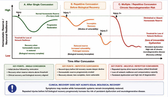
Figure 4: Loss of homeostatic reserve and increased vulnerability to repetitive concussion.
The CHM predicts that neurological recovery depends on restoration of biological homeostasis rather than resolution of clinical symptoms alone. Following a single concussion, homeostatic reserve declines but may progressively recover as structural, metabolic, proteostatic, neuroimmune, and network functions are restored. A subsequent concussion occurring before complete biological recovery is imposed on a nervous system with diminished homeostatic reserve and may therefore produce a disproportionately greater disturbance. Repetitive injuries can progressively lower resilience, prolong recovery, and increase the probability that compensatory mechanisms will fail, resulting in persistent network dysfunction and potentially increasing vulnerability to chronic neurodegeneration. The model therefore distinguishes symptomatic recovery from biological recovery and identifies incomplete restoration of homeostasis as a potential window of vulnerability to repetitive injury.
Perspective: A New Framework for Understanding Concussion
Concussion has traditionally been viewed as a transient biomechanical insult followed by multiple secondary pathological processes that are frequently investigated as distinct therapeutic targets [138]. Although this framework has generated important mechanistic insights, it provides only a limited explanation for how these processes interact to produce delayed and persistent neurological dysfunction.
In this review, we propose the Calpain Homeostasis Model as an alternative framework in which structural, metabolic, proteostatic, neuroimmune, and network abnormalities represent interacting failures of interconnected homeostatic systems. Mechanical injury initiates this process through calcium dysregulation and injury-associated signaling that progressively shift calpain activity from adaptive proteolytic remodeling toward persistent calpain-2-mediated maladaptive proteolysis. Once established, self-reinforcing feed forward mechanisms allow these disturbances to persist and amplify long after the initiating mechanical insult has resolved. These stages are neither discrete nor strictly sequential but overlap and mutually reinforce one another as pathology evolves. The CHM does not replace existing mechanisms of concussion pathology; rather, it provides a physiological framework that explains how these well-established mechanisms become integrated into a progressive process of failing neuronal homeostasis.
A central feature of the model is the progressive loss of biological resilience. Early after injury, compensatory mechanisms may preserve neuronal and network function despite ongoing cellular disturbances. As structural integrity, metabolic reserve, proteostatic capacity, neuroimmune regulation, and network organization progressively deteriorate, the capacity to restore homeostasis declines. The transition from reversible dysfunction to persistent neurological impairment therefore reflects not simply accumulation of molecular damage but progressive exhaustion of the systems that normally enable recovery.
This framework may explain several defining features of concussion, including persistent symptoms despite limited neuronal loss, the disproportionate consequences of repetitive injury, and increased vulnerability to chronic neurodegeneration [136,137]. In each case, outcome depends not only on the magnitude of the initiating insult but also on the pre-existing homeostatic state and the capacity of the nervous system to recover. Importantly, clinical recovery and biological recovery may therefore not be equivalent.
The model also suggests a different therapeutic objective. Rather than targeting individual downstream mediators [7-139], restoration of calpain homeostasis could potentially interrupt several interacting pathological processes while preserving mechanisms required for physiological remodeling. Selective inhibition of calpain-2, while sparing calpain-1-dependent functions, represents one strategy for testing this hypothesis. Therapeutic efficacy may depend not simply on time after injury but on intervention before homeostatic reserve has declined beyond the capacity for recovery.
The value of this framework ultimately depends on its experimental testability. The model predicts that biological recovery may lag behind symptomatic recovery; that vulnerability to repetitive concussion should increase when homeostatic restoration remains incomplete; that biomarkers reflecting different homeostatic domains should provide complementary information about disease stage; and that early restoration of calpain homeostasis should attenuate abnormalities across multiple downstream systems. These predictions provide experimentally and clinically testable criteria by which the CHM can be evaluated and refined.
Although developed in the context of concussion, elements of the model may extend to neurological disorders in which calpain dysregulation accompanies progressive failure of neuronal homeostasis. Determining whether similar relationships operate in other forms of acute and chronic neurodegeneration will require direct experimental validation rather than extrapolation from concussion alone.
Ultimately, the Calpain Homeostasis Model proposes that the long-term consequences of concussion arise not simply from the initial mechanical insult, but from the progressive inability of the nervous system to preserve its internal homeostasis. Whether this framework withstands the test of future experimentation will depend on its ability to generate new hypotheses, improve biomarker discovery, guide disease-modifying therapies, and deepen our understanding of how neuronal homeostasis is maintained—or lost—following brain injury.
Conflict of Interest
Drs. Baudry and Bi are co-founders if NeurAegis, Inc, a start-up biopharmaceutical company focusing on the development of selective calpain-2 inhibitors for the treatment of various neurodegenerative disorders.
ACKNOWLEDGEMENTS
The authors want to thank all the many collaborators, postdocs, graduate and undergraduate students and technicians who have worked on various aspects of the work discussed in the review. They also acknowledge the financial support from many grants from various Federal Agencies, including the NIH and the DoD which have made this work possible. A special thank you for Dr. Gary Lynch who was instrumental for getting us starting to work on calpains and not to be afraid of proposing bold general principles.
REFERENCES
- Hornburg K, Cassianni CE, Muhammad SM, Muñoz Eusse JE, Dixon RC. Concussion: A Comprehensive Review of Current Evidence. Curr Rev Musculoskelet Med. 2026; 19: 57.
- Mavroudis I, Kazis D, Petridis FE, Balmus IM, Papaliagkas V, Ciobica The Association Between Traumatic Brain Injury and the Risk of Cognitive Decline: An Umbrella Systematic Review and Meta-Analysis. Brain Sci. 2024; 14: 1188.
- Hadanny A, Efrati S. Persistent post-concussion syndrome: pathophysiology, diagnosis, current and evolving treatment strategies. Expert Rev Neurother. 2025; 25: 959-971.
- Graham NSN, Zimmerman KA, Hain J, Rooney E, Lee Y, Del Giovane M, et al. Biomarker evidence of neurodegeneration in mid-life former rugby players. Brain. 2025; 148: 2684-2697.
- Giza CC, Hovda DA. The new neurometabolic cascade of concussion. Neurosurgery. 2014; 75: S24-S33.
- Flavin WP, Bickart KC, Le Belle JE, Babikian T, Harris MB, Kamins J, et al. Evolution of the Neurometabolic Cascade of Traumatic Brain Injury. Neurol Clin. 2026; 44: 393-414.
- Heslot C, Azouvi P, Perdrieau V, Granger A, Lefèvre-Dognin C, Cogné M. A Systematic Review of Treatments of Post-Concussion Symptoms. J Clin Med. 2022; 11: 6224.
- Bielanin JP, Metwally SAH, Paruchuri SS, Sun D. An overview of mild traumatic brain injuries and emerging therapeutic targets. Neurochem Int. 2024; 172: 105655.
- Cook AM, Michas M, Robbins B. Update on Neuroprotection after Traumatic Brain Injury. CNS Drugs. 2025; 39: 473-484.
- Hossain I, Blennow K, Posti JP, Zetterberg H. Tau as a fluid biomarker of concussion and neurodegeneration. Concussion. 2022; 7: CNC98.
- Padmakumar S, Kulkarni P, Ferris CF, Bleier BS, Amiji MM. Traumatic brain injury and the development of parkinsonism: Understanding pathophysiology, animal models, and therapeutic targets. Biomed Pharmacother. 2022; 149: 112812.
- Baudry M, Bi X. Calpain-1 and Calpain-2 in the Brain: What Have We Learned from 45 Years of Research? Cells. 2025 Aug; 14: 1301.
- Croall DE, Ersfeld K. The calpains: modular designs and functional diversity. Genome Biol. 2007; 8: 218.
- GUROFF G. A NEUTRAL, CALCIUM-ACTIVATED PROTEINASE FROM THE SOLUBLE FRACTION OF RAT BRAIN. J Biol Chem. 1964; 239: 149-155.
- Baudry M, W Su, X Bi. The calpain proteolytic system., in Encyclopedia of Cell Biology, R. Bradshaw, Hart GW, Stahl PD, Editor. Elsevier. 2023; 852-864.
- Nixon RA. A “protease activation cascade” in the pathogenesis of Alzheimer’s disease. Ann N Y Acad Sci. 2000; 924: 117-131.
- Ray SK, Banik NL. Calpain and its involvement in the pathophysiology of CNS injuries and diseases: therapeutic potential of calpain inhibitors for prevention of neurodegeneration. Curr Drug Targets CNS Neurol Disord. 2003; 2: 173-189.
- Saito K, Elce JS, Hamos JE, Nixon RA. Widespread activation of calcium-activated neutral proteinase (calpain) in the brain in Alzheimer disease: a potential molecular basis for neuronal degeneration. Proc Natl Acad Sci U S A. 1993; 90: 2628-2632.
- Vanderklish PW, Bahr BA. The pathogenic activation of calpain: a marker and mediator of cellular toxicity and disease states. Int J Exp Pathol. 2000; 81: 323-339.
- Potz BA, Abid MR, Sellke FW. Role of Calpain in Pathogenesis of Human Disease Processes. J Nat Sci. 2016; 2: e218.
- Camins A, Verdaguer E, Folch J, Pallàs M. Involvement of calpain activation in neurodegenerative processes. CNS Drug Rev. 2006; 12: 135-148.
- Ivy G, Seubert P, Lynch G, Baudry M. Lesions of entorhinal cortex produce a calpain-mediated degradation of brain spectrin in dentate gyrus. II. Anatomical studies. Brain Res. 1988; 459: 233-240.
- Nelson WJ, Traub P. Proteolysis of vimentin and desmin by the Ca2+-activated proteinase specific for these intermediate filament proteins. Mol Cell Biol. 1983; 3: 1146-1156.
- McGinnis KM, Whitton MM, Gnegy ME, Wang KK. Calcium/calmodulin-dependent protein kinase IV is cleaved by caspase-3 and calpain in SH-SY5Y human neuroblastoma cells undergoing apoptosis. J Biol Chem. 1998; 273: 19993-20000.
- Lu X, Wyszynski M, Sheng M, Baudry M. Proteolysis of glutamate receptor-interacting protein by calpain in rat brain: implications for synaptic plasticity. J Neurochem. 2001; 77: 1553-1560.
- Lu X, Rong Y, Baudry M. Calpain-mediated degradation of PSD-95 in developing and adult rat brain. Neurosci Lett. 2000; 286: 149-153.
- Pain S, Monstero-Lastres A, Falet H, Brohard-Bohn B, Fraiz N, Bachelot-Loza C, et al. Calpain controls the balance between protein tyrosine kinase and tyrosine phosphatase activities during platelet activation. FEBS Lett. 1999; 453: 119-123.
- Guttmann RP, Sokol S, Baker DL, Simpkins KL, Dong Y, Lynch DR. Proteolysis of the N-methyl-d-aspartate receptor by calpain in situ. J Pharmacol Exp Ther. 2002; 302: 1023-1030.
- Saatman KE, Murai H, Bartus RT, Smith DH, Hayward NJ, Perri BR, et al. Calpain inhibitor AK295 attenuates motor and cognitive deficits following experimental brain injury in the rat. Proc Natl Acad Sci U S 1996; 93: 3428-3433.
- Bartus RT, Elliott PJ, Hayward NJ, Dean RL, Harbeson S, Straub JA, et al. Calpain as a novel target for treating acute neurodegenerative disorders. Neurol Res. 1995; 17: 249-258.
- Saez ME, Ramirez-Lorca R, Moron FJ, Ruiz A. The therapeutic potential of the calpain family: new aspects. Drug Discov Today. 2006; 11: 917-923.
- Wang Y, Bi X, Baudry M. Calpain-2 as a therapeutic target for acute neuronal injury. Expert Opin Ther Targets. 2018; 22: 19-29.
- Schallerer C, Neuschmid S, Ehrlich BE, McGuone D. Calpain in Traumatic Brain Injury: From Cinderella to Central Player. Cells. 2025; 14: 1253.
- Wang KK. Calpain and caspase: can you tell the difference? Trends Neurosci. 2000; 23: 20-26.
- Schulz DJ. Plasticity and stability in neuronal output via changes in intrinsic excitability: it’s what’s inside that counts. J Exp Biol. 2006; 209: 4821-4827.
- Wu HY, Lynch DR. Calpain and synaptic function. Mol Neurobiol. 2006; 33: 215-236.
- Storr SJ, Carragher NO, Frame MC, Parr T, Martin SG. The calpain system and cancer. Nat Rev Cancer. 2011; 11: 364-374.
- Baudry M, Bi X. Calpain-1 and Calpain-2: The Yin and Yang of Synaptic Plasticity and Neurodegeneration. Trends Neurosci. 2016; 39: 235-245.
- Abbott LF, Nelson SB. Synaptic plasticity: taming the beast. Nat Neurosci. 2000; 3 : 1178-1183.
- Goll DE, Thompson VF, Li H, Wei W, Cong J. The calpain system. Physiol Rev. 2003; 83: 731-801.
- Ono Y, Sorimachi H. Calpains: an elaborate proteolytic system. Biochim Biophys Acta. 2012; 1824: 224-236.
- Spinozzi S, Albini S, Best H, Richard I. Calpains for dummies: What you need to know about the calpain family. Biochim Biophys Acta Proteins Proteom. 2021; 1869: 140616.
- Wang Y, Zhu G, Briz V, Hsu YT, Bi X, Baudry M. A molecular brake controls the magnitude of long-term potentiation. Nat Commun. 2014; 5: 3051.
- Kawamoto EM, Vivar C, Camandola S. Physiology and pathology of calcium signaling in the brain. Front Pharmacol. 2012; 3: 61.
- Brini M, Calì T, Ottolini D, Carafoli E. Neuronal calcium signaling: function and dysfunction. Cell Mol Life Sci. 2014; 71: 2787-2814.
- Amrapali Vishwanath A, Comyn T, Mira RG, Brossier C, Pascual-Caro C, Faour M, et al. Mitochondrial Ca2+ efflux controls neuronal metabolism and long-term memory across species. Nat Metab. 2026; 8: 467-488.
- Giza CC, Hovda DA. The Neurometabolic Cascade of Concussion. J Athl Train. 2001; 36: 228-235.
- MacFarlane MP, Glenn TC. Neurochemical cascade of concussion. Brain Inj. 2015; 29: 139-153.
- Glading A, Bodnar RJ, Reynolds IJ, Shiraha H, Satish L, Potter DA, et al. Epidermal growth factor activates m-calpain (calpain II), at least in part, by extracellular signal-regulated kinase-mediated phosphorylation. Mol Cell Biol. 2004; 24: 2499-2512.
- Zadran S, Jourdi H, Rostamiani K, Qin Q, Bi X, Baudry M. Brain- derived neurotrophic factor and epidermal growth factor activate neuronal m-calpain via mitogen-activated protein kinase-dependent phosphorylation. J Neurosci. 2010; 30: 1086-1095.
- Wang Y, RA Hall, M Lee, A Kamgar-Parsi, X Bi, M Baudry, The tyrosine phosphatase PTPN13/FAP-1 links calpain-2, TBI and tau tyrosine phosphorylation. Sci Rep, 2017; 7: 11771.
- Wang Y, Liu Y, Bi X, Baudry M. Calpain-1 and Calpain-2 in the Brain: New Evidence for a Critical Role of Calpain-2 in Neuronal Death. Cells. 2020; 9: 2698.
- Nuthalapati P, Holmes SE, Altalib HH, Fesharaki-Zadeh A. Synaptic Pathology in Traumatic Brain Injury and Therapeutic Insights. Int J Mol Sci. 2025; 26: 9604.
- Dong YN, Waxman EA, Lynch DR. Interactions of postsynaptic density-95 and the NMDA receptor 2 subunit control calpain-mediated cleavage of the NMDA receptor. J Neurosci. 2004; 24: 11035-11045.
- Yuen EY, Ren Y, Yan Z. Postsynaptic density-95 (PSD-95) and calcineurin control the sensitivity of N-methyl-D-aspartate receptors to calpain cleavage in cortical neurons. Mol Pharmacol. 2008; 74: 360-370.
- Hou ST, Jiang SX, Smith RA. Permissive and repulsive cues and signalling pathways of axonal outgrowth and regeneration. Int Rev Cell Mol Biol. 2008; 267: 125-181.
- Yu L, Rostamiani K, Hsu YT, Wang Y, Bi X, Baudry M. Calpain-mediated regulation of stargazin in adult rat brain. Neuroscience. 2011; 178: 13-20.
- Mavroudis I, Petridis F, Ciobîc? A, Padurariu M, Papagiannopoulos S, Kazis D. Functional Neurological Symptoms After Mild Traumatic Brain Injury: A Scoping Review and Framework for Differentiating Functional and Organic Post-Concussion Presentations. Life (Basel). 2026; 16: 926.
- Ma S, Zuo Y. Synaptic modifications in learning and memory - A dendritic spine story. Semin Cell Dev Biol. 2022; 125: 84-90.
- Wang Y, Liu Y, Lopez D, Lee M, Dayal S, Hurtado A, et al. Protection against TBI-Induced Neuronal Death with Post-Treatment with a Selective Calpain-2 Inhibitor in Mice. J Neurotrauma. 2018; 35: 105-117.
- Wang Y, Liu Y, Nham A, Sherbaf A, Quach D, Yahya E, et al. Calpain-2 as a therapeutic target in repeated concussion-induced neuropathy and behavioral impairment. Sci Adv. 2020; 6: eaba5547.
- Baudry M, Reece T, Shahi R, Ta K, Bi X. Calpain-2 Inhibition or Deletion Enhances Levels of the Transcription Factor, MEIS2, and Stimulates Neurogenesis. Curr Neuropharmacol. 2026.
- Ehlers MD. Molecular morphogens for dendritic spines. Trends Neurosci. 2002; 25: 64-67.
- Ringger NC, O’Steen BE, Brabham JG, Silver X, Pineda J, Wang KK, et al. A novel marker for traumatic brain injury: CSF alphaII-spectrin breakdown product levels. J Neurotrauma. 2004; 21: 1443-1456.
- Pineda JA, Lewis SB, Valadka AB, Papa L, Hannay HJ, Heaton SC, et al. Clinical significance of alphaII-spectrin breakdown products in cerebrospinal fluid after severe traumatic brain injury. J Neurotrauma. 2007; 24: 354-366.
- Brophy GM, Pineda JA, Papa L, Lewis SB, Valadka AB, Hannay HJ, et al. alphaII-Spectrin breakdown product cerebrospinal fluid exposure metrics suggest differences in cellular injury mechanisms after severe traumatic brain injury. J Neurotrauma. 2009; 26: 471-479.
- Liu S, Yin F, Zhang J, Qian Y. The role of calpains in traumatic brain injury. Brain Inj. 2014; 28: 133-137.
- Olatona OA, Sterben SP, Kansakar SBS, Symes AJ, Liaudanskaya V. Mitochondria: the hidden engines of traumatic brain injury-driven neurodegeneration. Front Cell Neurosci. 2025; 19: 1570596.
- Wu J, Lipinski MM. Autophagy in Neurotrauma: Good, Bad, or Dysregulated. Cells. 2019; 8: 693.
- Lin PH, Kuo LT, Luh HT. The Roles of Neurotrophins in Traumatic Brain Injury. Life (Basel). 2021; 12: 26.
- Lifshitz, J., P.G. Sullivan, D.A. Hovda, T. Wieloch, and T.K. McIntosh, Mitochondrial damage and dysfunction in traumatic brain injury. Mitochondrion, 2004; 4: 705-713.
- Pekkurnaz, G. and X. Wang, Mitochondrial heterogeneity and homeostasis through the lens of a neuron. Nat Metab, 2022; 4: 802-812.
- Pandya, J.D., V.N. Nukala, and P.G. Sullivan, Concentration dependent effect of calcium on brain mitochondrial bioenergetics and oxidative stress parameters. Front Neuroenergetics, 2013; 5: 10.
- Dan Dunn, J., L.A. Alvarez, X. Zhang, and T. Soldati, Reactive oxygen species and mitochondria: A nexus of cellular homeostasis. Redox Biol, 2015; 6: 472-485.
- Liu, T., L. Wang, L. Tong, Z. Ren, B. Zhao, H. Liu, et al. PDZD8 Dysregulation Mediates RVLM Neuronal Hyperexcitation Via Activation of Ca(2+)-Calpain-2 Signaling in Stress-Induced Hypertension. Mol Neurobiol, 2025; 62: 12539-12558.
- Hu, D., X. Sun, Y. Shang, K. Lundberg, D.J. Adams, and X. Qi, A small-molecule stabilizer of the calpastatin-calpain-2 complex restores mitochondrial function and mitigates neurodegeneration. Sci Adv, 2026; 12: eaeb1174.
- Rao, W., L. Zhang, C. Peng, H. Hui, K. Wang, N. Su, L. Wang, S.H. Dai, Y.F. Yang, T. Chen, P. Luo, and Z. Fei, Downregulation of STIM2 improves neuronal survival after traumatic brain injury by alleviating calcium overload and mitochondrial dysfunction. Biochim Biophys Acta, 2015; 1852: 2402-2413.
- Garbincius, J.F. and J.W. Elrod, Mitochondrial calcium exchange in physiology and disease. Physiol Rev, 2022; 102: 893-992.
- Signoretti, S., A. Marmarou, B. Tavazzi, G. Lazzarino, A. Beaumont, and R. Vagnozzi, N-Acetylaspartate reduction as a measure of injury severity and mitochondrial dysfunction following diffuse traumatic brain injury. J Neurotrauma, 2001; 18: 977-991.
- Keshri, P.K. and S.P. Singh, Unraveling the AKT/ERK cascade and its role in Parkinson disease. Arch Toxicol, 2024; 98: 3169-3190.
- Spinelli, J.B. and M.C. Haigis, The multifaceted contributions of mitochondria to cellular metabolism. Nat Cell Biol, 2018; 20: 745-754.
- Kim, S., S.C. Han, A.J. Gallan, and J.P. Hayes, Neurometabolic indicators of mitochondrial dysfunction in repetitive mild traumatic brain injury. Concussion, 2017; 2: CNC48.
- Mbye, L.H., I.N. Singh, K.M. Carrico, K.E. Saatman, and E.D. Hall, Comparative neuroprotective effects of cyclosporin A and NIM811, a nonimmunosuppressive cyclosporin A analog, following traumatic brain injury. J Cereb Blood Flow Metab, 2009; 29: 87-97.
- Zhu, H. and E. Cohen, Regulation of the proteostasis network by the neuronal system. Front Mol Biosci, 2023; 10: 1290118.
- Swerdlow, N.S. and H.M. Wilkins, Mitophagy and the Brain. Int J Mol Sci, 2020; 21: 9661.
- Bao, H., X. Yang, Y. Zhuang, Y. Huang, T. Wang, M. Zhang, et al, The effects of poloxamer 188 on the autophagy induced by traumatic brain injury. Neurosci Lett, 2016; 634: 7-12.
- Geronimo-Olvera, C., T. Montiel, R. Rincon-Heredia, S. Castro-Obregon, and L. Massieu, Autophagy fails to prevent glucose deprivation/glucose reintroduction-induced neuronal death due to calpain-mediated lysosomal dysfunction in cortical neurons. Cell Death Dis, 2017; 8: e2911.
- Sarkar, C., J.W. Jones, N. Hegdekar, J.A. Thayer, A. Kumar, A.I. Faden, M.A. Kane, and M.M. Lipinski, PLA2G4A/cPLA2-mediated lysosomal membrane damage leads to inhibition of autophagy and neurodegeneration after brain trauma. Autophagy, 2020; 16: 466-485.
- Hernandez, M.L., M. Marone, K.M. Gorse, and A.D. Lafrenaye, Cathepsin B Relocalization in Late Membrane Disrupted Neurons Following Diffuse Brain Injury in Rats. ASN Neuro, 2022. 14: p. 17590914221099112.
- Zhang, S., Y. Qiu, Y. Feng, Y. Zhang, Y. Li, B. Wanget al, Calpain-2 Facilitates Autophagic/Lysosomal Defects and Apoptosis in ARPE-19 Cells and Rats Induced by Exosomes from RPE Cells under NaIO(3) Stimulation. Oxid Med Cell Longev, 2023: 3310621.
- Marti-Boltaina, A., D. Aluja, S. Delgado-Tomas, E. Miro-Casas, R. Rodriguez-Lecoq, M. Ruiz-Meana, et al, Calpain-2 inhibition prevents pressure overload–induced lysosomal cathepsin release and cardiac hypertrophy. Life Sciences, 2026; 403: 124614.
- Li, L., J. Tan, Y. Miao, P. Lei, and Q. Zhang, ROS and Autophagy: Interactions and Molecular Regulatory Mechanisms. Cell Mol Neurobiol, 2015; 35: 615-621.
- Zeng, Z., Y. Zhang, W. Jiang, L. He, and H. Qu, Modulation of autophagy in traumatic brain injury. J Cell Physiol, 2020; 235: 1973-1985.
- Lang, J., B. Sun, S. Feng, and G. Sun, Impaired autophagic flux in the human brain after traumatic brain injury. Neuroreport, 2024; 35: 387-398.
- Zhao, Q., Z. Guo, W. Deng, S. Fu, C. Zhang, M. Chen, et al, Calpain 2-mediated autophagy defect increases susceptibility of fatty livers to ischemia-reperfusion injury. Cell Death Dis, 2016; 7: e2186.
- Dikic, I., Proteasomal and Autophagic Degradation Systems. Annu Rev Biochem, 2017; 86: 193-224.
- Sakai, K., T. Fukuda, and K. Iwadate, Immunohistochemical analysis of the ubiquitin proteasome system and autophagy lysosome system induced after traumatic intracranial injury: association with time between the injury and death. Am J Forensic Med Pathol, 2014. 35: 38-44.
- Wang, K.K., Z. Yang, G. Sarkis, I. Torres, and V. Raghavan, Ubiquitin C-terminal hydrolase-L1 (UCH-L1) as a therapeutic and diagnostic target in neurodegeneration, neurotrauma and neuro-injuries. Expert Opin Ther Targets, 2017; 21: 627-638.
- Alam, A., E.P. Thelin, T. Tajsic, D.Z. Khan, A. Khellaf, R. Patani, et al, Cellular infiltration in traumatic brain injury. J Neuroinflammation, 2020; 17: 328.
- Xu, Y., F. Lin, G. Liao, J. Sun, W. Chen, and L. Zhang, Ripks and Neuroinflammation. Mol Neurobiol, 2024; 61: 6771-6787.
- Simon, D.W., M.J. McGeachy, H. Bayir, R.S.B. Clark, D.J. Loane, and P.M. Kochanek, The far-reaching scope of neuroinflammation after traumatic brain injury. Nat Rev Neurol, 2017; 13: 572.
- Obukohwo, O.M., O.A. Oreoluwa, U.O. Andrew, and U.E. Williams, Microglia-mediated neuroinflammation in traumatic brain injury: a review. Mol Biol Rep, 2024; 51: 1073.
- Gao, T., Z. Chen, H. Chen, H. Yuan, Y. Wang, X. Peng, et al, Inhibition of HMGB1 mediates neuroprotection of traumatic brain injury by modulating the microglia/macrophage polarization. Biochem Biophys Res Commun, 2018; 497: 430-436.
- Zhang, C., C. Liu, F. Li, M. Zheng, Y. Liu, L. Li, et al, Extracellular Mitochondria Activate Microglia and Contribute to Neuroinflammation in Traumatic Brain Injury. Neurotox Res, 2022; 40: 2264-2277.
- Sun, B., J. Zhang, Z. Li, J. Wang, C. Zhao, and X. Xu, Role of damage-associated molecular patterns in the pathogenesis and therapeutics of traumatic brain injury. Burns Trauma, 2025; 13: tkaf043.
- Myer, D.J., G.G. Gurkoff, S.M. Lee, D.A. Hovda, and M.V. Sofroniew, Essential protective roles of reactive astrocytes in traumatic brain injury. Brain, 2006; 129: 2761-2772.
- Ramlackhansingh, A.F., D.J. Brooks, R.J. Greenwood, S.K. Bose, F.E. Turkheimer, K.M. Kinnunen, et al, Inflammation after trauma: microglial activation and traumatic brain injury. Ann Neurol, 2011; 70: 374-383.
- Johnson, V.E., J.E. Stewart, F.D. Begbie, J.Q. Trojanowski, D.H. Smith, and W. Stewart, Inflammation and white matter degeneration persist for years after a single traumatic brain injury. Brain, 2013; 136: 28-42.
- Loane, D.J., A. Kumar, B.A. Stoica, R. Cabatbat, and A.I. Faden, Progressive neurodegeneration after experimental brain trauma: association with chronic microglial activation. J Neuropathol Exp Neurol, 2014; 73: 14-29.
- Witcher, K.G., C.E. Bray, T. Chunchai, F. Zhao, S.M. O’Neil, A.J. Gordillo, et al, Traumatic Brain Injury Causes Chronic Cortical Inflammation and Neuronal Dysfunction Mediated by Microglia. J Neurosci, 2021; 41: 1597-1616.
- George, K.K., B.P. Heithoff, O. Shandra, and S. Robel, Mild Traumatic Brain Injury/Concussion Initiates an Atypical Astrocyte Response Caused by Blood-Brain Barrier Dysfunction. J Neurotrauma, 2022; 39: 211-226.
- Rosa, J.M., V. Farre-Alins, M.C. Ortega, M. Navarrete, A.B. Lopez-Rodriguez, A. Palomino-Antolin, et al, TLR4 pathway impairs synaptic number and cerebrovascular functions through astrocyte activation following traumatic brain injury. Br J Pharmacol, 2021; 178: 3395-3413.
- Beschorner, R., K. Dietz, N. Schauer, M. Mittelbronn, H.J. Schluesener,K. Trautmann, R. Meyermann, et al, Expression of EAAT1 reflects a possible neuroprotective function of reactive astrocytes and activated microglia following human traumatic brain injury. Histol Histopathol, 2007; 22: 515-526.
- Seplovich, G., Y. Bouchi, J.P. de Rivero Vaccari, J.C.M. Pareja, A. Reisner, L. Blackwell, Y. Mechref, K.K. Wang, J.A. Tyndall, B. Tharakan, and F. Kobeissy, Inflammasome links traumatic brain injury, chronic traumatic encephalopathy, and Alzheimer’s disease. Neural Regen Res, 2025; 20: 1644-1664.
- Mamsa, R., K.S. Prabhavalkar, and L.K. Bhatt, Crosstalk between NLRP3 inflammasome and calpain in Alzheimer’s disease. Eur J Neurosci, 2023; 58: 3719-3731.
- Scott, G., H. Zetterberg, A. Jolly, J.H. Cole, S. De Simoni, P.O. Jenkins, et al, Minocycline reduces chronic microglial activation after brain trauma but increases neurodegeneration. Brain, 2018; 141: 459-471.
- Kalra, S., R. Malik, G. Singh, S. Bhatia, A. Al-Harrasi, S. Mohan, et al, Pathogenesis and management of traumatic brain injury (TBI): role of neuroinflammation and anti-inflammatory drugs. Inflammopharmacology, 2022; 30: 1153-1166.
- Das, M., S. Mohapatra, and S.S. Mohapatra, New perspectives on central and peripheral immune responses to acute traumatic brain injury. J Neuroinflammation, 2012; 9: 236.
- Li, L., R. Peng, C. Wang, X. Chen, D. Gheyret, S. Guan, et al, beta2 integrin regulates neutrophil trans endothelial migration following traumatic brain injury. Cell Commun Signal, 2025; 23: 70.
- Mayer, A.R., M.V. Mannell, J. Ling, C. Gasparovic, and R.A. Yeo, Functional connectivity in mild traumatic brain injury. Hum Brain Mapp, 2011; 32: 1825-1835.
- Li, F., L. Lu, S. Shang, L. Hu, H. Chen, P. Wang, et al, Disrupted functional network connectivity predicts cognitive impairment after acute mild traumatic brain injury. CNS Neurosci Ther, 2020; 26: 1083-1091.
- Amgalan, A., A.S. Maher, P. Imms, M.Y. Ha, T.A. Fanelle, and A. Irimia, Functional Connectome Dynamics After Mild Traumatic Brain Injury According to Age and Sex. Front Aging Neurosci, 2022. 14: 852990.
- Espana-Irla, G., E.M. Tinney, M. Ai, M. Nwakamma, and T.P. Morris, Functional Connectivity Patterns Following Mild Traumatic Brain Injury and the Association With Longitudinal Cognitive Function. Hum Brain Mapp, 2025; 46: e70237.
- Bonnelle, V., R. Leech, K.M. Kinnunen, T.E. Ham, C.F. Beckmann, X. De Boissezon, et al, Default mode network connectivity predicts sustained attention deficits after traumatic brain injury. J Neurosci, 2011; 31: 13442-13451.
- Bonnelle, V., T.E. Ham, R. Leech, K.M. Kinnunen, M.A. Mehta, R.J. Greenwood,et al, Salience network integrity predicts default mode network function after traumatic brain injury. Proc Natl Acad Sci U S A, 2012; 109: 4690-4695.
- Dunkley, B.T., K. Urban, L. Da Costa, S.M. Wong, E.W. Pang, and M.J. Taylor, Default Mode Network Oscillatory Coupling Is Increased Following Concussion. Front Neurol, 2018; 9: 280.
- Amir, J., J.K.R. Nair, R. Del Carpio-O’Donovan, A. Ptito, J.K. Chen, J. Chankowsky, et al, Atypical resting state functional connectivity in mild traumatic brain injury. Brain Behav, 2021; 11: e2261.
- Wu, Y.C., J. Harezlak, N.M.H. Elsaid, Z. Lin, Q. Wen, S.M. Mustafi, et al, Longitudinal white-matter abnormalities in sports-related concussion: A diffusion MRI study. Neurology, 2020; 95: e781-e792.
- Ellery, C.S., A.N. Avila, M.G. Papini, M. Fitzgerald, and S.C. Hellewell, Establishing the link between post-concussive symptoms and brain network dysfunction: A systematic scoping review of neuroimaging evidence. Neuroimage Clin, 2026; 49: 103956.
- Meehan, W.P., 3rd, J. Zhang, R. Mannix, and M.J. Whalen, Increasing recovery time between injuries improves cognitive outcome after repetitive mild concussive brain injuries in mice. Neurosurgery, 2012; 71: 885-891.
- Selwyn, R.G., S.J. Cooney, G. Khayrullina, N. Hockenbury, C.M. Wilson, S. Jaiswal, et al, Outcome after Repetitive Mild Traumatic Brain Injury Is Temporally Related to Glucose Uptake Profile at Time of Second Injury. J Neurotrauma, 2016; 33: 1479-1491.
- Brett, B.L., Y.C. Wu, S.M. Mustafi, A.J. Saykin, K.M. Koch, A.S. Nencka, et al, The Association Between Persistent White-Matter Abnormalities and Repeat Injury After Sport-Related Concussion. Front Neurol, 2019; 10: 1345.
- Churchill, N.W., M.G. Hutchison, D. Richards, G. Leung, S.J. Graham, and T.A. Schweizer, Neuroimaging of sport concussion: persistent alterations in brain structure and function at medical clearance. Sci Rep, 2017; 7: 8297.
- Tabor, J.B., B.L. Brett, L. Nelson, T. Meier, L.C. Penner, A.R. Mayer, et al, Role of biomarkers and emerging technologies in defining and assessing neurobiological recovery after sport-related concussion: a systematic review. Br J Sports Med, 2023; 57: 789-797.
- O’Brien, W.T., G. Spitz, B. Xie, B.P. Major, S. Mutimer, L.P. Giesler, et al, Biomarkers of Neurobiologic Recovery in Adults With Sport-Related Concussion. JAMA Netw Open, 2024. 7: e2415983.
- Vagnozzi, R., B. Tavazzi, S. Signoretti, A.M. Amorini, A. Belli, M. Cimatti, et al, Temporal window of metabolic brain vulnerability to concussions: mitochondrial-related impairment--part I. Neurosurgery, 2007; 61: 379-88; discussion 388-389.
- Prins, M.L., D. Alexander, C.C. Giza, and D.A. Hovda, Repeated mild traumatic brain injury: mechanisms of cerebral vulnerability. J Neurotrauma, 2013; 30: 30-38.
- Kochanek, P.M., T.C. Jackson, R.M. Jha, R.S.B. Clark, D.O. Okonkwo, H. Bayir, S.M. Poloyac, et al, Paths to Successful Translation of New Therapies for Severe Traumatic Brain Injury in the Golden Age of Traumatic Brain Injury Research: A Pittsburgh Vision. J Neurotrauma, 2020; 37: 2353-2371.
- Kabadi, S.V. and A.I. Faden, Neuroprotective strategies for traumatic brain injury: improving clinical translation. Int J Mol Sci, 2014; 15: 1216-1236.