Therapeutic Potential of Immunomodulatory Hydrogels in Infected and Non-Infected Wounds: A Comprehensive Review
- 1. Department of Microbiology, University of Jhang, Pakistan
Abstract
Wound healing is a precisely orchestrated cascade involving coordinated interactions among immune cells, cytokines, growth factors, and extracellular matrix (ECM). Although acute wounds heal through orderly phases, infected and chronic wounds exhibit dysregulation with persistent inflammation, impaired immune resolution, and compromised regeneration. Conventional dressings provide passive barrier functions but limited therapeutic intervention. Immunomodulatory hydrogels represent sophisticated platforms that actively reprogram the wound immune microenvironment. These three-dimensional hydrophilic networks deliver bioactive agents, modulate immune responses, provide antimicrobial protection, and mimic native ECM. By controlling inflammation, promoting macrophage polarization toward pro-regenerative phenotypes, and stimulating angiogenesis and re-epithelialization, these materials address fundamental healing barriers. In infected wounds, they integrate antimicrobial and biofilm-disruptive mechanisms while preserving immune homeostasis. In non-infected lesions, they facilitate inflammatory resolution and promote regenerative repair. This review critically examines immunomodulatory hydrogel design, mechanisms, preclinical evidence, clinical translation, and future directions.
Keywords
• Wound healing; Immunomodulatory hydrogels; Macrophage polarization; Infected wounds; Chronic wounds; Biomaterials; Immune regulation; Drug delivery
Citation
Manzoor S (2026) Therapeutic Potential of Immunomodulatory Hydrogels in Infected and Non-Infected Wounds: A Comprehensive Review. Arch Emerg Med Crit Care 10(1): 1078.
INTRODUCTION
Wound healing restores tissue integrity through four overlapping phases: hemostasis, inflammation, proliferation, and remodeling, coordinated by immune cells, growth factors, and ECM [1,2]. Despite skin’s regenerative capacity, many wounds become chronic—diabetic foot ulcers, venous leg ulcers, and pressure injuries impose significant global burdens [3]. These wounds exhibit persistent inflammation, impaired angiogenesis, excessive protease activity, and diminished cellular responsiveness. Infected wounds add microbial colonization and biofilm formation that subvert host immunity and prolong healing [4,5].
The immune system coordinates healing. Innate immune cells—neutrophils and macrophages—eliminate pathogens and secrete cytokines that activate fibroblasts, endothelial cells, and epithelial cells [6]. Macrophages are pivotal due to their phenotypic plasticity: pro-inflammatory (M1) macrophages orchestrate early defense, while pro regenerative (M2) macrophages facilitate angiogenesis, ECM deposition, and remodeling. Non-healing wounds show dysregulated M1 predominance [7,8].
Traditional wound care relies on passive dressings (gauze, foams, films) that maintain moisture but inadequately address biological dysfunctions. Advanced therapies (antimicrobials, growth factors, negative pressure) have benefits but are constrained by narrow therapeutic indices and delivery challenges [9,10]. Hydrogels have emerged as promising biomaterial platforms. These hydrophilic networks absorb water, provide moist environments, conform to wound geometries, and mimic ECM [11]. Their versatility enables precise tuning of mechanical properties, degradation, and bioactivity [12].
Hydrogels now serve as active therapeutic platforms for localized delivery of growth factors, anti-inflammatory drugs, antimicrobials, nucleic acids, and cells [10]. Immunomodulatory hydrogels represent a paradigm shift—they actively control immune cell activity and inflammatory milieu. Rather than suppressing inflammation, they normalize immune balance by regulating macrophage polarization [9]. In infected wounds, they incorporate antimicrobial effects alongside immune control [13,14]. In non-infected wounds, they recreate regenerative microenvironments [15]. Stimuli responsive “smart” hydrogels react to pH, ROS, enzymes, or temperature changes, enabling dynamic treatment adjustment [16].
Despite promise, obstacles remain: biocompatibility, manufacturing scalability, regulatory pathways, and understanding hydrogel-immune interactions [17]. This review comprehensively examines therapeutic prospects of immunomodulatory hydrogels for infected and non infected wounds (Figure 1).
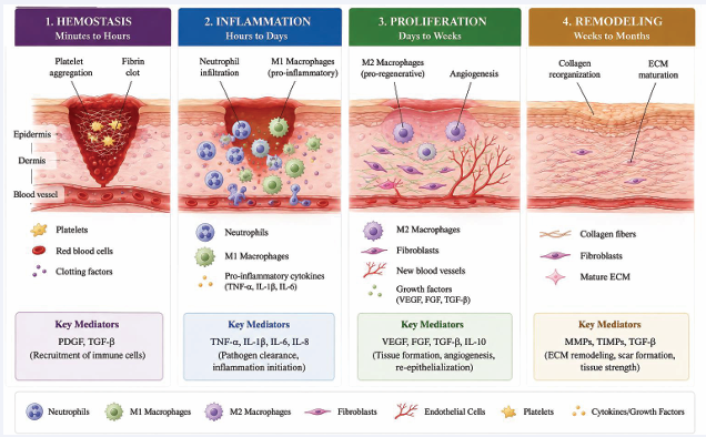
Figure 1: Schematic of wound healing phases and immune cell dynamics. Four-panel illustration depicting hemostasis (platelet aggregation, fibrin clot, PDGF/TGF-β), inflammation (neutrophil infiltration, M1 macrophages, TNF-α/IL-1β/IL-6), proliferation (M2 macrophages, angiogenesis, fibroblast activation, VEGF/FGF/TGF-β), and remodeling (collagen reorganization, ECM maturation).
BIOLOGY OF WOUND HEALING AND IMMUNE REGULATION
Following injury, hemostasis begins with vasoconstriction and platelet aggregation, forming a fibrin clot. Platelets release PDGF and TGF-β, recruiting immune cells [18,19]. The inflammatory phase dominates the first days: neutrophils clear pathogens via phagocytosis and ROS. Macrophages then become master regulators, transitioning from M1 (TNF-α, IL-1β) to M2 (VEGF, FGF, IL-10, TGF-β), promoting angiogenesis, fibroblast activation, and ECM deposition [6,7,20]. The proliferative phase involves granulation tissue, angiogenesis, and re-epithelialization. Remodeling involves collagen reorganization over weeks to months [21,22].
DAMPs and PAMPs activate PRRs including TLRs, triggering NF-κB and pro-inflammatory cytokine transcription [20,23]. Macrophage polarization is regulated by interconnected pathways: M1 via IFN-γ/LPS through JAK/STAT1 and NF-κB; M2 via IL-4/IL-13 through STAT6 or IL-10 through STAT3. Chronic wounds show M1 predominance with impaired M2 transition [7,8].
Pathological wounds show sustained inflammation, failed macrophage polarization, diminished growth factor responsiveness, and biofilm formation. Elevated pro inflammatory cytokines, ROS, and MMP activity create unfavorable microenvironments [24,25]. Biofilms protect bacteria and sustain TLR activation, causing frustrated phagocytosis and chronic inflammation [26,27] (Figure 2).
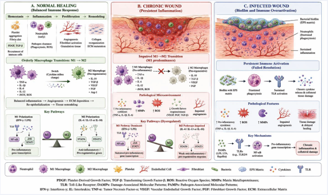
Figure 2: Immune dysregulation in chronic and infected wounds. (A) Normal healing: orderly M1→M2 transition with balanced inflammation. (B) Chronic wound: persistent M1, elevated cytokines/MMP/ROS, impaired angiogenesis. (C) Infected wound: biofilm with EPS matrix, frustrated phagocytosis, sustained TLR activation, collateral damage.
PATHOPHYSIOLOGY OF CHRONIC AND INFECTED WOUNDS
Chronic wounds are arrested in inflammation with persistent cytokines (TNF-α, IL-1β, IL-6), excessive ROS, elevated MMP activity, reduced growth factors, impaired angiogenesis, and cellular senescence [24,25]. Diabetic wounds involve AGE-RAGE activation of NF-κB, impaired neutrophil function, and reduced growth factor responsiveness [28,29].
Biofilms (present in ~80% chronic wounds) provide physical protection and create microenvironments promoting bacterial persistence [5,4]. Continuous PAMP release sustains inflammation without bacterial clearance, causing frustrated phagocytosis and chronic inflammation [26,27].
HYDROGELS AS ADVANCED BIOMATERIALS
Hydrogels absorb water, maintain moist environments, conform to wounds, and provide barriers. Their porous structure enables exudate management and oxygen/ nutrient diffusion [10-13] (Table 1).
Table 1: Classification and Physicochemical Properties of Hydrogel Systems
|
Hydrogel Type |
Representative Polymers |
Crosslinking |
Modulus (kPa) |
Pore Size (μm) |
Degradation |
Advantages |
Limitations |
|
Natural |
Collagen, chitosan, HA, alginate |
Physical/chemical |
1-50 |
50-300 |
Days-weeks |
Biocompatible, bioactive |
Batch variability, weak mechanics |
|
Synthetic |
PEG, PVA, PCL, PLGA |
Chemical |
5-200 |
10-200 |
Weeks-months |
Tunable, reproducible |
Biologically inert |
|
Hybrid |
GelMA, HAMA, PEG- collagen |
Photo/enzymatic |
10-150 |
20-250 |
Weeks |
Bioactive + tunable |
Complex synthesis |
|
Injectable |
Pluronic, PNIPAAm, alginate |
Thermo/ionic |
0.1-50 |
50-200 |
Days-weeks |
Minimally invasive |
Premature gelation |
|
Self-healing |
Schiff base, disulfide |
Dynamic covalent |
1-50 |
50-300 |
Weeks-months |
Autonomous repair |
Complex synthesis |
|
Conductive |
PEDOT:PSS, polypyrrole |
Oxidative |
10-200 |
50-250 |
Weeks-months |
Electrical stimulation |
Toxicity concerns |
Natural polymers (collagen, chitosan, HA, alginate) offer bioactivity but have batch variability and weak mechanics [11,30]. Synthetic polymers (PEG, PVA, PLGA) provide tunable properties but lack bioactivity [10,31].Hybrid systems (GelMA, HAMA) combine advantages [30,31]. Hydrogels now encapsulate and release growth factors, antimicrobials, anti-inflammatory agents, nucleic acids, and cells in controlled fashion [10,13]. Release kinetics are tuned via crosslinking density, degradation rate, and architecture [10,11].
IMMUNOMODULATORY HYDROGELS: CONCEPTS AND MECHANISMS
Immunomodulatory hydrogels actively regulate immune cell activities, inflammatory signals, and tissue repair pathways, promoting transition from pro inflammatory to pro-regenerative milieus [9,32].
Macrophage polarization is a key design goal [7,23]. Approaches include sustained cytokine delivery (IL-4, IL-13), ECM-derived peptides, small molecule immunomodulators, physical property manipulation, and MSC-derived secretome [13,14]. IL-4/IL-13 activate STAT6; some hydrogel compositions directly interact with macrophage receptors [15,33] (Table 2).
Table 2: Immunomodulatory Agents Incorporated into Hydrogels
|
Agent Class |
Examples |
Mechanism |
Pathways |
Preclinical Outcomes |
Clinical Status |
|
Growth Factors |
VEGF, PDGF, FGF, EGF, TGF-β |
Angiogenesis, proliferation |
PI3K/Akt, MAPK/ ERK |
↑Angiogenesis 2-3×, ↑Closure 40-70% |
FDA-approved PDGF |
|
Anti-inflammatory Cytokines |
IL-4, IL-10, IL-13 |
M2 polarization |
JAK/STAT, NF-κB |
↑M2/M1 3-5×, ↓TNF-α 50-80% |
Preclinical-Phase II |
|
Small Molecules |
Curcumin, resveratrol |
NF-κB inhibition, ROS scavenging |
NF-κB, Nrf2 |
↓ROS 50-70%, ↑Closure 30-50% |
Preclinical |
|
Immunomodulatory Peptides |
RGD, YIGSR |
Cell adhesion, integrin signaling |
FAK/Src, PI3K/Akt |
↑M2 polarization 2-3× |
Preclinical |
|
Nucleic Acids |
siRNA, miRNA |
Gene silencing |
RNAi, RISC |
↓Target genes 70-90% |
Preclinical |
|
Extracellular Vesicles |
MSC exosomes |
Paracrine signaling |
Multiple |
↑M2 3-5×, ↑Closure 50-80% |
Preclinical-Phase I |
|
Living Cells |
MSCs, macrophages |
Paracrine secretion |
Multiple |
↑Closure 50-80% |
Clinical trials |
Hydrogels regulate neutrophil, dendritic cell, and lymphocyte migration and function via chemoattractants and adhesion ligands [20,23]. Neutrophil modulation prevents excessive activation and tissue damage [23,34]. Agents are incorporated via physical entrapment, covalent conjugation, nanoparticle encapsulation, or affinity binding [10,11]. Stimuli-responsive systems enable dynamic adaptation [14,12]. Hydrogels modulate NF-κB (via inhibitors or ROS scavenging), JAK/STAT (via cytokine release), and TGF-β/Smad pathways. Inflammasome (NLRP3) is another target [7,20,23,35].
STIMULI-RESPONSIVE SMART HYDROGEL SYSTEMS
ROS-cleavable linkers (thioketal, boronate ester) release antioxidants in oxidative stress. Preclinical studies show 60-80% ROS reduction, 50-70% cytokine reduction, and 30-50% accelerated closure [16,17,36].
Infected wounds are acidic (pH 5.0-6.0). Polymers with ionizable groups enable selective antimicrobial release. Bacterial burden reduced by 3-5 log [12,14,26,27].
MMP-cleavable peptides release payloads in high protease environments. MMP-2/9 are elevated 10-100× in chronic wounds. Wound closure improves 40-60% [24 27].
Phenylboronic acid or glucose oxidase systems release insulin/growth factors in diabetic wounds [15]. pH/ROS dual-responsive systems target infected chronic wounds [14,31] (Table 3).
Table 3: Stimuli-Responsive Hydrogel Systems
|
Type |
Chemical Mechanism |
Trigger |
Payloads |
Release Profile |
Preclinical Efficacy |
Status |
|
pH-Responsive |
Ionizable groups, acid-labile bonds |
pH 5.0-6.0 |
Antimicrobials, anti-inflammatories |
10-20% burst, 70-90% over 5-14 days |
↑Bacterial reduction 3-5 log |
Clinical trials |
|
ROS-Responsive |
Thioketal, boronate ester |
H?O? 50-200 μM |
Antioxidants, anti-inflammatories |
5-15% burst, ROS-triggered over 3-10 days |
↓ROS 60-80%, ↑Closure 50-70% |
Preclinical-Phase I |
|
MMP-Responsive |
MMP-cleavable peptides |
MMP-2/9 10- 100× |
Growth factors, anti-inflammatories |
Minimal burst, MMP-dependent over 7-21 days |
↑Closure 40-60% |
Preclinical |
|
Bacterial Enzyme |
Bacterial protease peptides |
Infection |
Antimicrobials |
Infection-triggered over 1-7 days |
↓Bacteria 4-6 log |
Preclinical |
|
Glucose-Responsive |
Phenylboronic acid, glucose oxidase |
Glucose >200 mg/dL |
Insulin, growth factors |
Pulsatile over 7-28 days |
↑Angiogenesis 2-3× |
Preclinical |
|
Temperature-Responsive |
LCST polymers |
37-39°C |
Multiple |
Temperature-modulated over 3-14 days |
↑Closure 30-50% |
Some clinical use |
ANTIMICROBIAL AND ANTI-BIOFILM STRATEGIES
Agents include metal nanoparticles (silver, gold), antimicrobial peptides (LL-37), antibiotics, NO donors, and natural compounds [26,27]. Localized release achieves high concentrations with minimal systemic exposure. Silver nanoparticles reduce bacterial burden by 4-7 log [17,36].
Strategies include EPS degradation (dispersin B, DNase), NO release, iron chelation, quorum sensing inhibition, and photothermal approaches [5,26]. Combination hydrogels show synergistic biofilm eradication (>90% reduction) [17,33]. Optimal systems achieve pathogen clearance (4-6 log reduction) while maintaining IL-10/TNF-α ratios >1.0, improving closure 2-3 fold [15,33] (Table 4).
Table 4: Antimicrobial and Anti-Biofilm Hydrogel Strategies
|
Strategy |
Active Agent |
Mechanism |
Spectrum |
Biofilm Efficacy (Log) |
Immune Modulation |
Status |
|
Metal Nanoparticles |
AgNPs, AuNPs, ZnO |
ROS, membrane disruption |
Broad |
2-5 log |
AgNPs: M1 |
FDA-approved some |
|
Antimicrobial Peptides |
LL-37, Defensins |
Membrane disruption |
Broad, including MRSA/VRE |
3-5 log |
LL-37: M2 |
Preclinical-Phase II |
|
Antibiotics |
Vancomycin, Gentamicin |
Cell wall/protein synthesis |
Narrow |
1-3 log |
Minimal |
Clinical use |
|
Nitric Oxide |
NO donors |
Biofilm dispersion, killing |
Broad |
4-6 log |
M2 polarization |
Preclinical-Phase I |
|
Biofilm Enzymes |
Dispersin B, DNase |
EPS degradation |
Broad |
3-5 log |
Minimal |
Preclinical |
|
Quorum Sensing Inhibitors |
Furanones, AHL analogs |
QS antagonism |
Gram-specific |
2-4 log |
May reduce inflammation |
Preclinical |
|
Natural Antimicrobials |
Chitosan, Honey |
Multiple |
Broad |
2-4 log |
Chitosan: M2 |
Some clinically approved |
THERAPEUTIC APPLICATIONS
Hydrogels address pathogen elimination and inflammation control in infected wounds. Applications include diabetic foot infections, surgical site infections, burn infections, and P. aeruginosa biofilms [26,27,15,36]. In chronic non-infected wounds, hydrogels deliver growth factors and anti-inflammatory cytokines to address sterile inflammation, impaired angiogenesis, and cellular dysfunction [13,14]. Wound closure improves 2-4 fold in diabetic models [37,38]. For acute wounds and surgical incisions, hydrogels fine-tune inflammation and accelerate proliferation via pro-resolving mediators [19,21]. Applications include anti-adhesion, antimicrobial/ immunomodulatory dressings, and burn care [17,36].
PRECLINICAL EVIDENCE
In vitro studies show hydrogels improve viability, proliferation, and migration of fibroblasts, keratinocytes, and endothelial cells. IL-4/IL-10 systems induce M2 shift within 24-72 hours [13,14]. Antimicrobial hydrogels show 15-30 mm inhibition zones and 3-6 log bacterial reduction [26,27].
In diabetic models, hydrogels show accelerated closure (40-70%), enhanced granulation (2-3×), angiogenesis (1.5-2.5×), collagen (1.5-2×), and re-epithelialization (50-80%) [15,37]. M1 markers reduced 50-80%; M2 markers increased 3-5× [17,33]. Infected models show bacterial reduction 3-6 log with combination systems; biofilm reduction 70-95% [5,26,27]. Burn and pressure ulcer models show reduced systemic inflammation (30-50%) and infection rates (60-80%) with faster re-epithelialization [25,37]. Porcine models confirm accelerated healing (30-50%), reduced scarring, and improved biomechanical properties [11,39] (Table 5).
Table 5: Representative Preclinical Studies
|
Hydrogel System |
Model |
Agents |
Key Outcomes |
Reference |
|
GelMA-IL4/IL13 |
db/db mouse |
IL-4, IL-13 |
Closure 85% vs 45%; ↑M2/M1 4.2× |
[15] |
|
Chitosan/AgNP/IL-10 |
Rat S. aureus |
AgNP, IL-10 |
Bacterial reduction 4.8 log; Closure 72% vs 28% |
[13] |
|
PEG-MMP/PDGF |
Mouse pressure ulcer |
PDGF-BB |
Closure 92% vs 48%; ↑Tensile strength 2.1× |
[25] |
|
Alginate/MSC-EV |
Rat burn |
MSC exosomes |
Closure 68% vs 32%; ↑M2/M1 3.5× |
[38] |
|
PVA/Curcumin/NO |
Mouse P. aeruginosa biofilm |
Curcumin, NO |
Biofilm reduction 4.2 log; ↓ROS 71% |
[17] |
|
Pluronic/AgNP/IL-4 |
Rabbit surgical infection |
AgNP, IL-4 |
Infection 8% vs 42%; ↓Bacteria 5.2 log |
[36] |
|
HAMA/VEGF/PDGF |
Pig diabetic |
VEGF, PDGF |
Closure 82% vs 41%; ↑Vessel density 3.1× |
[40] |
|
Collagen/miR-146a |
Aged mouse |
miR-146a |
Closure 88% vs 54%; ↓NF-κB 72% |
[14] |
CLINICAL TRANSLATION
Conventional moisture-retentive hydrogels (alginate, PVA, polyacrylamide, gelatin) and antimicrobial hydrogels (silver, PHMB) have FDA clearance. Few have active immunomodulatory functionality [10,11,13,30].
Chronic wound trials: PDGF-eluting hydrogel in DFU (n=120) showed 65% closure vs 32% at 12 weeks; amputation 5% vs 15% [28,29]. IL-10 hydrogel in VLU (n=85) showed 58% closure vs 29%; MMP-9 decreased 60-70% [24,25]. Infected wound trials: Silver/curcumin hydrogel in infected DFU (n=95) showed bacterial reduction 3.2 log vs 1.1 log; closure 58% vs 31% [17,36] (Table 6).
Table 6: Clinical Studies and Translational Outcomes
|
Product |
Wound Type |
Design |
n |
Key Outcomes |
Limitations |
|
PDGF-BB/HA hydrogel |
DFU |
RCT |
120 |
Closure 65% vs 32%; Amputation 5% vs 15% |
High cost ($300); Variable stability |
|
IL-10/Chitosan hydrogel |
VLU |
Double-blind RCT |
85 |
Closure 58% vs 29%; Pain reduction 4.2 vs 1.8 |
Short half-life; Cold chain |
|
AgNP/Curcumin hydrogel |
Infected DFU |
Open-label RCT |
95 |
Bacterial reduction 3.2 vs 1.1 log; Closure 58% vs 31% |
Silver accumulation; Variable stability |
|
MSC-embedded hydrogel |
Chronic DFU |
Phase I |
15 |
Closure 67% at 12 weeks |
High cost ($10,000+); Variable potency |
|
LL-37/PEG hydrogel |
Infected surgical |
Phase I/II |
30 |
Infection 3% vs 17%; Hospital stay 8.2 vs 12.5 days |
Peptide degradation; High cost |
|
Exosome-loaded hydrogel |
DFU |
Phase I |
20 |
Closure 55% at 8 weeks (highest dose) |
Scalability; Regulatory undefined |
Translational barriers include manufacturing (batch variability, sterilization degradation, GMP complexity) [11,30], stability (cold chain required for growth factors/ cytokines) [10,13], regulatory (combination product classification increases timeline 3-7 years and cost >$50M) [10,30], and reimbursement (high cost $50-500/dressing requires cost-effectiveness demonstration) [28,29] (Table 7).
Table 7: Critical Comparison of Hydrogel Systems
|
Category |
Advantages |
Limitations |
Scalability* |
Regulatory |
Future Directions |
|
Natural |
Biocompatible, bioactive |
Batch variability, weak mechanics |
4 |
510(k) |
Blends, crosslinking |
|
Synthetic |
Tunable, reproducible |
Biologically inert |
5 |
510(k)/PMA |
Bioactive functionalization |
|
Growth Factor |
Potent efficacy |
High cost, stability |
2-3 |
Combination |
Controlled release |
|
Cell-Based |
Regenerative potential |
High complexity |
1-2 |
Biologic + device |
Off-the-shelf cells, exosomes |
|
Antimicrobial |
Addresses infection |
Resistance, toxicity |
4 |
510(k)/PMA |
Biofilm-specific strategies |
|
Smart |
Precision delivery |
Complex design |
2-3 |
Combination |
Multi-stimuli, wearables |
|
Exosome/EV |
Cell-free, stable |
Scalability challenges |
2-3 |
Biologic |
Engineered exosomes |
*Scalability: 1 (very low) to 5 (high)
DESIGN STRATEGIES AND MATERIAL ENGINEERING
Natural polymers (collagen, chitosan, HA, alginate) offer bioactivity [11,30]. Synthetic polymers (PEG, PVA) provide tenability [10,31]. Hybrid systems (GelMA, HAMA) combine advantages [30,31]. Physical entrapment (20-50% burst), covalent conjugation (5-15% burst), nanoparticle encapsulation, or affinity binding enable controlled release [10,11]. Carriers include liposomes, polymeric nanoparticles, and MSNs [10,13].
Softer matrices (0.1-1 kPa) promote M2 polarization; stiffer matrices favor M1 [11,40]. Degradation half-lives range from days (acute) to weeks (chronic) [24,25]. Injectable hydrogels enable minimally invasive delivery. Self-healing hydrogels autonomously repair mechanical damage [15,39]. Conductive hydrogels enable electrical stimulation [17,36].
CHALLENGES AND FUTURE DIRECTIONS
Achieving precise modulation without over/under suppression is challenging. Patient-specific variability (age, comorbidities) affects responses [9,32,28,20]. Synthetic/modified hydrogels may induce immune responses. Degradation products must be non-toxic [11,30]. GMP production of complex formulations is challenging. Quality-by-design approaches may accelerate scale-up [10,11,13,40]. Combination product classification increases complexity. Smart hydrogel systems present additional regulatory challenges [10,14,30,31].
FUTURE PERSPECTIVES
Hydrogels with diagnostic sensors monitoring wound state (pH, temperature, ROS, bacterial presence) enable real-time adjustment. Patient stratification based on biomarkers will guide formulation selection [12,14,20,28]. AI/ML accelerate hydrogel design by predicting optimal formulations from high-throughput screening data [10,11,31,40]. MSC-embedded hydrogels show 50-80% improved healing [38,39]. EV-loaded hydrogels offer cell free alternatives [14,38]. Bioprinting enables complex wound dressings with spatially patterned cells and growth factors [38,40]. Integration with flexible electronics enables closed-loop therapeutic systems with automated treatment adjustment [12,14,17,36-50].
CONCLUSION
Immunomodulatory hydrogels represent a transformative paradigm in wound management, offering multifunctional capabilities beyond passive dressings. By actively engaging wound microenvironments through immune regulation, tissue repair promotion, and microbial burden control, they address multifactorial pathophysiology. Preclinical evidence demonstrates improved closure (40-70% acceleration), collagen deposition (1.5-2×), angiogenesis (2-3×), reepithelialization (50-80%), reduced inflammation (50 80% cytokine reduction), and bacterial burden (3-6 log reduction). Emerging clinical data support translational potential. Challenges remain in precision modulation, manufacturing scalability, and regulatory navigation. Future personalized, multifunctional, and intelligent platforms— integrated with stem cells, EVs, AI, or smart wearables— promise further advances. Immunomodulatory hydrogels offer an innovative, adaptive approach to improving healing outcomes and quality of life.
REFERENCES
- Eming SA, Wynn TA, Martin P. Inflammation and metabolism in tissue repair and regeneration. Science. 2017; 356: 1026-1030.
- Rodrigues M, Kosaric N, Bonham CA, Gurtner GC. Wound healing: a cellular perspective. Physiol Rev. 2019; 99: 665-706.
- Sen CK. Human wound and its burden: updated 2020 compendium of estimates. Adv Wound Care. 2021; 10: 281-292.
- Frykberg RG, Banks J. Challenges in the treatment of chronic wounds. Adv Wound Care. 2015; 4: 560-582.
- Malone M, Bjarnsholt T, McBain AJ. The prevalence of biofilms in chronic wounds: a systematic review and meta-analysis. Wound Repair Regen. 2017; 25: 744-757.
- Wynn TA, Vannella KM. Macrophages in tissue repair, regeneration, and fibrosis. Immunity. 2016; 44: 450-462.
- Mantovani A, Biswas SK, Galdiero MR, Sica A, Locati M. Macrophage plasticity and polarization in tissue repair and regeneration. Immunity. 2013; 39: 633-645.
- Gordon S, Martinez FO. Alternative activation of macrophages: mechanism and functions. Immunity. 2010; 32: 593-604.
- He Y, Cen Y, Tian M. Immunomodulatory hydrogels for skin wound healing: cellular targets and design strategy. J Mater Chem B. 2024; 12: 2435-2458.
- Li J, Mooney DJ. Designing hydrogels for controlled drug delivery. Nat Rev Mater. 2016; 1: 16071.
- Annabi N, Rana D, Shirzaei Sani E. Engineering biomaterials for wound healing. Adv Mater. 2017; 29: 1700399.
- Peng Y. Stimuli-responsive hydrogels for skin wound healing and regeneration. Emerg Mater. 2021; 4: 9-30.
- Zhu Y, Ma Z, Kong L, He Y, Chan HF, Li H. Modulation of macrophages by bioactive glass/sodium alginate hydrogel enhances skin regeneration. Biomaterials. 2020; 230: 119614.
- Huang X, Zhang W, Yang X, Chen L. Bioinspired nanovesicles from injectable hydrogels facilitate diabetic wound healing via macrophage polarization. J Nanobiotechnol. 2023; 21: 358.
- Chen L, Wang Z, Liu L. Injectable self-healing hydrogel with antimicrobial and immunomodulatory properties for diabetic wound healing. ACS Nano. 2022; 16: 2739-2754.
- Zhang Q. ROS-responsive hydrogels in wound repair. Polymers. 2019; 11: 420.
- Wang C, Wang M, Xu T. Engineering bioactive self-healing antibacterial hydrogels with ROS-scavenging activity for chronic wound healing. ACS Nano. 2019; 13: 5700-5714.
- Gurtner GC, Werner S, Barrandon Y, Longaker MT. Wound repair and regeneration. Nature. 2008; 453: 314-321.
- Martin P, Nunan R. Cellular and molecular mechanisms of repair in acute and chronic wound healing. Br J Dermatol. 2015; 173: 370-378.
- Larouche J, Sheoran S, Maruyama K, Martino MM. Immune regulation of skin wound healing: mechanisms and novel therapeutic targets. Adv Wound Care. 2018; 7: 209-231.
- Singer AJ, Clark RA. Cutaneous wound healing. N Engl J Med. 1999; 341: 738-746.
- Takeo M, Lee W, Ito M. Wound healing and skin regeneration. Cold Spring Harb Perspect Med. 2015; 5: a023267.
- Krzyszczyk P, Schloss R, Palmer A, Berthiaume F. The role of macrophages in acute and chronic wound healing and interventions to promote pro-wound healing phenotypes. Front Physiol. 2018; 9: 419.
- Zhao R, Liang H, Clarke E, Jackson C, Xue M. Inflammation in chronic wounds. Int J Mol Sci. 2016; 17: 2085.
- Veith AP, Henderson K, Spencer A, Sligar AD, Baker AB. Therapeutic strategies for enhancing angiogenesis in wound healing. Adv Drug Deliv Rev. 2019; 146: 97-125.
- Zhou J, Yao D, Qian Z, Hou S, Li L, Fan Y, et al. Bacteria-responsive intelligent wound dressings: simultaneous in situ detection and inhibition of bacterial infection for accelerated wound healing. Biomaterials. 2018; 161: 11-23.
- Qian Y, Zhang Y, Qi J, Fan W, Wang Y. Immune regulation in tissue repair and biomaterial-based wound dressings. Acta Biomater. 2020; 112: 1-17.
- Guo S, DiPietro LA. Factors affecting wound healing. J Dent Res. 2010; 89: 219-229.
- Pastar I, Stojadinovic O, Yin NC, Ramirez H, Nusbaum AG, Sawaya A, et al. Epithelialization in wound healing: a comprehensive review. Adv Wound Care. 2014; 3: 445-464.
- Kharaziha M, Baidya A, Annabi N. Rational design of hydrogels for wound healing. Adv Mater. 2021; 33: 2100176.
- Xu Z, Guo Y, Wang Y, Chen Y, Luo H. Smart hydrogels for wound healing and tissue regeneration. J Mater Chem B. 2021; 9: 548-563.
- Li X, Liu X, Josey B. Microenvironment-responsive immunomodulatory hydrogels for tissue repair. Adv Funct Mater. 2023; 33: 2207654.
- Shi Q, Luo X, Huang Y. A multifunctional hydrogel dressing with immunomodulatory and angiogenic capabilities for chronic wound healing. Biomaterials. 2023; 292: 121876.
- Sridharan R, Cameron AR, Kelly DJ, Kearney CJ, O’Brien FJ. Biomaterial based modulation of macrophage phenotype: a frontier in tissue engineering. Mater Today. 2015; 18: 313-325.
- Spiller KL, Koh TJ. Macrophage-based therapeutic strategies in regenerative medicine. Adv Drug Deliv Rev. 2017; 122: 74-83.
- Zhao X, Guo B, Wu H, Liang Y, Ma PX. Injectable antibacterial conductive nanocomposite cryogels with rapid shape recovery for noncompressible hemorrhage and wound healing. Nat Commun. 2018; 9: 2784.
- Dash BC, Xu Z, Lin L. Stem cells and engineered scaffolds for regenerative wound healing. Bioeng Transl Med. 2018; 3: 23-41.
- Liang Y, Li Z, Huang Y, Yu R, Guo B. Dual-dynamic-bond cross-linked antibacterial adhesive hydrogel sealants with on-demand removability for post-wound closure. ACS Nano. 2022; 16: 5522-5536.
- Chen L, Wang Z, Liu L. Injectable self-healing hydrogel with antimicrobial and immunomodulatory properties for diabetic wound healing. ACS Nano. 2022; 16: 2739-2754.
- Shen Z, Zhang C, Wang T, Xu J. Advances in functional hydrogel wound dressings: a review. Polymers. 2023; 15: 2000.
- Julier Z, Park AJ, Briquez PS, Martino MM. Promoting tissue regeneration by modulating the immune system. Acta Biomater. 2017; 53: 13-28.
- Mosser DM, Edwards JP. Exploring the full spectrum of macrophage activation. Nat Rev Immunol. 2008; 8: 958-969.
- Bainbridge P. Wound healing and the role of fibroblasts. J Wound Care. 2013; 22: 407-412.
- Schultz GS, Sibbald RG, Falanga V. Wound bed preparation: a systematic approach to wound management. Wound Repair Regen. 2003; 11: S1-S28.
- Discher DE, Mooney DJ, Zandstra PW. Growth factors, matrices, and forces combined and artificial cellular microenvironments. Science. 2009; 324: 1673-1677.
- Place ES, Evans ND, Stevens MM. Complexity in biomaterials design. Nat Mater. 2009; 8: 457-470.
- Drury JL, Mooney DJ. Hydrogels for tissue engineering: scaffold design variables and applications. Biomaterials. 2003; 24: 4337-4351.
- Hoffman AS. Hydrogels for biomedical applications. Adv Drug Deliv Rev. 2012; 64: 18-23.
- Tibbitt MW, Anseth KS. Hydrogels as extracellular matrix mimics for 3D cell culture. Biotechnol Bioeng. 2009; 103: 655-663.
- Slaughter BV, Khurshid SS, Fisher OZ, Khademhosseini A, Peppas NA. Hydrogels in regenerative medicine. Adv Mater. 2009; 21: 3307-3329.