Loading

MEG Study with Pico-Tesla External TMS on Children with Dystonia

Original Research | Open Access | Volume 1 | Issue 1

  • 1. Department of Medical Physics, Democritus University of Thrace, Greece
  • 2. Department of Electrical Engineering, Democritus University of Thrace, Greece
+ Show More - Show Less
Corresponding Authors
Photios Anninos, Laboratory of Medical Physics, School of Medicine, Democritus University of Thrace, University Campus, Alexandroupoli 68100, Greece, Tel/ Fax:302551030392.
Abstract

Magnetoencephalographic (MEG) recordings were obtained in children with dystonia, in order to look for possible effects using external pico-Tesla transcranial magnetic stimulation (pT-TMS). We used a whole-head 122 channel MEG system. The study was consisted of 8 children with dystonia (7 male, 1 female) ranging from 9-12 years of age (mean: 10 ± 1.3). The pT-TMS was applied to the above children with proper field characteristics (magnetic field amplitude: (1-7.5pT) and frequency: the alpha-rhythm of each child 8-13Hz).We have found a significant effect of an increase in the 2-7Hz frequencies range toward the patients’ alpha-rhythm which is ranged from 8-13Hz and were above the baseline frequencies. The children were not complained using the pT-TMS device and the results were statistically significant at 5 out of 8 (62.5%) children with dystonia. This method of the pT-TMS has some prospective effects to be considered as a non invasive safe and efficient modality in managing the symptoms in children with dystonia.

Keywords

•    MEG
•    Children with dystonia
•    pT-TMS
•    Brain frequencies

Citation

Anninos P, Adamopoulos A, Kotinisor A, Tsagas N (2017) MEG Study with Pico-Tesla External TMS on Children with Dystonia. Arch Paediatr Dev Pathol 1(1): 1004.

INTRODUCTION

Transcranial magnetic stimulation (TMS) is a non-invasive technique which has a variety of diagnostic and therapeutic uses in clinical conditions [1-3]. The signal which we can get with the Biomagnetic measurements which were performed using a wholehead 122-channel MEG device is called Magnetoencephalogram (MEG) and is regarded as the most efficient method for recording brain activity in real time. It has unique sensitivity to CNS disorders and normal brain functions and also is a wellestablished non-invasive method, for investigating human brain activity with whole head neurophysiological measurements. The MEG offers functional mapping information and measurement of brain activity in real time, unlike CT, MRI and fMRI which only provide structural, anatomical and metabolic information. In addition with the MEG the brain is seen in “action” rather than viewed as still image. Furthermore, the MEG has the ability to resolve msec temporal activity which is associated with the information processing which is the main task of the human brain. Finally, the MEG measures weak magnetic fields which are generated at the scalp surface by the underlying electrical activity in the brain and it is very significant for diagnostic purposes. Furthermore, TMS is mainly useful to establish with MEG map causal brain-behavior relations in motor and non-motor cortical areas. Shelley and Trimble [4], suggested that MEG, data fusion models, TMS, evoked potentials, intracranial electrophysiology, and EEG complemented by fMRI and PET would definitely help in further understanding the broader connection between brain and motor behavior.

Albanese et al., [5] in a review article reported that the use of repetitive TMS (rTMS) has been assumed to reduce abnormal cortical excitability, thus improving dystonia. Recent studies reported clinical, neurophysiologic, and imaging improvements after rTMS in patients with focal hand dystonia, axial dystonia or cervical dystonia [6-8]. Additionally, it was found that transcranial direct current stimulation of the motor cortex may improve hand motor control in musicians or children with dystonia [9,10]

Anninos et al. [11], suggested that pico-Tesla (pT) (1pT= 10-12 T) external transcranial magnetic stimulation (pT-TMS) to patients has some quantifiable benefits. Using external weak pT-TMS they were able successfully to attenuate seizure activity in a cohort of over 100 patients with various forms of epilepsy. Specifically, using an electronic device [13], they were able to increase frequencies of the (2-7Hz) posterior dominant rhythms of the brain activity towards frequencies of less than or equal to those frequencies of the alpha frequency range (8- 13Hz) of each patient [13-23]. One possible electrophysiological explanation for the efficacy of pT-TMS has been provided by the proposed “Neural Net Model” [19] that suggests that pT-TMS causes a temporally modulated neuronal inhibition in regions exhibiting abnormal activity in the frequency range of 2-7Hz. This hypothesis is in accordance with data presented by other investigators [24-26].

There are no studies in the literature reported the effects of pT- TMS on children with dystonia using MEG. Thus, the aim of this research was to identify any change in the state between brain and motor behavior in children with dystonia involving involuntary movements, muscle contractions and other movements disorders like twisting body movements, tremor and unusual or awkward postures.

METHODS AND RESULTS

Diamagnetic measurements were performed using a whole-head 122-channel MEG device (Neuromag-122, Neuromag Ltd. Helsinki, Finland). Recordings were taken in an electromagnetically shielded room. The spontaneous MEG recordings were taken with sampling frequency rate of 256Hz and associated Nyquist frequency of 128Hz. The MEG signal was filtered with cut-off frequencies at 0.3 and 40Hz.

All the children with dystonia were 8 (7 male, 1 female) ranging from 9-12 years of age (mean: 10 ± 1.3) and they were without medications during their examinations. The research was approved by our University Research Committee (project number: 80347). In all patients informed consent for the methodology and the aim of the study was obtained prior to the procedure. All children were referred to our lab by practicing neurologists. In this study we set out to show the effect of pTTMS in children with dystonia using the MEG recordings before and after pT-TMS. In our study we haven’t included healthy subjects as controls because this research was already published by Troebinger et al.[27].

All the children with dystonia were at rest with their eyes closed in order to avoid artifacts and to enhance alpha rhythm during MEG recordings. The head was stabilized within the whole-head 122-channel MEG helmet with plastic patches during MEG recordings. Four indicator coils were attached to the head of each patient in order to determine the exact position of the head with respect to the MEG device sensors. The exact positions of the coils were determined using a three dimensional head position indicator (HPI) digitizer. All MEG data tracings were visually inspected carefully off-line for movement artifacts and periods contaminated with movement artifacts were cut off. The time taken for each recording was 2min in order to ensure alertness for each subject. With this in mind, the time frame of our clinical investigations was in the following way:

1st day: MEG measurements in our lab. Application of pT-TMS and MEG recordings afterwards.

2nd day: The MEG spectrum was almost normal in the majority of the children with absence most of the abnormal posterior frequencies at (2-7Hz). Examination by two clinicians, confirmed our findings of normalization of our MEG recordings and patient symptoms

10th day: MEG recordings and evaluation by the same clinicians. Most of the children reported a progressive deterioration of their pretreatment status.

To confirm that the responses to pT-TMS treatment were reproducible we advised the relatives of all patients to apply the pT-TMS electronic device treatment nightly at home (23:00pm).

The pT-TMS electronic device is a modified helmet containing up to 122 coils which are arranged in 5 array groups, so as to cover the 7 brain regions (frontal, vertex, right and left temporal, right and left parietal and occipital) for each subject. It is designed to create pT-TMS range modulations of magnetic flux (intensity: 1-7.5 pT), in the alpha frequency range (8-13Hz) of every child with dystonia. The pT-TMS device was configured for each patient to generate a square wave activity as is stated in [11,12]. A software program was developed in our lab in order to detect the amplitude of the primary dominant frequency of the power spectra of the MEG recordings obtained from each patient with dystonia and channel after the application of Fast Fourier Transform (FFT). Then we looked at (alpha rhythm: 8-13Hz) for calibration of the electronic device and for frequencies at (2-7Hz) for the analysis.

In Table (1) shows the brain regions and the corresponding channels in each brain region. In Table (2) shows the symptoms in each of the children with dystonia evaluated by two clinicians according to the American Classification of the Unified Dystonia Rating Scale, (UDRS) [28], one month after daily pT-TMS treatment at home, following the instructions given to the relatives of all patients .It was observed an improvement in the dystonia symptoms at 5 out of 8 children with dystonia (62.5%). Figure (1)
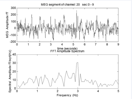
Figure 1 A) The MEG obtained from a dystonia child B) After the application of FFT on the above MEG we get primary dominant frequency of 3Hz.

Figure 1

A) The MEG obtained from a dystonia child

B) After the application of FFT on the above MEG we get primary dominant frequency of 3Hz.

shows the MEG record obtained from a child with dystonia and the application of FFT on the above MEG record which gives the primary dominant frequency at 3Hz.

DISCUSSION

In our research we have attempted to influence the patients with dystonia with the pT-TMS electronic device(13).The coils of the device were constructed to emit back to the brain of the patients magnetic fields with proper field characteristics( magnetic field amplitude: (1-7.5pT) and frequency: the alpharhythm of each child 8-13Hz). This resulted to decrease of the maximal magnetic power emitted from these areas and an attenuation of the dystonia activity observed by the clinicians. As a final point to confirm that the responses to pT-TMS were reproducible we have advised the relatives [11], of all children with dystonia to apply the pT-TMS treatment nightly at home (23:00 pm).

After one month of pT-TMS treatment at home all the children with dystonia were evaluated again with MEG recordings and interview by the same clinicians and their benefit from this treatment it is shown in Table (2).

The mechanism by which the application of the pT-TMS has some beneficial effects in the children with dystonia is unidentified. Although, one possible explanation is that these magnetic fields (pT-TMS) have been shown to influence the activity of the pineal gland (PG) which regulates the endogenous opioid functions [29], and the dopaminergic modulation [30], GABA [31]. Moreover, the PG is a regulator of our immune system through the action on the thymus gland generating the infection fighting T-cells which are needed to neutralize foreign invaders such as viruses and bacteria. If the thymus gland shrinks with the age or due to other disorders its ability to generate T-cells is sapping.

In conclusion, this method of the pT-TMS has some possible effects to be considered as a non invasive safe and efficacious modality in managing the symptoms of children with dystonia where we have 62.5% effect of using the pT-TMS. Nevertheless further investigation with more patients are required in order to estimate the possible effect of pT-TMS and its significant contribution for managing the symptoms of children with dystonia.

Table 1: It is shown the brain regions and the corresponding channels.

Brain Regions Channels
Frontal 17-42
Occipital 75-86,91-96 ,101-110
Vertex 13-16,49-54,61-66,73,74,89,90,99,100 , 117-122
Right Temporal 1-14 , 111-120
Right Parietal 5-6,11-16,97-100,109,110 , 115-122
Left Temporal 43-50 ,55-62,67-74
Left Parietal 47-52,59-64,71-74,79,80,87-90

Table 2: The symptoms of the 8 dystonia children before and after pT-TMS as were evaluated by interview by clinicians according to the American Classification of the Unified Dystonia Rating Scale, (UDRS), one month after daily pT-TMS treatment at home (F:Female; M:Male).

Patient Code Age Sex Symptoms before pT-TMS Symptoms after pT-TMS
  1 9 F Focal dystonia where one part of the body is effected(UDRS:3) She has a good effect (UDRS:0)
  2 9 M Generaliged dystonia where both legs and other regions of the body are effected (UDRS:3) No effect. (UDRS:3)
  3 9 M Generalized dystonia where both legs and other regions of the body are effected (UDRS:3) He was feeling better (UDRS: 0)
  4 9 M Focal dystonia where one part of the body is effected(UDRS:3) He was feeling better (UDRS: 0)
  5 10 M Focal dystonia where one part of the body is effected(UDRS:3) No effect(UDRS:3)
  6 10 M Multifocal dystonia where at least two regions of the body that are not connected to each other are affected as for example in this case one arm on one side as well as one leg of the other side of the body (UDRS:3) He has effect in both sides of his body (UDRS: 0)
  7 12 M Segmental dystonia where two or more regions of the body are affected and the regions somehow are connected to each other as for example neck and shoulder (UDRS:3) No effect(UDRS:3)
  8 12 M Focal dystonia where one part of the body is effected(UDRS:3) Very good effect(UDRS:0)


SOURCE OF FUNDING

Funding for this work was provided by a collaboration of GGET (General Secretariat of Research and Technology, GR) and ERGO AEBE, INC, GR under the research program titled “Foundation of a Laboratories Network and purchase of a Multichannel Biomagnetometer SQUID (Superconducting Quantum Interference Device), in order to develop an expert system for automatic acquisition, analysis, evaluation and exploitation of MEG signals that are emitted from different organs of the human body” (Grant Number: 80623).

REFERENCES

1. Oberman LM, Pascual-Leone A, Rotenberg A. Modulation of corticospinal excitability by transcranial magnetic stimulation in children and adolescents with autism spectrum disorder. Front Hum Neurosci. 2014; 8: 627.

2. Barker AT, Jalinous R, Freeston IL. Non-invasive magnetic stimulation of human motor cortex. Lancet. 1985; 1: 1106-1107.

3. Michelucci R, Valzania F, Passarelli D, Santangelo M, Rizzi R, Buzzi AM, et al. Rapid rate transcranial magnetic stimulation and hemispheric language dominance, Usefulness and safety in epilepsy. Neurology. 1994; 44: 1697-1700.

4. Shelley BP, Trimble MR. “All that spikes is not fits”, mistaking the woods for the trees: the interictal spikes--an “EEG chameleon” in the interface disorders of brain and mind: a critical review. Clin EEG Neurosci. 2009; 40: 245-261.

5. Albanese A, Romito LM, Calandrella D. Therapeutic advances in dystonia. Mov Disord. 2015; 30: 1547-1556.

6. Bharath RD, Biswal BB, Bhaskar MV, Gohel S, Jhunjhunwala K, Panda R, et al. Repetitive transcranial magnetic stimulation induced modulations of resting state motor connectivity in writer’s cramp. Eur J Neurol. 2015; 22: 796-805.

7. Kimberley TJ, Borich MR, Schmidt RL, Carey JR, Gillick B. Focal hand dystonia, individualized intervention with repeated application of repetitive transcranial magnetic stimulation. Arch Phys Med Rehabil. 2015; 96: S122-S128.

8. Pirio RS, Tinaz S, Chen R. Repetitive transcranial magnetic stimulation in cervical dystonia, effect of site and repetition in a randomized pilot trial. PLoS One. 2015; 10: e0124937.

9. Furuya S, Nitsche MA, Paulus W, Altenmuller E. Surmounting retraining limits in musicians’ dystonia by transcranial stimulation. Ann Neurol. 2014; 75: 700-707.

10. Young SJ, Bertucco M, Sanger TD. Cathodal transcranial direct current stimulation in children with dystonia, a sham-controlled study. J Child Neurol. 2014; 29: 232-239.

11. Anninos P, Adamopoulos A, Kotini A, Tsagas N. MEG evaluation of pico-Tesla external TMS on multiple sclerosis patients. Mult Scler Relat Disord. 2016; 8: 45-53.

12. Anninos PA, Tsagas N, Sandyk R, Derpapas K. Magnetic stimulation in the treatment of partial seizures. Int J Neurosci. 1991; 60: 141-171.

13. Anninos PA, Tsagas N. Electronic apparatus for treating epileptic individuals. USA patent 5453072. 1995.

14. Anninos P, Adamopoulos A, Kotini A, Tsagas N, Tamiolakis D, Prassopoulos P. MEG evaluation of Parkinson’s diseased patients after external magnetic stimulation. Acta Neurol Belg. 2007; 107: 5-10.

15. Anninos P, Adamopoulos A, Kotini A, Tsagas N. Combined MEG and pTTMS study in Parkinson’s disease. J Integr Neurosci. 2016; 20: 1-18.

16. Anninos P, Kotini A, Anninou N, Adamopoulos A, Papastergiou A, Tsagas N. MEG recordings of patients with CNS disorders before and after external magnetic stimulation. J Integr Neurosci. 2008; 7: 17-27.

17. Anninos P, Kotini A, Tamiolakis D, Tsagas N. Transcranial magnetic stimulation A case report and review of the literature. Acta Neurol Belg. 2006; 106: 26-30.

18. Anninos PA, Adamopoulos AV, Kotini A, Tsagas N. Nonlinear analysis of brain activity in magnetic influenced Parkinson patients. Brain Topogr. 2000; 13: 135-144.

19. Anninos PA, Beek B, Csermely TJ, Harth EM, Pertile G. Dynamics of neural structures. J Theor Biol. 1970; 26: 121-148.

20. Anninos PA, Tsagas N, Adamopoulos A. A brain model theory for epilepsy and the mechanism for treatment with experimental verification using SQUID measurements. In: Cotterill RM, editors. Models of brain function. New York: Cambridge University Press. 1989: 405-421.

21. Anninos P, Adamopoulos A, Kotini A. MEG as a Medical Diagnostic Tool in the Greek Population. Acta Medica (Hradec Kralove). 2015; 58: 71- 78.

22. Anninos P, Kotini A, Adamopoulos A, Tsagas N. Magnetic stimulation can modulate seizures in epileptic patients. Brain Topogr. 2003; 16: 57-64.

23. Anninos PA, Tsagas N, Jacobson JI, Kotini A. The biological effects of magnetic stimulation in epileptic patients. Panminerva Med. 1999; 41: 207-215.

24. Ossenkopp KP, Cain DP. Inhibitory effects of acute exposure to low intensity 60Hz magnetic fields on electrically kindled seizures in rats. Brain Res. 1988; 442: 255-260.

25. John ER. Mechanisms of memory in representational systems. New York: Academic Press; 1967.

26. Kaczmarek LK, Adey WR. Weak electric gradients change ionic and transmitter fluxes in cortex. Brain Res 1974; 66: 537-540.

27. Troebinger L, Anninos P, Barnes G. Neuromagnetic effects of picoTesla stimulation. Physiol Meas. 2015; 36: 1901-1912.

28. Comella CL, Leurgans S, Wuu J, Stebbins GT, Chmura T; Dystonia Study Group. Rating Scales for Dystonia. A. Multicenter Assessment. Mov Discord. 2003; 18: 303-312.

29. Lissoni P, Esposti D, Esposti G, Mauri R, Resentini M, Morabito F, et al. A clinical study on the relationship between the pineal gland and the opioid system. J Neural Transm. 1986; 65: 63-73.

30. Brandbury AJ, Kelly ME, Smith JA. Melatonin action in the mid-brain can regulate dopamine function both behaviourally and biochemically. In: Brown GM, Wainwright SD, editors. The Pineal gland, endocrine aspects. Oxford: Pergamon Press. 1985: 327-332.

31. Nitsche MA, Lampe C, Antal A, Liebetanz D, Lang N, Tergau F, et al. Dopaminergic modulation of long-lasting direct current-induced cortical excitability changes in the human motor cortex. Eur J Neurosci. 2006; 23: 1651-1657.

Anninos P, Adamopoulos A, Kotinisor A, Tsagas N (2017) MEG Study with Pico-Tesla External TMS on Children with Dystonia. Arch Paediatr Dev Pathol 1(1): 1004.

Received : 14 Mar 2017
Accepted : 19 Apr 2017
Published : 21 Apr 2017
Journals
Annals of Otolaryngology and Rhinology
ISSN : 2379-948X
Launched : 2014
JSM Schizophrenia
Launched : 2016
Journal of Nausea
Launched : 2020
JSM Internal Medicine
Launched : 2016
JSM Hepatitis
Launched : 2016
JSM Oro Facial Surgeries
ISSN : 2578-3211
Launched : 2016
Journal of Human Nutrition and Food Science
ISSN : 2333-6706
Launched : 2013
JSM Regenerative Medicine and Bioengineering
ISSN : 2379-0490
Launched : 2013
JSM Spine
ISSN : 2578-3181
Launched : 2016
Archives of Palliative Care
ISSN : 2573-1165
Launched : 2016
JSM Nutritional Disorders
ISSN : 2578-3203
Launched : 2017
Annals of Neurodegenerative Disorders
ISSN : 2476-2032
Launched : 2016
Journal of Fever
ISSN : 2641-7782
Launched : 2017
JSM Bone Marrow Research
ISSN : 2578-3351
Launched : 2016
JSM Mathematics and Statistics
ISSN : 2578-3173
Launched : 2014
Journal of Autoimmunity and Research
ISSN : 2573-1173
Launched : 2014
JSM Arthritis
ISSN : 2475-9155
Launched : 2016
JSM Head and Neck Cancer-Cases and Reviews
ISSN : 2573-1610
Launched : 2016
JSM General Surgery Cases and Images
ISSN : 2573-1564
Launched : 2016
JSM Anatomy and Physiology
ISSN : 2573-1262
Launched : 2016
JSM Dental Surgery
ISSN : 2573-1548
Launched : 2016
Annals of Emergency Surgery
ISSN : 2573-1017
Launched : 2016
Annals of Mens Health and Wellness
ISSN : 2641-7707
Launched : 2017
Journal of Preventive Medicine and Health Care
ISSN : 2576-0084
Launched : 2018
Journal of Chronic Diseases and Management
ISSN : 2573-1300
Launched : 2016
Annals of Vaccines and Immunization
ISSN : 2378-9379
Launched : 2014
JSM Heart Surgery Cases and Images
ISSN : 2578-3157
Launched : 2016
Annals of Reproductive Medicine and Treatment
ISSN : 2573-1092
Launched : 2016
JSM Brain Science
ISSN : 2573-1289
Launched : 2016
JSM Biomarkers
ISSN : 2578-3815
Launched : 2014
JSM Biology
ISSN : 2475-9392
Launched : 2016
Archives of Stem Cell and Research
ISSN : 2578-3580
Launched : 2014
Annals of Clinical and Medical Microbiology
ISSN : 2578-3629
Launched : 2014
JSM Pediatric Surgery
ISSN : 2578-3149
Launched : 2017
Journal of Memory Disorder and Rehabilitation
ISSN : 2578-319X
Launched : 2016
JSM Tropical Medicine and Research
ISSN : 2578-3165
Launched : 2016
JSM Head and Face Medicine
ISSN : 2578-3793
Launched : 2016
JSM Cardiothoracic Surgery
ISSN : 2573-1297
Launched : 2016
JSM Bone and Joint Diseases
ISSN : 2578-3351
Launched : 2017
JSM Bioavailability and Bioequivalence
ISSN : 2641-7812
Launched : 2017
JSM Atherosclerosis
ISSN : 2573-1270
Launched : 2016
Journal of Genitourinary Disorders
ISSN : 2641-7790
Launched : 2017
Journal of Fractures and Sprains
ISSN : 2578-3831
Launched : 2016
Journal of Autism and Epilepsy
ISSN : 2641-7774
Launched : 2016
Annals of Marine Biology and Research
ISSN : 2573-105X
Launched : 2014
JSM Health Education & Primary Health Care
ISSN : 2578-3777
Launched : 2016
JSM Communication Disorders
ISSN : 2578-3807
Launched : 2016
Annals of Musculoskeletal Disorders
ISSN : 2578-3599
Launched : 2016
Annals of Virology and Research
ISSN : 2573-1122
Launched : 2014
JSM Renal Medicine
ISSN : 2573-1637
Launched : 2016
Journal of Muscle Health
ISSN : 2578-3823
Launched : 2016
JSM Genetics and Genomics
ISSN : 2334-1823
Launched : 2013
JSM Anxiety and Depression
ISSN : 2475-9139
Launched : 2016
Clinical Journal of Heart Diseases
ISSN : 2641-7766
Launched : 2016
Annals of Medicinal Chemistry and Research
ISSN : 2378-9336
Launched : 2014
JSM Pain and Management
ISSN : 2578-3378
Launched : 2016
JSM Women's Health
ISSN : 2578-3696
Launched : 2016
Clinical Research in HIV or AIDS
ISSN : 2374-0094
Launched : 2013
Journal of Endocrinology, Diabetes and Obesity
ISSN : 2333-6692
Launched : 2013
Journal of Substance Abuse and Alcoholism
ISSN : 2373-9363
Launched : 2013
JSM Neurosurgery and Spine
ISSN : 2373-9479
Launched : 2013
Journal of Liver and Clinical Research
ISSN : 2379-0830
Launched : 2014
Journal of Drug Design and Research
ISSN : 2379-089X
Launched : 2014
JSM Clinical Oncology and Research
ISSN : 2373-938X
Launched : 2013
JSM Bioinformatics, Genomics and Proteomics
ISSN : 2576-1102
Launched : 2014
JSM Chemistry
ISSN : 2334-1831
Launched : 2013
Journal of Trauma and Care
ISSN : 2573-1246
Launched : 2014
JSM Surgical Oncology and Research
ISSN : 2578-3688
Launched : 2016
Annals of Food Processing and Preservation
ISSN : 2573-1033
Launched : 2016
Journal of Radiology and Radiation Therapy
ISSN : 2333-7095
Launched : 2013
JSM Physical Medicine and Rehabilitation
ISSN : 2578-3572
Launched : 2016
Annals of Clinical Pathology
ISSN : 2373-9282
Launched : 2013
Annals of Cardiovascular Diseases
ISSN : 2641-7731
Launched : 2016
Journal of Behavior
ISSN : 2576-0076
Launched : 2016
Annals of Clinical and Experimental Metabolism
ISSN : 2572-2492
Launched : 2016
Clinical Research in Infectious Diseases
ISSN : 2379-0636
Launched : 2013
JSM Microbiology
ISSN : 2333-6455
Launched : 2013
Journal of Urology and Research
ISSN : 2379-951X
Launched : 2014
Journal of Family Medicine and Community Health
ISSN : 2379-0547
Launched : 2013
Annals of Pregnancy and Care
ISSN : 2578-336X
Launched : 2017
JSM Cell and Developmental Biology
ISSN : 2379-061X
Launched : 2013
Annals of Aquaculture and Research
ISSN : 2379-0881
Launched : 2014
Clinical Research in Pulmonology
ISSN : 2333-6625
Launched : 2013
Journal of Immunology and Clinical Research
ISSN : 2333-6714
Launched : 2013
Annals of Forensic Research and Analysis
ISSN : 2378-9476
Launched : 2014
JSM Biochemistry and Molecular Biology
ISSN : 2333-7109
Launched : 2013
Annals of Breast Cancer Research
ISSN : 2641-7685
Launched : 2016
Annals of Gerontology and Geriatric Research
ISSN : 2378-9409
Launched : 2014
Journal of Sleep Medicine and Disorders
ISSN : 2379-0822
Launched : 2014
JSM Burns and Trauma
ISSN : 2475-9406
Launched : 2016
Chemical Engineering and Process Techniques
ISSN : 2333-6633
Launched : 2013
Annals of Clinical Cytology and Pathology
ISSN : 2475-9430
Launched : 2014
JSM Allergy and Asthma
ISSN : 2573-1254
Launched : 2016
Journal of Neurological Disorders and Stroke
ISSN : 2334-2307
Launched : 2013
Annals of Sports Medicine and Research
ISSN : 2379-0571
Launched : 2014
JSM Sexual Medicine
ISSN : 2578-3718
Launched : 2016
Annals of Vascular Medicine and Research
ISSN : 2378-9344
Launched : 2014
JSM Biotechnology and Biomedical Engineering
ISSN : 2333-7117
Launched : 2013
Journal of Hematology and Transfusion
ISSN : 2333-6684
Launched : 2013
JSM Environmental Science and Ecology
ISSN : 2333-7141
Launched : 2013
Journal of Cardiology and Clinical Research
ISSN : 2333-6676
Launched : 2013
JSM Nanotechnology and Nanomedicine
ISSN : 2334-1815
Launched : 2013
Journal of Ear, Nose and Throat Disorders
ISSN : 2475-9473
Launched : 2016
JSM Ophthalmology
ISSN : 2333-6447
Launched : 2013
Journal of Pharmacology and Clinical Toxicology
ISSN : 2333-7079
Launched : 2013
Annals of Psychiatry and Mental Health
ISSN : 2374-0124
Launched : 2013
Medical Journal of Obstetrics and Gynecology
ISSN : 2333-6439
Launched : 2013
Annals of Pediatrics and Child Health
ISSN : 2373-9312
Launched : 2013
JSM Clinical Pharmaceutics
ISSN : 2379-9498
Launched : 2014
JSM Foot and Ankle
ISSN : 2475-9112
Launched : 2016
JSM Alzheimer's Disease and Related Dementia
ISSN : 2378-9565
Launched : 2014
Journal of Addiction Medicine and Therapy
ISSN : 2333-665X
Launched : 2013
Journal of Veterinary Medicine and Research
ISSN : 2378-931X
Launched : 2013
Annals of Public Health and Research
ISSN : 2378-9328
Launched : 2014
Annals of Orthopedics and Rheumatology
ISSN : 2373-9290
Launched : 2013
Journal of Clinical Nephrology and Research
ISSN : 2379-0652
Launched : 2014
Annals of Community Medicine and Practice
ISSN : 2475-9465
Launched : 2014
Annals of Biometrics and Biostatistics
ISSN : 2374-0116
Launched : 2013
JSM Clinical Case Reports
ISSN : 2373-9819
Launched : 2013
Journal of Cancer Biology and Research
ISSN : 2373-9436
Launched : 2013
Journal of Surgery and Transplantation Science
ISSN : 2379-0911
Launched : 2013
Journal of Dermatology and Clinical Research
ISSN : 2373-9371
Launched : 2013
JSM Gastroenterology and Hepatology
ISSN : 2373-9487
Launched : 2013
Annals of Nursing and Practice
ISSN : 2379-9501
Launched : 2014
JSM Dentistry
ISSN : 2333-7133
Launched : 2013
Author Information X