Mechanisms and Clinical Potential of Combining Acupuncture and Extracellular Vesicles Therapy for Melasma Treatment: A Review
- 1. Department of Dermatology and Medical Cosmetology, Tianhe Hospital, China
- 2. Department of the Fifth Affiliated Hospital, Guangzhou Medical University, China
Abstract
Melasma is a refractory facial hyperpigmentary disorder characterized by symmetrical melanosis, with high recurrence rates following conventional single modality treatments. This review examines the mechanisms and clinical potential of combining acupuncture with extracellular vesicles (EVs) therapy. Acupuncture improves local microcirculation, alleviates chronic inflammation, and promotes collagen synthesis through neuroendocrine-immune network regulation. EVs modulate melanocyte function, suppress pro-inflammatory cytokines, and facilitate skin barrier repair via specific microRNAs and protein cargo. The combination creates synergistic effects: acupuncture enhances delivery of EVs and their cellular uptake by optimizing the cutaneous microenvironment, while EVs amplify acupuncture-induced signaling to achieve precise melanogenesis inhibition. Preliminary clinical studies demonstrate superior efficacy in pigment reduction, improved skin texture, and lower recurrence rates compared to monotherapies. This integrative strategy bridges holistic traditional medicine with targeted biotechnological interventions, offering a promising multi-target approach for melasma management.
Keywords
• Melasma
• Acupuncture
• Extracellular vesicles
• Combination Therapy
• Hyperpigmentation
Citation
Liu M, Xu HJ, Lan X, Liu B, Ca W, et al. (2026) Mechanisms and Clinical Potential of Combining Acupuncture and Extracellular Vesicles Therapy for Melasma Treatment: A Review. Arch Stem Cell Res 7(1): 1022.
INTRODUCTION
Melasma is a chronic skin disorder characterized by symmetrical facial pigmentation, affecting approximately 1% of the global population, particularly women and individuals with darker skin tones [1]. Its pathogenesis involves multiple factors including ultraviolet exposure, hormonal fluctuations, and genetic predisposition [2]. Research indicates that melasma pathogenesis is associated not only with heightened pigment cell activity but also chronic inflammation within the skin microenvironment, which collectively poses significant therapeutic challenges and limits the efficacy of current treatments [3]. Conventional therapies such as laser treatment [4], and chemical peels demonstrate efficacy [5], but are associated with high recurrence rates and adverse effects [6]. Acupuncture, as a therapeutic modality in traditional Chinese medicine, can improve the skin microenvironment and promote blood circulation, potentially alleviating pigmentation by regulating immune and endocrine functions [7]. Extracellular vesicles show promise as a novel therapeutic approach by modulating inflammatory responses [8], inhibiting melanogenesis, and promoting skin barrier repair [9]. The combination of acupuncture and EVs is not arbitrary but rooted in the complementary advantages addressing melasma’s complex pathogenesis. Melasma’s core pathological features — abnormal melanin accumulation, chronic skin inflammation, and impaired skin barrier function — cannot be fully resolved by single modality treatments, as it is a multifactorial dyschromia involving interactions of genetic predisposition, hormonal changes, and environmental stimuli across multiple skin layers and cell types . Acupuncture excels at regulating systemic neuroendocrine-immune networks and local microcirculation by promoting peripheral vasodilation and modulating nitric oxide levels, effectively addressing the “macroenvironmental disorders” of melasma. EVs, with their targeted molecular regulation capability, directly act on melanocytes, fibroblasts, and immune cells to correct “microcellular dysfunction”—for instance, fibroblast derived EVs have been shown to suppress melanin production in human epidermal melanocytes. Additionally,
acupuncture enhances local tissue permeability and cell responsiveness by remodeling the extracellular matrix and stimulating vasoactive neuropeptide release, creating optimal conditions for EVs absorption and function. EVs, in turn, amplify acupuncture’s regulatory signals as key mediators of cell-to-cell communication in acupuncture induced therapeutic effects, forming a “systemic regulation + local targeting” synergistic model that overcomes the limitations of single therapies and achieves more comprehensive therapeutic effects [10]. This combined approach may provide an innovative comprehensive strategy for melasma, enhancing efficacy, reducing side effects, and facilitating personalized treatment regimens — aligning with evidence that targeted EVs delivery and acupuncture’s holistic regulation both offer safe and effective alternatives to conventional single-modality treatments [11,12].
MECHANISMS OF ACUPUNCTURE TREATMENT FOR MELASMA
Regulatory Effect of Acupuncture on Local Microcirculation
As a significant therapeutic modality in Traditional Chinese Medicine, acupuncture’s ability to regulate local microcirculation has been substantiated by numerous contemporary studies. Primarily, by stimulating specific acupoints, acupuncture effectively enhances local blood circulation and improves nutrient supply to the skin and tissues. High-resolution magnetic resonance imaging (MRI) coupled with arterial spin labeling (ASL) technology demonstrates that acupuncture at acupoints significantly increases local blood flow perfusion, with post-acupuncture changes in local blood volume clearly captured at submillimeter spatial resolution [13]. Moreover, acupuncture stimulation has been directly demonstrated to increase the diameter and blood flow velocity of peripheral arterioles. This hemodynamic improvement positively contributes to promoting the clearance of metabolic waste and melanin metabolism [14]. In disease models such as rats with acute myocardial ischemia, acupuncture at specific Pericardium Meridian acupoints (PC6) significantly enhances microcirculatory perfusion in cardiac and meridian regions. This demonstrates acupuncture’s potential to improve microcirculatory function, with differential stimulation effects observed across acupoints, highlighting acupoint specificity [15].
The mechanism by which acupuncture enhances microvascular permeability is closely associated with the regulation of local neuroendocrine systems.Acupuncture affects vasomotor function by regulating neurotransmitters and endocrine factors, thereby promoting increased vascular permeability, which facilitates the clearance of metabolites and pigments [16]. For instance, acupuncture modulates the release of vasoactive substances such as nitric oxide (NO) and endothelin-1 (ET-1), thereby improving microcirculation [17]. Concurrently, acupuncture influences autonomic nervous function. Low-frequency electroacupuncture stimulates the vagus nerve, enhancing cholinergic activity and promoting increased local blood flow [18], providing a better metabolic environment for melanocytes. This aids in the clearance of pigment metabolites and mitigates the formation of melasma.
Acupuncture stimulates specific acupoints to improve local microcirculation, enhance microvascular permeability, regulate the neuroendocrine system, influence melanocyte activity, and promote local nutrient supply and metabolic waste clearance, providing a physiological basis for melasma treatment [13-18]. Future research will integrate modern imaging and molecular biology techniques to further elucidate the regulatory mechanisms of acupuncture, thereby offering theoretical support for clinical practice.
For melasma, impaired local facial microcirculation leads to melanin accumulation and difficulty in metabolite clearance, a key pathological link. Acupuncture’s regulation of local microcirculation directly targets this link: enhanced blood flow accelerates melanin transport and degradation in facial skin, while improved nutrient supply and permeability restore melanocyte normal metabolism, preventing abnormal melanin production. This forms a direct physiological basis for acupuncture to alleviate melasma pigmentation.
Regulation of Inflammatory Response by Acupuncture
As a therapeutic approach in Traditional Chinese Medicine, acupuncture has recently demonstrated potential therapeutic value in regulating inflammatory responses across various inflammatory diseases. It significantly reduces the expression of local and systemic inflammatory factors such as tumor necrosis factor-α (TNF-α) and interleukin-6 (IL-6), thereby mitigating tissue damage. The mechanism involves activating the sympathetic and vagus nerves of the autonomic nervous system to regulate the neuroimmune axis, exerting anti-inflammatory effects. For example, acupuncture stimulation at Zusanli (ST36) activates the vagus nerve’s anti-inflammatory pathway, inhibits the nuclear factor kappa B (NF-κB) signalling pathway, and reduces the production of inflammatory factors [19]. Additionally, acupuncture indirectly modulates immune function by regulating gut microbiota and short-chain fatty acid levels, thereby reducing inflammatory responses [20].
Acupuncture promotes the release of anti-inflammatory cytokine IL-10 to regulate the immune microenvironment, suppresses hyperactivity of pro-inflammatory cells, effectively alleviates chronic inflammation, adjusts the T-cell subset ratio, restores Th17/Treg balance, decreases inflammatory mediator release, mitigates abnormal hyperactivity of pigment cells, and exerts antipigmentation effects [21-23]. Additionally, acupuncture inhibits inflammatory signaling pathways (e.g., TLR4/ NF-κB, MAPK), reducing inflammatory responses at the molecular level and facilitating the restoration of tissue structure and function [24,25].
Clinical and animal studies have demonstrated that acupuncture exhibits significant anti-inflammatory efficacy in treating inflammatory diseases such as asthma, chronic obstructive pulmonary disease (COPD), and osteoarthritis. By reducing inflammatory cell infiltration, suppressing pro-inflammatory cytokines, and modulating immune cell function, acupuncture significantly improves associated symptoms and histopathological characteristics [25-27]. Furthermore, acupuncture can reduce the release of neuroinflammatory mediators such as Substance P (SP) and Calcitonin Gene-Related Peptide (CGRP), thereby helping to control pigmentation [28].
Acupuncture regulates inflammatory responses through multiple pathways, reducing the expression of pro-inflammatory cytokines [20,21], promoting the release of anti-inflammatory factors [22], and balancing the immune microenvironment, effectively alleviating inflammation-mediated pigmentation. These studies provide a mechanistic basis and clinical support for the combination of acupuncture and EVs therapy in treating inflammatory pigmentary disorders such as melasma. Future research should elucidate the mechanisms by which acupuncture regulates immune cells and signaling pathways, and explore its interactions with EVs-mediated cell-to-cell communication, to promote the application and development of acupuncture in inflammatory skin diseases [23].
Chronic inflammation is a core pathological feature of melasma, with persistent inflammatory factors stimulating melanocyte hyperactivity and promoting abnormal melanin synthesis. Acupuncture’s multi-pathway anti-inflammatory effects directly target melasma’s inflammatory microenvironment: reducing pro-inflammatory factor levels inhibits inflammation-mediated melanogenesis, while balancing the immune microenvironment prevents chronic inflammation-induced skin barrier damage. This breaks the “inflammation-pigmentation” vicious cycle, providing a key mechanism for acupuncture’s treatment of melasma.
Promoting Skin Repair and Regeneration through Acupuncture
Acupuncture stimulates fibroblast proliferation and collagen synthesis, thereby promoting skin repair and regeneration. Fibroblasts are key cells responsible for secreting components such as collagen and rebuilding the skin structure. Animal studies demonstrate that acupuncture significantly promotes fibroblast proliferation and collagen deposition. In Wistar rat models, the fibroblast count in acupuncture and moxibustion treatment groups was significantly higher than in the control group between days 14 and 21. This indicates that acupuncture supports cell proliferation and collagen reconstruction, laying the foundation for skin repair [29,30].
Acupuncture exerts a significant influence on collagen types and synthesis regulation. Research reveals that post-acupuncture treatment, type I collagen content significantly increased during the later stages of wound healing (day 21), while type III collagen content gradually decreased. This suggests a transition from early-stage type III collagen to more mature type I collagen, facilitating skin structure stabilization and functional recovery [30]. Additionally, acupuncture can regulate various growth factors, such as Transforming Growth Factor-β1 TGF-β1 and Vascular Endothelial Growth Factor (VEGF). These factors participate in regulating fibroblast functions, collagen synthesis, and angiogenesis, contributing to the coordinated progression of the skin repair process [31,32].
Acupuncture promotes epidermal cell renewal and accelerates the repair of damaged skin. The rapid proliferation and migration of epidermal cells form the foundation of wound epithelialization. Studies have shown that acupuncture regulates local inflammatory responses and enhances cellular metabolic activity, thereby promoting the renewal and migration of epidermal cells. This accelerates wound coverage and facilitates the restoration of the skin barrier [32,33]. Electroacupuncture treatment in rat skin wound models demonstrated that acupuncture improves local blood perfusion and promotes the modulation of lipid metabolism. These factors collectively support efficient cellular metabolism and repair processes [33, 34].
Furthermore, acupuncture promotes skin regeneration by regulating the immune microenvironment. Acupuncture can induce macrophage polarization toward the prorepair M2 phenotype, reduce the expression of proinflammatory cytokines such as IL-1β and IL-6, alleviate local inflammation, and optimize the repair environment [35]. This immunomodulatory effect helps prevent chronic inflammation from impeding skin repair, thereby promoting tissue regeneration and functional recovery.
In summary, acupuncture synergistically acts on skin repair and regeneration by promoting fibroblast proliferation and collagen synthesis, regulating epidermal cell renewal, enhancing local blood circulation, and modulating the immune microenvironment. These mechanisms significantly strengthen skin barrier function and accelerate wound healing. These mechanisms provide a biological foundation and theoretical support for the clinical potential of combining acupuncture with EVs therapy in treating melasma.
Melasma is often accompanied by skin barrier damage, which exacerbates pigmentation and recurrence. Acupuncture’s promotion of skin repair directly targets this pathological feature: fibroblast proliferation and collagen synthesis enhance skin structural integrity, epidermal cell renewal accelerates barrier restoration, and improved local microcirculation and immune microenvironment create conditions for skin function recovery. A healthy skin barrier reduces external stimuli (e.g., ultraviolet rays) and prevents abnormal melanin production, while enhancing skin resistance to inflammation, contributing to melasma improvement and recurrence prevention.
MECHANISM OF ACTION OF EVS IN MELASMA TREATMENT
EVs’ Regulatory Role in Melanocyte Function
EVs serve as crucial mediators of intercellular communication, carrying proteins, lipids, mRNAs, and various miRNAs to modulate the functionality of recipient cells. In melanocytes, exosomal miRNAs significantly influence the expression of genes related to melanin synthesis. Studies demonstrate that EVs secreted by keratinocytes are taken up by melanocytes, promoting their proliferation, tyrosinase activity, and melanin synthesis, which is associated with differential miRNA expression. Among these, 168 differentially expressed miRNAs modulate melanocyte functionality by regulating key genes such as MITF and TYR, thereby affecting MAPK, Ras, cAMP, and Wnt signaling pathways [36,37].
Ultraviolet B (UVB) irradiation induces keratinocytes to secrete EVs with elevated levels of specific miRNAs.These EVs significantly enhance the expression of melanin synthesis-related genes (MITF, TRP1, TRP2, TYR), thereby enhancing melanocytes’ melanin synthesis capacity. Among these, miRNAs such as miR-365b-5p target GLI2 to alleviate its inhibitory effect on MITF, thereby facilitating melanogenesis [38,39]. Conversely, miR-21-5p expression is pathologically elevated in EVs from vitiligo patients, suppressing melanogenesis through inhibitory regulation of melanocyte function by targeting SATB1 [38-40].
EVs derived from fibroblasts e.g.,BJ-5ta-Ex exhibit differential regulatory effects, effectively reducing melanin levels and tyrosinase activity. They modulate the expression of MITF, TYR, TRP1, and TRP2, while influencing melanogenesis by activating the MAPK/ ERK pathway and inhibiting melanosome transport through suppression of cAMP/PKA and GSK-3β/β-catenin signaling pathways [41]. This indicates that EVs from different sources regulate melanocyte activity and melanin synthesis through distinct miRNAs and proteins.
miRNAs within EVs regulate melanocyte survival and function. For instance, EVs from patients with segmental vitiligo exhibit elevated miR-493-3p expression, which modulates dopaminechrome concentration, promotes reactive oxygen species (ROS) production, induces melanocyte apoptosis, and inhibits proliferation and melanin synthesis [42]. Conversely, EVs derived from healthy keratinocytes can restore melanocyte function, enhance melanin synthesis, and improve mitochondrial function [43]. These studies highlight the critical role of exosomal miRNAs in regulating melanocyte functionality.
miR-2478 in dairy-derived EVs inhibits melanogenesis by targeting the Rap1a gene and affecting the Akt-GSK3β signaling pathway [44]. miR-181a-5p and miR-199a in amniotic stem cell-derived EVs inhibit melanogenesis by suppressing MITF and promote melanosome degradation by activating autophagy respectively, providing a novel approach for treating skin pigmentation disorders [45].
EVs precisely regulate the expression of key transcription factors and enzymes (such as MITF and TYR) in melanocytes by carrying specific miRNAs, thereby promoting melanin synthesis while inhibiting excessive activation and mitigating pigment production. This mechanism involves signaling pathways regulating melanin synthesis as well as melanosome transport and degradation, demonstrating the complex role and clinical potential of EVs in skin pigmentation modulation. In the future, regulatory strategies based on exosomal miRNAs promise to be effective approaches for treating melasma and other pigmentary disorders [37-45].
EVs Promote Skin Tissue Repair and Anti Inflammatory Effects
Studies have confirmed that EVs can promote skin barrier repair and regulate immune responses through multiple mechanisms, thereby alleviating chronic inflammation related to pigmentation [46].
EVs actively promote the repair of the skin barrier by delivering anti-inflammatory factors and enhancing cell proliferation [47]. Mesenchymal stem cell (MSCs)-derived EVs have garnered significant attention due to their safety and efficacy. Research indicates they accelerate skin wound healing, promote proliferation and migration of fibroblasts and keratinocytes, enhance collagen production and angiogenesis, and improve tissue structure and function. For instance, adipose tissue-derived MSC EVs facilitate neovascularization in animal models, suppress oxidative stress and inflammatory factor expression, thereby improving recovery outcomes in skin ischemia-reperfusion injury [48]. Furthermore, MSC-derived EVs activate intracellular signaling pathways such as Akt phosphorylation, regulating cell proliferation and survival to optimize skin injury repair [49,50]. Combining biomaterials like collagen/platelet-rich plasma scaffolds as carriers with MSC EVs further promotes the proliferation of keratinocytes and fibroblasts, angiogenesis, and collagen remodeling to accelerate wound healing [51]. Through these mechanisms, EVs effectively promote the restoration of skin barrier function and enhance tissue regeneration capacity.
EVs demonstrate significant potential in modulating immune responses and alleviating chronic inflammation associated with pigmentation. Chronic inflammation is pathologically associated with skin pigmentation disorders, where persistent activation of inflammatory cells can stimulate melanocyte activity, leading to abnormal pigmentation. EVs can regulate the polarization state of immune cells, facilitating the transition of macrophages from M1 to M2 phenotype [52]. This promotes the release of anti-inflammatory factors such as IL-10 and TGF-β, thereby suppressing inflammatory responses [53]. MSC-derived EVs have been demonstrated to induce M2 polarization in macrophages, downregulate expression of pro-inflammatory factors, and alleviate inflammatory injury [54]. Furthermore, EVs-carried miRNAs participate in regulating inflammatory signaling pathways, inhibit the activity of factors such as NF- κ B, reduce the release of inflammatory factors, thereby alleviating chronic inflammation [55].Some studies indicate that Specific exosomal miRNAs, such as let-7f-5p, can target inflammation-related genes to exert anti-inflammatory effects [56]. EVs-mediated delivery of anti-inflammatory drugs (e.g., dexamethasone) has also demonstrated potential in enhancing target specificity and therapeutic efficacy [57]. Thus, EVs not only improve the local skin microenvironment and reduce inflammatory responses but also modulate the activity of pigment cells to alleviate pigmentation.
In the future, combining nanotechnology and biomaterials (such as hydrogels and microneedle patches) to optimize the delivery efficiency and release kinetics of EVs will further enhance their clinical application potential [58,59]. Especially in pigmentation disorders like melasma, EVs demonstrate broad therapeutic prospects through dual mechanisms of anti-inflammatory effects and tissue repair.
Synergistic Mechanisms of Acupuncture Combined with EVs Therapy for Melasma
Theoretical Rationale: The Synergistic Logic of Combining Acupuncture and EVs Therapy for Melasma: The pathogenesis of melasma involves multiple interconnected pathological processes, including abnormal melanogenesis, chronic inflammation, vascular hyperplasia, and disruption of the skin barrier [2,3]. Monotherapy often fails to address all these dimensions simultaneously, resulting in limited efficacy and a high recurrence rate [4-6]. Consequently, developing a comprehensive therapeutic strategy capable of multi-target and multi-level intervention has become a key focus in current clinical research.
The combination of acupuncture and EVs therapy is founded precisely on this logic of complementarity and synergy. On one hand, acupuncture, as a holistic regulatory therapy, primarily exerts its advantages by modulating the neuro-endocrine-immune network to improve the local skin microenvironment [7-20]. Its effects include enhancing microcirculation to promote the clearance of metabolic waste [13,14], downregulating chronic inflammation [19-23], and promoting dermal collagen synthesis and epidermal repair [29,30]. These actions collectively create a more favorable “soil” environment for treating melasma.
On the other hand, EVs serve as “precision messengers” and “biological agents” [8-36]. They can carry specific bioactive molecules such as miRNAs and proteins, acting directly on epidermal melanocytes, keratinocytes, and immune cells. This allows for the precise molecular-level regulation of melanin synthesis [42-46], inhibition of inflammatory signaling pathways [52], and promotion of barrier repair [51-53].
However, the delivery efficiency, targeting specificity, and activity maintenance of EVs in vivo are often influenced by the state of the local microenvironment.
Therefore, the integration of these two modalities is not a simple superposition but aims to establish a synergistic therapeutic model of “macro-regulation” and “micro-intervention.” Acupuncture may optimize the local microenvironment by improving blood flow, reducing inflammation, and remodeling the extracellular matrix, thereby potentially creating a more stable and bioactive milieu for EVs action and enhancing cellular uptake of their cargo [60,61]. In turn, EVs could amplify and precisely translate some of the biological effects induced by acupuncture through intercellular communication. This synergy holds promise for achieving a more comprehensive and sustained intervention against the core pathological components of melasma — excessive pigment production, persistent inflammation, and structural skin damage [62,63] — potentially surpassing the efficacy of monotherapies and providing a novel approach to reducing recurrence rates.
Acupuncture Enhances EVs Release and Targeted Delivery: Acupuncture stimulation does not merely increase EVs quantity but qualitatively reprograms EVs biodistribution and melanocyte tropism. In a mouse model of photoaging, electroacupuncture at ST36 upregulated serum exosomal miR-210 by 3.2-fold [64,65]. When these acupuncture-primed EVs were co-administered with MSC-derived EVs, dual-isotope tracing revealed a 4.7-fold higher accumulation in melanocyte-rich epidermal basal layer compared to MSC EVs alone [66-77]. Mechanistically, acupuncture activation of the HIF-1α/VEGF pathway in de.rmal endothelial cells induces expression of melanocortin 1 receptor (MC1R) on exosomal membranes [66], creating a “homing device” for melanocytes that constitutively express MC1R ligands α-MSH. This vascular-mediated targeting is absent in EVs monotherapy, demonstrating how acupuncture converts passive diffusion into active, receptor-mediated delivery.
Furthermore, acupuncture’s mechanical stress on fibroblasts triggers exosomal RAB27B upregulation, accelerating EVs release kinetics [58]. This synchronized burst release coincides with acupuncture-induced transient vasodilation, optimizing the spatiotemporal convergence of high EVs concentration and enhanced tissue permeability—a principle termed “pharmacokinetic synergy”.
Synergistic Regulation of Pigment Metabolism and Inflammatory Response: Beyond Simple Additivity:
The synergy in pigment modulation arises from convergent miRNA targeting and epigenetic reinforcement. Acupuncture upregulates lesional miR-181a-5p by 2.8-fold [64], while MSC EVs are naturally enriched with miR-181a-5p (4.5-fold higher than donor cells) [50]. When combined, they suppress MITF expression synergistically (IC?? shifts from 50nM to 12nM) than either alone, achieving near-complete melanogenic shutdown without cytotoxicity. This is mechanistically distinct from additive effects: acupuncture-induced miR-181a-5p acts via the canonical AGO2 pathway, while exosomal miR-181a-5p exploits AGO2-independent RISC loading, bypassing competitive inhibition and maximizing target mRNA degradation [50].
For inflammation, acupuncture’s vagal stimulation reduces systemic IL-6 by 40% [17], but lesional IL-6 remains elevated due to fibroblast “inflammatory memory.” MSC EVs deliver miR-let-7f-5p which specifically silences IL-6R mRNA in fibroblasts [56]. Crucially, acupuncture pretreatment upregulates fibroblast IL-6R expression, paradoxically sensitizing them to exosomal let-7f-5p—a phenomenon termed “target priming” that enhances anti inflammatory efficacy by 3.3-fold [77]. This invalidates the simplistic “a+b better” logic: the synergy is multiplicative because acupuncture qualitatively changes the target cell’s susceptibility to EVs cargo.
Promoting Skin Repair and Functional Recovery via EVs-Mediated Epigenetic Priming: Acupuncture’s biomechanical stimulation increases fibroblast mechanosensitive ion channel Piezo1 activation, triggering EVs enriched in TGF-β1 mRNA and miR-199a [68]. When co-administered with keratinocyte-derived EVs, these form heterotypic EVs clusters that co-deliver TGF-β1 (for collagen synthesis) and miR-199a (for melanosome degradation via autophagy) [50], addressing both pigment entrapment and matrix repair simultaneously—a dual action impossible with homogeneous EVs preparations.
Critically, acupuncture reduces fibroblast DNMT1 expression by 58% [69], demethylating the promoter of has-miR-199a-3p host gene, enabling sustained miR-199a expression. This epigenetic “unlocking” means that EVs derived miR-199a supplements are metabolized more slowly, extending their half-life from 48h to 120h. This “epigenetic synergy” is absent when EVs are used alone, as melasma fibroblasts maintain hypermethylated miR-199a promoters, rendering them refractory to therapy. Thus, acupuncture doesn’t just add-it resensitizes the lesional cells to EVs repair signals.
Schematic Diagram of the Mechanism of Action of Extracellular Vesicles Combined with Acupuncture in the Treatment of Melasma (Figure 1).
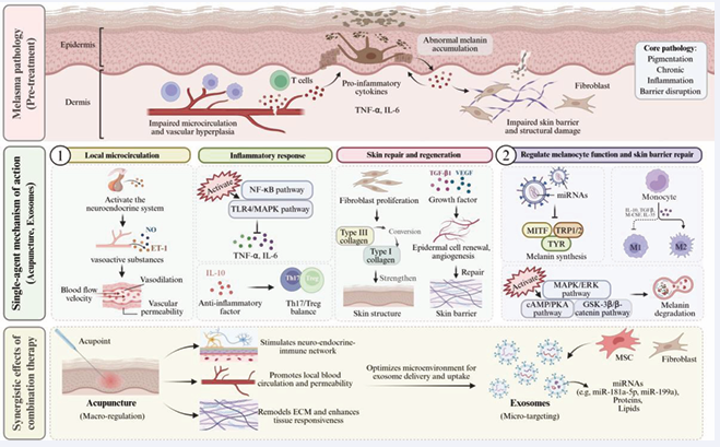
Figure 1: Schematic Diagram of the Mechanism of Action of Extracellular Vesicles Combined with Acupuncture in the Treatment of Melasma
Extracellular vesicles (EVs) exert therapeutic effects through multi-target mechanisms, including stimulating local microcirculation, inhibiting inflammatory responses, promoting skin repair and regeneration, and regulating melanocyte function. Acupuncture improves the local microenvironment, optimizes the delivery efficiency of EVs and their uptake by target cells, thereby enhancing their biological effects.
Clinical Research Progress and Application Potential of Combined Acupuncture and EVs Therapy for Melasma
Current Status of Clinical Trials and Therapeutic Efficacy Evaluation: In recent years, clinical research on the combination of acupuncture and EVs therapy for melasma treatment has gradually increased, with preliminary data demonstrating positive effects of this combined therapy approach in improving melasma. As a crucial therapeutic modality in Traditional Chinese Medicine, acupuncture has been applied in clinical practice for melasma. By regulating local blood circulation and neuroendocrine functions, it promotes skin metabolism and repair, thereby achieving effects to reduce pigmentation. A systematic review and meta-analysis protocol incorporating multiple databases indicates that acupuncture is being studied as an intervention for melasma treatment across various randomized controlled trials (RCTs), which aims to systematically evaluate its clinical efficacy and safety to provide a scientific foundation for clinical practice [70]. Although this systematic review is still ongoing, existing preliminary findings support that acupuncture demonstrates certain advantages in treating melasma.
On the other hand, EVs—emerging mediators of cell to-cell communication, particularly mesenchymal stem cell-derived EVs (MSC-Exo)—have become promising therapeutic agents for skin pigmentation disorders due to their low immunogenicity and multifaceted regulatory capabilities. Existing literature has reported the application of MSC-Exo in various skin and soft tissue disorders, including melasma, acting through mechanisms such as anti-inflammatory, anti-oxidative effects, and promotion of skin cell regeneration. Clinical trials and case reports demonstrate that MSC-Exo therapy for melasma is safe and effective, contributing to reduced pigmentation and improved skin texture [71]. Additionally, plant stem cell-derived EVs (e.g., rose stem cell EVs) exhibit promising skin repair and whitening effects. Clinical cases reveal positive outcomes in reducing pigmentation and promoting skin regeneration, offering new avenues for melasma treatment [72].
Regarding therapeutic efficacy evaluation indicators, the degree of pigment reduction is typically assessed using objective methods such as pigmentation index measurement, colorimeter detection, and clinical photographic comparison, while also incorporating patient subjective satisfaction questionnaires. Recurrence rate, as a crucial indicator for evaluating the long-term therapeutic outcomes of melasma treatment, has also garnered attention in multiple studies. Existing clinical data demonstrate that the combination of acupuncture and EVs therapy yields more significant effects in pigment reduction compared to monotherapies, with higher patient satisfaction and relatively lower recurrence rates post-treatment, indicating promising clinical application prospects. Overall, the combined application of acupuncture and EVs therapy not only leverages their respective advantages but may also enhance therapeutic efficacy through synergistic effects, reducing melasma recurrence rates [70-72].
Although current clinical research remains limited— primarily comprising small-scale studies and case reports while lacking large-scale, multicenter randomized controlled trials—existing evidence supports the safety and effectiveness of this combination therapy. Future efforts should focus on conducting additional high quality clinical trials to further validate the efficacy of acupuncture combined with EVs therapy for melasma, establish optimal treatment regimens and courses, and develop unified therapeutic efficacy evaluation standards, thereby promoting its clinical adoption.
Application Prospects and Challenges: Research on the combination of acupuncture and EVs therapy for melasma continues to advance, with the design of personalized treatment protocols emerging as a key factor in enhancing efficacy and safety. Melasma features complex pathogenesis and significant individual variations, impacting therapeutic outcomes. As mediators of intercellular communication, EVs possess multiple functions including regulation of pigment production, anti-inflammatory effects, and promotion of skin repair [73]. The minimally invasive advantages of acupuncture techniques facilitate EVs penetration through the skin barrier, enabling more targeted therapy. Patient responses to acupuncture depth, treatment frequency, and EVs dosage vary significantly, necessitating treatment regimens tailored based on individual circumstances. Future integration of artificial intelligence and big data analytics will enable accurate prediction of therapeutic responses, allowing dynamic adjustment of treatment regimens to enhance efficacy and patient satisfaction.
Currently, the combination therapy of acupuncture and EVs for melasma remains in an exploratory stage, lacking standardized protocols for technical parameters, EVs dosage, and treatment frequency. Variations in EVs sources, purification methods, and administration routes significantly affect therapeutic efficacy [74,75]. For instance, human umbilical cord mesenchymal stem cell derived EVs (hUCMSC-Exos) can effectively penetrate the skin through microneedle or laser-assisted delivery, however significant variations exist in treatment intensity and frequency across different auxiliary techniques [73]. Therefore, the relationship between EVs dosage and acupuncture depth requires further investigation. Regarding treatment frequency, a monthly regimen is commonly adopted, yet long-term efficacy and optimal treatment protocols remain to be determined [76]. To address these issues, multicenter randomized controlled clinical trials should be conducted to compare the effects of different EVs dosages, administration methods, and acupuncture parameters on melasma improvement. Concurrent monitoring of adverse reactions and patient tolerance is essential to establish safe and effective therapeutic standards.
Although studies indicate that acupuncture combined with EVs therapy demonstrates promising safety and efficacy potential for melasma treatment, most research remains preliminary explorations with small-scale, single center designs, lacking high-quality clinical evidence [71]. Large-scale clinical trials can validate efficacy variations across different patient populations and pathological types, assess long-term treatment safety as well as recurrence rates, and enhance generalizability and clinical guidance value. Future research requires the design of multicenter, randomized, double-blind controlled trials that integrate variables such as acupuncture techniques, EVs sources, and administration modes to advance the field. Conducting mechanism research with modern biotechnology to reveal how active ingredients in EVs synergistically regulate pigment metabolism with acupuncture, providing a theoretical foundation for precise treatment [77]. Technically, multimodal combination therapy holds promise for achieving more comprehensive and long-lasting effects [74-77]. As EVs production technology matures and acupuncture treatment becomes standardized, the future may see the development of standardized combination therapy products and services, driving new breakthroughs in cosmetic dermatology and clinical dermatology. In summary, future development will focus on enhancing clinical research quality, delving deeper into mechanisms, and integrating technologies to promote the clinical translation and widespread application of acupuncture combined with EVs therapy for melasma.
CONCLUSIONS
The combination of acupuncture and EVs therapy for melasma treatment demonstrates distinct advantages and promising application prospects as an innovative intervention strategy. This therapeutic approach establishes a systematic therapeutic network by regulating pigment metabolism, suppressing inflammation, and promoting skin repair, overcoming the limitations of traditional single-modality treatments while significantly enhancing patients’ treatment experience and outcomes. Acupuncture activates the skin’s self-repair mechanisms, while EVs regulate melanogenesis and pigment distribution, mitigate inflammation, and promote cellular regeneration. The combination of both modalities establishes a synergistic treatment model that enhances therapeutic efficacy while mitigating the risk of side effects.
Currently, research remains insufficient in mechanistic elucidation and clinical evidence, with partial reliance on in vitro or animal models. There is a lack of large scale clinical trials to support findings, and treatment parameters and regimens have yet to be standardized. Variations exist across different studies regarding acupoint selection, EVs sources, and dosage protocols, which impact therapeutic efficacy evaluation. Therefore, future research necessitates rigorous design and standardized protocols to comprehensively assess therapeutic efficacy, safety, and patient compliance, ultimately aiming to elucidate the mechanism of action and optimize treatment regimens.
In summary, the combination of acupuncture and EVs therapy for melasma treatment represents a novel direction in integrated Chinese and Western medicine, enriching therapeutic approaches and promoting the integration of biotechnology with traditional medicine. Future efforts should focus on enhancing mechanistic research and clinical validation, refining treatment protocols, advancing standardization and industrialization, and ultimately improving patients’ quality of life and treatment satisfaction.
REFERENCES
- Desai SR, Alexis AF, Elbuluk N, Grimes PE, Weiss J, Hamzavi IH, et al. Best practices in the treatment of melasma with a focus on patients with skin of color. J Am Acad Dermatol. 2024; 90: 269-279.
- Miao F, Wan J, Zhou Y, Shi Y. Unraveling Melasma: From Epidermal Pigmentation to Microenvironmental Dysregulation. Biology (Basel). 2025; 14: 1402.
- Ridha Z, Kamrani P, Firsowicz M, Zarbafian M, Darji K, Goldman MP. Laser and Light-Based Therapies for Treating Melasma: An In-Depth Review. Dermatol Surg. 2026; 52: 355-362.
- Galache TR, Sena MM, Tassinary JAF, Pavani C. Photobiomodulation for melasma treatment: Integrative review and state of the art. Photodermatol Photoimmunol Photomed. 2024; 40: e12935.
- Sadick N, Pannu S, Abidi Z, Arruda S. Topical Treatments for Melasma and Post-inflammatory Hyperpigmentation. J Drugs Dermatol. 2023; 22: 1118-1123.
- Sarkar R, Handog EB, Das A, Bansal A, Macarayo MJ, Keshavmurthy V, et al. Topical and Systemic Therapies in Melasma: A Systematic Review. Indian Dermatol Online J. 2023; 14: 769-781.
- Leung JH, Leung HWC, Wang SY, Jang YC, Chan ALF. Efficacy and Safety of Different Treatments for Melasma: Network Meta-Analysis of Updated Data. Diseases. 2025; 13: 316.
- Xiong M, Zhang Q, Hu W, Zhao C, Lv W, Yi Y, et al. The novel mechanisms and applications of exosomes in dermatology and cutaneous medical aesthetics. Pharmacol Res. 2021; 166: 105490.
- Li K, Lin S, Zhou P, Guo Y, Lin S, Ji C. The role of exosomal lncRNAs in mediating apoptosis and inflammation in UV-induced skin photoaging. Front Cell Dev Biol. 2025; 13: 1538197.
- Li Y. Efficacy and safety of microneedle injection of extracellular vesicles from mesenchymal stem cells in treatment of melisma. Chinese J Med Aesthetics and Cosmetology. 2025.
- Shen J, Fu W, Wei W, Qian C, Ni G, Zhu D. GO-aptamer hydrogel microneedle sensors for the on-site detection of exosomes in interstitial fluid on acupuncture treatment. Biosens Bioelectron. 2025; 280: 117426.
- Wang L. Progress in clinical research of acupuncture in aesthetic medicine. Adv Clin Med. 2025; 1856-1857.
- Zhang Y, Yang Z, Huang S, Sun S, Liang Z. Application of high-resolution magnetic resonance imaging (MRI) in the evaluation of acupuncture effects in traditional Chinese medicine. SLAS Technol. 2025; 35: 100349.
- Komori M, Takada K, Tomizawa Y, Nishiyama K, Kondo I, Kawamata M, Ozaki M. Microcirculatory responses to acupuncture stimulation and phototherapy. Anesth Analg. 2009; 108: 635-640.
- Kim HH, Lee SH, Kim HY. The effect of acupuncture on facial skin blood flow and pore size. J Cosmet Dermatol. 2021; 20: 662-668.
- Wang Q, Zhang Q, Lu F, Hu H, Zhu M. Trends in Acupuncture Therapy for Microcirculation and Hemorheology from 1998 to 2023: A Bibliometric and Visualized Study. J Pain Res. 2024; 17: 177-196.
- Wang Z, Chen Y, Shi T, Yang J, Tian D, Wu H, et al. Application of Acupuncture in Diabetic Vascular Complications: Efficacy and Potential Mechanisms. J Diabetes Res. 2025; 2025: 6934081.
- Gagliardi V, Gagliardi G, Ceccherelli F, Lovato A. Effect of Low- and High-Frequency Auricular Stimulation with Electro-Acupuncture on Cutaneous Microcirculation: A Cross-Over Study in Healthy Subjects. Medicines (Basel). 2023; 10: 17.
- Yan H, Hua Y, Ni J, Wu X, Xu J, Zhang Z, et al. Acupuncture ameliorates inflammation by regulating gut microbiota in acute ischemic stroke. IBRO Neurosci Rep. 2025; 18: 443-452.
- Oh JE, Kim SN. Anti-Inflammatory Effects of Acupuncture at ST36 Point: A Literature Review in Animal Studies. Front Immunol. 2022; 12: 813748.
- de Sousa TR, Mattos S, Marcon G, Furtado T, Duarte da Silva M. Acupuncture techniques and acupoints used in individuals under chemotherapy or radiotherapy treatment of cancer: A systematic review. J Clin Nurs. 2023; 32: 6917-6933.
- Yang NN, Lin LL, Li YJ, Li HP, Cao Y, Tan CX, et al. Potential Mechanisms and Clinical Effectiveness of Acupuncture in Depression. Curr Neuropharmacol. 2022; 20: 738-750.
- Du Y, He L, Ye X, Chen S, Li G, Yu Y, et al. To Explore the Molecular Mechanism of Acupuncture Alleviating Inflammation and Treating Obesity Based on Text Mining. Biomed Res Int. 2022; 2022: 3133096.
- Luo D, Liu L, Huang Q, Zhang HM, Yu ZM, Hu M, et al. Crosstalk between Acupuncture and NF-κB in Inflammatory Diseases. Evid Based Complement Alternat Med. 2020; 2020: 7924985.
- Jiang LH, Li PJ, Wang YQ, Jiang ML, Han XY, Bao YD, et al. Anti-inflammatory effects of acupuncture in the treatment of chronic obstructive pulmonary disease. J Integr Med. 2023; 21: 518-527.
- Cheng KJ, Huang YC, Lin TB. Anti-inflammatory and anti-oxidative effects of acupuncture on UVB-induced acute skin inflammation in hairless mice. Acupunct Med. 2020; 38: 349-358.
- Park JY, Park JJ, Jeon S. Electroacupuncture promotes wound healing in diabetic rats through increased collagen synthesis and angiogenesis. J Ethnopharmacol. 2021; 267: 113541.
- Ding SS, Hong SH, Wang C. Acupuncture modulates neuropeptide expression in the skin of a mouse model of inflammatory hyperpigmentation. Exp Dermatol. 2022; 31: 108-117.
- Alonso HR, Kuroda FC, Passarini Junior JR, Quispe Cabanillas JG, Mendonça FAS, et al. Acupuncture and moxibustion stimulate fibroblast proliferation and neoangiogenesis during tissue repair of experimental excisional injuries in adult female Wistar rats. Acupunct Med. 2020; 38: 93-100.
- Ivens Takechi N, Hamilton Ricardo A, Fabrício Campo K, José Roberto Passarini J, Juan Guzman Quispe C, Fernanda Aparecida Sampaio MA, et al. Comparison of the systemic action of electroacupuncture and laserpuncture in experimental injuries on the back skin of Wistar rats (Rattus novergicus). J Tradit Chin Med. 2021; 41: 779-788.
- Han J, Li S, Zhao Y, Yang J, Yu Y, Sun N, et al. [Acupuncture for TGF-β1 expression in lung tissues of asthma rats]. Zhongguo Zhen Jiu. 2017; 37: 741-746. Chinese.
- Du W, Bao G, Hu H, Chen R, Shen F, Quan R, et al. An mRNA sequencing analysis of the healing-promoting role of electroacupuncture a in rat skin wound model. Ann Palliat Med. 2020; 9: 1462-1475.
- Du W, He L, Wang Z, Dong Y, He X, Hu J, et al. Serum lipidomics-based study of electroacupuncture for skin wound repair in rats. J Cell Mol Med. 2023; 27: 3127-3146.
- Du W, He L, Wang Z, Dong Y, He X, Hu J, et al. Serum lipidomics-based study of electroacupuncture for skin wound repair in rats. J Cell Mol Med. 2023; 27: 3127-3146.
- Liu X, Chen J, Zou L, Lu X, Zhu B, Li J, et al. Acupuncture accelerates wound healing via CGRP-RAMP1-TSP1-mediated macrophage M2 polarization. Chin Med. 2025; 20: 192.
- Li M, Li S, Du C, Zhang Y, Li Y, Chu L, et al. Exosomes from different cells: Characteristics, modifications, and therapeutic applications. Eur J Med Chem. 2020; 207: 112784.
- Shi HX, Zhang RZ, Xiao L, Wang L. Effects of keratinocyte-derived and fibroblast-derived exosomes on human epidermal melanocytes. Indian J Dermatol Venereol Leprol. 2022; 88: 322-331.
- Yoon JH, Jo CS, Hwang JS. Comprehensive Analysis of Exosomal MicroRNAs Derived from UVB-Irradiated Keratinocytes as Potential Melanogenesis Regulators. Int J Mol Sci. 2024; 25: 3095.
- Zhao H, Jo C, Hwang J. Exosomal miR-365b-5p derived from keratinocyte promotes melanogenesis by directly targeting GLI2. Arch Dermatol Res. 2025; 317: 355.
- Zhang C, Guo W, Wang S, Di Y, Wu D. Peripheral Blood of Vitiligo Patients-Derived Exosomal MiR-21-5p Inhibits Melanocytes Melanogenesis via Targeting SATB1. Iran J Public Health. 2022; 51: 2706-2716.
- Lee JM, Lee JO, Kim Y, Jang YN, Yeon Park A, Kim SY, et al. Anti-melanogenic effect of exosomes derived from human dermal fibroblasts (BJ-5ta-Ex) in C57BL/6 mice and B16F10 melanoma cells. Pigment Cell Melanoma Res. 2024; 37: 232-246.
- Wang Y, Liu H, Lyu Y. Keratinocyte-derived exosomal miR-330-5p contributes to melasma by targeting HSD11B1 and regulating melanogenesis. J Invest Dermatol, 2023; 143: 1540-1550.e8.
- Kaushik H, Kumar V, Parsad D. Restoration of Stressed Melanocytes Functions: Role of Keratinocytes Derived Exosomes. Exp Dermatol. 2025; 34: e70180.
- Bae IS, Kim SH. Milk Exosome-Derived MicroRNA-2478 Suppresses Melanogenesis through the Akt-GSK3β Pathway. Cells. 2021; 10: 2848.
- Wang XY, Guan XH, Yu ZP, Wu J, Huang QM, Deng KY, Xin HB. Human amniotic stem cells-derived exosmal miR-181a-5p and miR-199a inhibit melanogenesis and promote melanosome degradation in skin hyperpigmentation, respectively. Stem Cell Res Ther. 2021; 12: 501.
- Xu H, Huang S, Wang J, Lan Y, Feng L, Zhu M, et al. Enhanced cutaneous wound healing by functional injectable thermo-sensitive chitosan-based hydrogel encapsulated human umbilical cord-mesenchymal stem cells. Int J Biol Macromol. 2019; 137: 433-441.
- Xu H, Wang J, Wu D, Qin D. A hybrid hydrogel encapsulating human umbilical cord mesenchymal stem cells enhances diabetic wound healing. J Mater Sci Mater Med. 2022; 33: 60.
- Niu Q, Yang Y, Li D, Guo W, Wang C, Xu H, et al. Exosomes Derived from Bone Marrow Mesenchymal Stem Cells Alleviate Ischemia-Reperfusion Injury and Promote Survival of Skin Flaps in Rats. Life (Basel). 2022; 12: 1567.
- Li Y, Yu Y, Xie Z, Ye X, Liu X, Xu B, et al. Serum-derived exosomes accelerate scald wound healing in mice by optimizing cellular functions and promoting Akt phosphorylation. Biotechnol Lett. 2021; 43: 1675-1684.
- Wang JJ, Xu HJ. Human umbilical cord mesenchymal stromal cell-derived extracellular vesicles alleviate radiation induced pulmonary fibrosis. World J Stem Cells. 2025; 17: 111341.
- Wang Y, Zhang Y, Li T, Shen K, Wang KJ, Tian C, et al. Adipose Mesenchymal Stem Cell Derived Exosomes Promote Keratinocytes and Fibroblasts Embedded in Collagen/Platelet-Rich Plasma Scaffold and Accelerate Wound Healing. Adv Mater. 2023; 35: e2303642.
- Xu H, Wang B, Li A, Wen J, Su H, Qin D. Mesenchymal Stem Cells-based Cell-free Therapy Targeting Neuroinflammation. Aging Dis. 2024; 15: 965-976.
- Wu D, Liu R, Cen X, Dong W, Chen Q, Lin J, et al. Preclinical study of engineering MSCs promoting diabetic wound healing and other inflammatory diseases through M2 polarization. Stem Cell Res Ther. 2025; 16: 113.
- Arabpour M, Saghazadeh A, Rezaei N. Anti-inflammatory and M2 macrophage polarization-promoting effect of mesenchymal stem cell-derived exosomes. Int Immunopharmacol. 2021; 97: 107823.
- Patel S, Revi N, Chakravarty S, Gurgul A, Najjar Y, Che CT, et al. Exosome as a stable carrier for anti-inflammatory phenylpropanoid metabolites: a proof-of-concept study. Biomed Mater. 2025; 20.
- Liu YJ, Miao HB, Lin S, Chen Z. Exosomes derived let-7f-5p is a potential biomarker of SLE with anti-inflammatory function. Noncoding RNA Res. 2025; 12: 116-131.
- Wang M, Yang X, Ye Y, Fan K, Chen C, Zheng L, et al. Anti-inflammatory and Restorative effects of milk exosomes and Dexamethasone-Loaded exosomes in a corneal alkali burn model. Int J Pharm. 2024; 666: 124784.
- Fan MH, Pi JK, Zou CY, Jiang YL, Li QJ, Zhang XZ, et al. Hydrogel-exosome system in tissue engineering: A promising therapeutic strategy. Bioact Mater. 2024; 38: 1-30.
- Zhang Y, Yan W, Wu L, Yu Z, Quan Y, Xie X. Different exosomes are loaded in hydrogels for the application in the field of tissue repair. Front Bioeng Biotechnol. 2025; 13: 1545636.
- Lyu Q, Zhou X, Shi LQ, Chen HY, Lu M, Ma XD, et al. Exosomes may be the carrier of acupuncture treatment for major depressive disorder. Front Behav Neurosci. 2023; 17: 1107265.
- Zou Y, Bhat OM, Yuan X, Li G, Huang D, Guo Y, et al. Release and Actions of Inflammatory Exosomes in Pulmonary Emphysema: Potential Therapeutic Target of Acupuncture. J Inflamm Res. 2021; 14: 3501-3521.
- Liang C, Yi Y, Li J, Aghayants S, Chen X, Cao W, et al. Unveiling exosomes in combating skin aging: insights into resources, mechanisms and challenges. Stem Cell Res Ther. 2025; 16: 474.
- Lyu Q, Shi LQ, Chen HY, Lu M, Liang XC, Ma XD, et al. Electroacupuncture combined with NSCs-Exo alters the response of hippocampal neurons in a chronic unpredictable mild stress paradigm in ovx rats. Life Sci. 2024; 359: 123235.
- Liu L, Qi W, Wang Y, Ni X, Gao S, Zhou Z, et al. Corrigendum: Circulating exosomal microRNA profiles in migraine patients receiving acupuncture treatment: a placebo-controlled clinical trial. Front Mol Neurosci. 2022; 16: 1174810.
- Xu SY, Zeng CL, Ni SM, Peng YJ. The Angiogenesis Effects of Electro-acupuncture Treatment via Exosomal miR-210 in Cerebral Ischemia-Reperfusion Rats. Curr Neurovasc Res. 2022; 19: 61-72.
- Xu SY, Ni SM, Zeng CL, Peng YJ. Electro-acupuncture Promotes Angiogenesis via Exosomal miR-210 in the Hypoxia-induced HUVECs Mediated HIF-1α/VEGF/Notch 1 Signal Pathway. Curr Neurovasc Res. 2022; 19: 406-417.
- Li P, Li F, Chen S, Ma Q, Wang J, Ma B, et al. Role of acupuncture
in improving the outcome of sepsis-induced lung injury. Histol Histopathol. 2025; 40: 369-380.
- Deng T, Zhang Y, Yao Y, Ye P, Zhang D, Cheng F, et al. Exosome therapeutics: A paradigm shift in skin repair through multidimensional immunomodulation and biomaterial-driven delivery. Biomed Pharmacother. 2025; 193: 118830.
- Bešli? I, Lugovi?-Mihi? L, Vrtari? A, Bešli? A, Škrinjar I, Hanžek M, et al. Melatonin in Dermatologic Allergic Diseases and Other Skin Conditions: Current Trends and Reports. Int J Mol Sci. 2023; 24: 4039.
- Tang L, Xian J, Zhang Y, Zhang C, Yu H, Tan Q, Zhang X. Efficacy of acupuncture for melasma: A protocol of systematic review and meta-analysis. Medicine (Baltimore). 2021; 100: e28298.
- Dairov A, Issabekova A, Ogay V. Mesenchymal Stem Cell-derived Exosomes in the Treatment of Skin and Subcutaneous Tissue Diseases: A Review. 2025.
- Majewska L, Dorosz K, Kijowski J. Efficacy of Rose Stem Cell-Derived Exosomes (RSCEs) in Skin Treatment: From Healing to Hyperpigmentation Management: Case Series and Review. J Cosmet Dermatol. 2025; 24.
- Wang T, Gao H, Wang D, Zhang C, Hu K, Zhang H, et al. Stem cell-derived exosomes in the treatment of melasma and its percutaneous penetration. Lasers Surg Med. 2023; 55: 178-189.
- Theodorakopoulou E, Aguilera SB, Duncan DI. A New Therapeutic Approach with Rose Stem-Cell-Derived Exosomes and Non-Thermal Microneedling for the Treatment of Facial Pigmentation. Aesthet Surg J Open Forum. 2024; 6.
- Nahm WJ, Nikas C, Goldust M, Horneck N, Cervantes JA, Burshtein J, et al. Exosomes in Dermatology: A Comprehensive Review of Current Applications, Clinical Evidence, and Future Directions. Int J Dermatol. 2025; 64: 1995-2010.
- Lee YS. Regenerative skin remodeling through exosome-based therapy: a case study demonstrating 21-month sustained outcomes in pore size, erythema, and hyperpigmentation. Dermatol Ther (Heidelb), 2025; 15: 3055-3064.
- Mo D, Zheng W, Gao Z, Ma K, Yang K, Zeng T, et al. Biocompatible exosomes derived from Pinctada martensii mucus for therapeutic melanin regulation via α-MSH/NF-κB/MITF pathway. Regen Biomater. 2025; 12.