Homonymous Hemimacular Ganglion Cell Layer Loss Detectable by SD-OCT: A Biomarker of Retrochiasmal Visual Pathway Lesion
- 1. Department of Neurology, Institute of Clinical and Experimental Medicine, Linköping University Hospital, Sweden
- 2. Department of Radiology, Institute of Clinical and Experimental Medicine, Linköping University Hospital, Sweden
- 3. Department of Radiology, Institute of Clinical and Experimental Medicine, Linköping University Hospital, Sweden
- 4. Department of Radiology, Institute of Clinical and Experimental Medicine, Linköping University Hospital, Sweden
- 5. Department of Clinical Neuroscience, Karolinska Institute, Sweden
ABSTRACT
n the retina, Ganglion Cell Layer (GCL) and their axons Retinal Nerve Fiber Layer (RNFL) representing the first-order neurons of the visual pathway can be affected by retrograde degeneration of the Central Nervous System (CNS) lesions. Thickness of GCL and RNFL can be quantified close to histological level using Spectral-Domain Optical Coherence Tomography (SD-OCT). We report two patients, one with previous ischemic stroke affecting left medial occipital lobe with permanent right homonymous hemianopia, and one with Multiple Sclerosis (MS) and a MS plaque at the right optic radiation but no homonymous hemianopia. Both patients had homonymous hemimacular GCL loss irrespective of visual field defect. Homonymous hemimacular GCL loss is consistent with ipsilateral retrochiasmal visual pathway lesions and represents imaging biomarker of retrochiasmal lesions which can be detected precisely and easily by OCT. This imaging biomarker can be of value in diagnosis, prognosis and clinical trials of developing novel therapies.
KEYWORDS
Macular ganglion cell layer ; Homonymous visual field effect ;Spectral-domain optical coherence tomography ; Retinal nerve fiber layer; Retrograde degeneration ;Retrochiasmal visual pathway ; Biomarker ; Ischemic stroke ; Multiple sclerosis ; Optical coherence tomography; macular ganglion cell layer; retinal nerve fiber layer; retrograde degeneration; visual pathway.
CITATION
Huang-Link YM, Petré B, Lindehammar H, Al-Hawasi A, Link H (2015) Homonymous Hemimacular Ganglion Cell Layer Loss Detectable by SD-OCT: A Biomarker of Retrochiasmal Visual Pathway Lesion. JSM Biomar 2(1): 1006.
ABBREVIATIONS
CSF: Cerebrospinal Fluid; GCL-IPL: Ganglion Cell Layer—Inner Plexiform Layer; SD-OCT: Spectral-Domain Optical Coherence Tomography; RNFL: Retinal Nerve Fiber Layer; MRI: Magnetic Resonance Imaging; VEP: Visual Evoked Potentials; VF: Visual Field
INTRODUCTION
Optical Coherence Tomography (OCT) using the intensity of back-reflected infrared light provides non-contact, real-time, high-resolution imaging of the retina. Spectral-Domain OCT (SD-OCT) with higher speed and a broader wavelength improves image resolution to a few micrometers and visualizes the retinal microstructure with 10 distinct layers close to histologic level [1]. The first-order neuron of the visual pathway, equal to the Ganglion Cell Layer (GCL) in the macula and their unmyelinated axons in the optic disc, equal to the Retinal Nerve Fiber Layer (RNFL), provide a natural window to study the Central Nervous System (CNS). Thinning of macular GCL and peripapillary RNFL can occur through retrograde trans-synaptic degeneration of second-order neurons in the brain, as detected by magnetic resonance imaging (MRI). The thickness of GCL and RNFL may reflect global CNS damage [2, 3], and they have been suggested as biomarkers of brain atrophy [4].
Lesions of the retrochiasmal visual pathway resulting from stroke [5] are characterized by a homonymous visual field (VF) defect. In contrast, lesions of the visual pathway caused by neuro-inflammation like multiple sclerosis (MS) may not lead to persistent visual field defects [6, 7]. Impaired low-contrast letter acuity [8] and color vision may be present in MS [9]. Irrespective of location and cause of lesion in retrochiasmal visual pathway, and degree of visual dysfunction, SD-OCT can visualize distinct homonymous hemimacular GCL loss which corresponds to ipsilateral lesions behind the chiasm. This unique pattern of GCL loss is a hallmark of primary CNS lesion and CNS damage-related visual disability. Here, we report 2 patients with homonymous hemimacular GCL loss on SD-OCT and lesion in ipsilateral retrochiasmal visual pathway on MRI, in presence or absence of homonymous VF defect.
CASES
Case 1 is a 56-year-old woman who, as a teenager, suffered from toxoplasma chorioret. It was diagnosed by ophthalmoscopy. Her visual acuity had been 4/20 in the left eye and 20/20 in the right eye. At age 20, she suffered from a left hemispheric ischemic stroke with right homonymous hemianopia as sequel. At age 54, she developed a loss of pain and temperature affecting her left arm and leg over a few days. A brain MRI showed an old ischemic lesion of the left medial occipital lobe (Figure 1A). Spinal cord MRI showed one spinal cord lession C3 (Figure 1B), diagnosed as myelitis. Cerebrospinal fluid (CSF) showed slight mononuclear pleocytosis, a normal CSF/serum albumin ratio, no oligoclonal IgG bands and negative serology for viral and bacterial infection. The anti-aquaporin-4 antibody was negative on two occasions, tested 6 months apart. The sensory symptoms disappeared after 6 months without therapy. Follow-up with brain and spinal cord MRI and repeated CSF analysis 1 and 2 years after onset, respectively, showed no new findings on MRI and normal CSF.
Ophthalmological examinations revealed an unchanged optic disk for many years, with a normal fundus in the right eye (Figure C) and retinochoroidal scars in the left macula on ophthalmoscopy (Figure 1D). Intraocular pressure was normal (right 14, left 15 mm Hg). SD-OCT on three occasions during the last 1.5 years showed unchanged nasal hemimacular GCL loss of the right eye (Figure 1E), atrophy of the left macula (Figure 1F), and bilaterally reduced RNFL thickness (Figures 1G and H). Automated Visual Field (VF) with the Humphrey instrument showed right homonymous hemianopia (Figure 1I). Visual Evoked Potentials (VEP) showed normal latency of P100 (positive downwards) bilaterally (100 microseconds), normal amplitude in the right eye (15 microvoltages), and lower amplitude in the left eye (7 microvoltages)
Case 2 is a 34-year-old woman with a 10-year history of multiple sclerosis (MS) without a history of optic neuritis. At age 24, she had vertigo, nausea, ataxia, and diplopia lasting several days. The CSF showed no mononuclear pleocytosis but oligoclonal IgG bands. MRI revealed multiple MS-like plaques in cerebral hemispheres, brain stem, and spinal cord. One lesion was located in the right optic radiation (Figure 2A). The patient has been on interferon-beta therapy. She had four normal childbirths at ages 25, 27, 30, and 33. She had one MS relapse after her first childbirth. During her second and fourth pregnancies, she was treated with intravenous immunoglobulin (IVIg) once per month. The last relapse was 6 months after fourth childbirth, when she had vertigo, nausea, and diplopia lasting several days without a sequel. In

Figure 1: Images from a 56-year-old woman with a history of toxoplasma chorioretin, it is in the left eye, and an ischemic stroke in the left occipital lobe. There is a T1-MRI lesion in the left medial part of the occipital lobe (A, arrow), corresponding to right homonymous hemianopia. A T2 lesion in the spinal cord at C3 (B, arrow) is also seen. Fundus photography shows a normal appearance in the right eye (C) and an inactive retinochoroid scar in the left eye (D, arrow). A color-coded thickness map (red = thicker, blue = thinner) from SD-OCT showed loss of nasal hemimacular ganglion cells + inner plexiform layers (GCL-IPL) in the right eye (E) and diffuse macular atrophy of the left eye (F). A color-coded thickness map of optic disc showed slightly reduced retinal nerve fiber layers (RNFL) in the right eye (G, 76 µm) and moderately reduced RNFL thickness in the left eye (H, 70 µm). Automated visual fields (I) showed right homonymous hemianopia, consistent with the T1-MRI lesion in the contralateral visual cortex.
In addition, she experienced difficulties with vision and balance in the dark. EDSS was 0. The patient had a high JCV titer, and therapy was switched from IVIg to rituximab 8 months after last childbirth.
Brain MRI during the last relapse revealed gadolinium enhancement of a previous lesion located in the left frontal lobe (Figure 2B). The lesion in the right optic radiation on the MRI remained unchanged. Ophthalmological examinations revealed normal-appearing optic discs (Figures 2C and D). Visual acuity was 20/20 in both eyes. Intraocular pressure and color vision were normal. VEP showed a delayed latency of P100 at 122 microseconds (ms) with normal amplitude in both eyes. SD-OCT on four occasions during the last 1.5 years showed right homonymous hemimacular GCL-IPL loss (Figures 2E and F). Peripapillary RNFL thickness was normal in right eye (Figure 2G) and reduced temporally in left
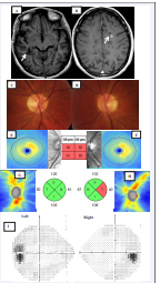
Figure 2: Images from a 34-year-old woman with a 10-year history of MS. A T1-MRI lesion on the right optic radiation (A) is consistent with ipsilateral homonymous hemimacular loss. There are multiple MS-like plaques, including one enhanced lesion (B, arrow). Fundus photography showed normal fundus in both eyes (C and D). The color-coded thickness map (red = thicker, blue = thinner) from SD-OCT showed hemimacular loss of GCL-IPL (E and F). A color-coded thickness map of optic disc showed marginally reduced RNFL thickness in the left eye (G, right 80 µm; and H, left 76 µm). Automated Visual Fields (VF) (I) showed a slightly enlarged blind spot in the left eye and normal in the right eye.
eye (Figure 2H). VF showed an enlarged central blind spot in the left and normal in the right eye (Figure 2I).
RESULTS AND DISCUSSION
We demonstrate that SD-OCT can visualize specific patterns of homonymous hemimacular GCL loss, functioning as an imaging biomarker for primary lesions in the retrochiasmal visual pathway. SD-OCT with a B-scan of 512 x 128 pixels at the macular cube acquires a greater amount of data at a higher speed and better resolution and enables the generation of the microstructure of the retina close to the histological level [2]. Homonymous hemimacular GCL loss detected by SD-OCT corresponds to an ipsilateral lesion behind the chiasm as detected by MRI. Case 1 had an infarction in the occipital lobe. VF defects of homonymous hemianopia persisted, corresponding to the remaining ipsilateral ischemic lesion in visual cortex. Case 2 had MS. Ophthalmologic examination revealed a slightly enlarged blind spot in one eye and moderately delayed VEP latency bilaterally. Other visual functions were intact; fundus findings were unremarkable. Homonymous hemimacular GCL loss on SD-OCT is consistent with ipsilateral MS plaque revealed by MRI in optic radiation.
Homonymous hemianopia has been used as hallmark for retrochiasmal visual pathway damage [10]. Peripheral homonymous hemianopia caused by stroke [11] or surgical lobectomy [12] is consistent with persistent lesions in visual tract and visual cortex. Such lesions can be detected using diffusion-tensor tractography [13]. Selective hemimacular thinning of each eye was also reported in a patient with neuromyelitis optica who had an ipsilateral optic tract lesion confirmed by diffusion-tensor tractography [14]. In a longitudinal study with SD-OCT, we recently observed one MS patient who developed hemimacular GCL-IPL loss over 4 months in relation to MS relapse [15]. One MS plaque was detected in ipsilateral optic radiation by MRI, although the patient had no new visual dysfunction. Additional thinning of hemimacular GCL-IPL was registered in another patient with subclinical progressive MS who had had an ipsilateral optic radiation lesion on MRI for several years [15]. This patient also had unchanged visual dysfunction. In a longitudinal study of ON, we observed GCL-IPL thinning earlier than RNFL thinning and were not affected by optic disc swelling [16]. We believe that hemimacular GCL-IPL loss detected by SD-OCT is a sensitive and specific imaging biomarker for lesions in retrochiasmal visual pathway, even in absence of VF defects.
CONCLUSION
These two cases illustrate existence of retrograde degeneration of visual pathway. Homonymous hemimacular GCL-IPL loss visualized by SD-OCT is consistent with ipsilateral lesions behind chiasm detectable by MRI, irrespective of VF defect. Such selective GCL-IPL loss can thus eventually serve as imaging biomarker to localize visual pathway lesions, monitoring disease progression and prognosis.
ACKNOWLEDGEMENT
The authors gratefully acknowledge financial support from Linköping University and the County Council of Östergötland, Sweden.
Conflict of Interest
The authors declare no conflict of interest. The study was approved by the Ethics Committee Review Board of the University in Linköping, Sweden (reference number 2013/141-31).
REFERENCES
- Schuman JS. Spectral domain optical coherence tomography for glaucoma (an AOS thesis). Trans Am Ophthalmol Soc. 2008; 106: 426-458.
- Dörr J, Wernecke KD, Bock M, Gaede G, Wuerfel JT, Pfueller CF, et al. Association of retinal and macular damage with brain atrophy in multiple sclerosis. PLoS One. 2011; 6: e18132.
- Saidha S, Sotirchos ES, Oh J, Syc SB, Seigo MA, Shiee N, et al. Relationships between retinal axonal and neuronal measures and global central nervous system pathology in multiple sclerosis. JAMA Neurol. 2013; 70: 34-43.
- Gabilondo I, Martínez-Lapiscina EH, Martínez-Heras E, Fraga-Pumar E, Llufriu S, Ortiz S, et al. Trans-synaptic axonal degeneration in the visual pathway in multiple sclerosis. Ann Neurol. 2014; 75: 98-107.
- Zhang X, Kedar S, Lynn MJ, Newman NJ, Biousse V. Homonymous hemianopia in stroke. J Neuroophthalmol. 2006; 26: 180-183.
- Balcer LJ, Miller DH, Reingold SC, Cohen JA. Vision and vision-related outcome measures in multiple sclerosis. Brain. 2015; 138: 11-27.
- Plant GT, Kermode AG, Turano G, Moseley IF, Miller DH, MacManus DG, et al. Symptomatic retrochiasmal lesions in multiple sclerosis: clinical features, visual evoked potentials, and magnetic resonance imaging. Neurology. 1992; 42: 68-76.
- Millington RS, Yasuda CL, Jindahra P, Jenkinson M, Barbur JL, Kennard C, et al. Quantifying the pattern of optic tract degeneration in human hemianopia. J Neurol Neurosurg Psychiatry. 2014; 85: 379-386.
- Martínez-Lapiscina EH, Ortiz-Pérez S, Fraga-Pumar E, Martínez-Heras E, Gabilondo I, Llufriu S, et al. Colour vision impairment is associated with disease severity in multiple sclerosis. Mult Scler. 2014; 20: 1207-1216.
- Fraser JA, Newman NJ, Biousse V. Disorders of the optic tract, radiation, and occipital lobe. Handb Clin Neurol. 2011; 102: 205-221.
- Ogawa K, Ishikawa H, Suzuki Y, Oishi M, Kamei S. Clinical study of the visual field defects caused by occipital lobe lesions. Cerebrovasc Dis. 2014; 37: 102-108.
- McDonald CR, Hagler DJ Jr, Girard HM, Pung C, Ahmadi ME, Holland D, et al. Changes in fiber tract integrity and visual fields after anterior temporal lobectomy. Neurology. 2010; 75: 1631-1638.
- Piper RJ, Yoong MM, Kandasamy J, Chin RF. Application of diffusion tensor imaging and tractography of the optic radiation in anterior temporal lobe resection for epilepsy: a systematic review. Clin Neurol Neurosurg. 2014; 124: 59-65.
- Romero RS, Gutierrez I, Wang E, Reder AT, Bhatti MT, Bernard JT, et al. Homonymous hemimacular thinning: a unique presentation of optic tract injury in neuromyelitis optica. J Neuroophthalmol. 2012; 32: 150-153.
- Huang-Link YM, Al-Hawasi A, Eveman I. Retrograde degeneration of visual pathway: hemimacular thinning of retinal ganglion cell layer in progressive and active multiple sclerosis. J Neurol. 2014; 261: 2453-2456.
- Huang-Link YM, Al-Hawasi A, Lindehammar H. Acute optic neuritis: retinal ganglion cell loss precedes retinal nerve fiber thinning. Neurol Sci. 2014.


