Epiploic Appendagitis: An Uncommon Cause of Abdominal Pain
- 1. Department of Internal Medicine, Ohio State University, USA
Citation
Langan M, Marcantoni V, Ungureanu C (2014) Epiploic Appendagitis: An Uncommon Cause of Abdominal Pain. JSM Clin Case Rep 2(1): 1016.
CLINICAL IMAGE
Epiploic appendages are fat filled sacs extending from the serosal surface of the colon into the peritoneum. There are approximately 100 appendices along the length of the colon, with most clustering in the cecal and sigmoid region [1,2]. Epiploic appendagitis occurs when epiploic appendages undergo torsion or have spontaneous thrombosis of the draining veins and subsequently become infarcted [1,2,3]. Presentation can be seen at any age but is most common in the second to fifth decades of life [3]. A 34 year old male presented to the emergency room with a three day history of left lower quadrant abdominal pain. The pain was sudden in onset, sharp, stabbing and pressure-like in character with an intensity of 8/10. Associated symptoms included nausea and lightheadedness. It worsened with food consumption and external pressure. There were no alleviating factors. He denied fever, chills, bloating, constipation, diarrhea, melena, hematochezia, dysuria, or hematuria. He had no previous history of nephrolithiasis, colon cancer or diverticulitis.
On physical exam, he was afebrile, normotensive, but appeared ill and pale. On abdominal exam, he had exquisite left lower quadrant pain on palpation with voluntary guarding but no rebound tenderness. Laboratory findings did not show leukocytosis (Figure 1,2).
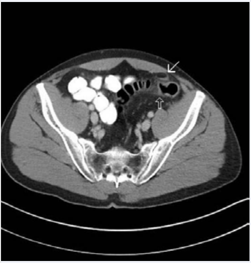
Figure 1 Abdominal CT showing epiploic appendagitis (white arrow) and adjacent colitis (black arrow).
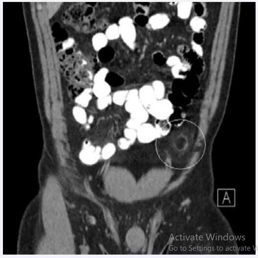
Figure 2 Coronal view of abdominal CT scan showing inflamed appendage (circled).
He had a mild thrombocytopenia. An abdomen/ pelvis CT scan revealed a curvilinear soft tissue density surrounding area of fat with adjacent inflammatory fat stranding, suggestive of epiploic appendagitis as well as surrounding inflammation of the adjacent colon.
Treatment was initiated with ibuprofen for five days. The patient improved and was symptom-free after this intervention. This case illustrates a presentation seen in primary care and urgent care settings with an unlikely but benign etiology. Epiploic appendagitis commonly presents with sharp lower quadrant abdominal pain which can be mistaken for an acute abdomen. This may lead to misdiagnosis with acute diverticulitis or appendicitis [1].
CT is the most common modality to encounter acute epiploic appendagitis [2,4]. The most common CT features include an oval fat-density lesion 1.5-3.5 cm in diameter which abuts the anterior wall of the sigmoid colon with surrounding inflammatory changes of the bowel and mesenteric fat [2]. Primary differential considerations on imaging include acute omental infarct (which isusually located in the right abdomen, simulating appendicitis) and acute uncomplicated diverticulitis [2]
A conservative approach with anti-inflammatory agents is usually sufficient for treatment of epiploic appendagitis as it is usually a self-limiting condition [1,3].


