Imaging of Urological Cancer Morphology by Using Probe-Based Confocal Laser Endomicroscopy with New Contrast Agent-Preliminary Study for New TURBT Technique-
- 1. Department of Urology, Meiji University of Integrative Medicine, Japan
- 2. Department of Urology, Kyoto Prefectural University of Medicine, Japan
- 3. Department of Pathology, Kyoto Prefectural University of Medicine, Japan
Abstract
Objective: To observe the nucleus of tumor cell using probe-based confocal laser endomicroscopy (pCLE), we examined ethacridine as new contrast agent.
Recently, usefulness of pCLE by using fluorescein for detection of urothelial carcinomawas reported. Fluorescein did not reveal cell nucleus and cytoplasm. In this study, we examined ethacridine for pCLE to observe the nuclei of urothelial carcinoma cells ex vivo.
Methods: 3 samples were obtained from TURBT specimens. Samples from freshly excised urothelial cancerous lesion were retrieved from resected tissue. The samples were placed on the glass slide, immersed immediately in ethacridine solution (0.01% in saline) for 60 secs, and subsequently rinsed in normal saline solution, and pCLE imaging was performed. After observation, the samples were fixed by formalin on the glass slide, followed by placement of a cover glass, and stained by hematoxylin-eosin. The imaging of pCLE was compared with histologic findings under the same horizontal view.
Results: All 3 patients with high-grade urothelial carcinoma, stained nuclei were observed. The morphological patterns of pCLE were like those of histopathological study.
Conclusion: pCLE with ethacridine can clearly detect the cancerous lesion. pCLE by using intravesical ethacridine instillation might be useful and less invasive.
KEYWORDS
• Endomicroscopy
• Laser optical fiber
• Urothelial carcinoma
• Ethacridine
Citation
Naya Y, Konishi E, Takaha N, Oishi M, Ueda T, et al. (2017) Imaging of Urological Cancer Morphology by Using Probe-Based Confocal Laser Endomicroscopy with New Contrast Agent-Preliminary Study for New TURBT Technique. JSM Clin Oncol Res 5(1): 1051.
ABBREVIATIONS
pCLE: probe Based Confocal Laser Endomicroscopy; NBI: Narrow Band Imaging; PDD: Photodynamic Diagnosis; TURBT: Transurethral Resection of Bladder Tumor; UC: Urothelial Carcinoma
INTRODUCTION
Bladder cancer is the 8th most commonly newly diagnosed cancer in the world [1]. For diagnosis, cystoscopy by using white light is performed as a standard procedure; however, white light can lead to missing lesions that are present but not visible [2]. Photodynamic diagnosis (PDD) and narrow band imaging (NBI) are developed as a new technique of cystoscopy. The value of PDD and NBI for improved outcome in relation to recurrence remains to be controversial [3-5]. Recently, several authors reported the usefulness of probe based confocal laser endomicroscopy (pCLE) by using fluorescein for detection of urothelial carcinoma during endoscopic procedure [6-9].
ne correspondence. Next study will be needed for clinical use of pCLE using ethacridine Fluorescein revealed tissue structures of urothelial carcinoma without imaging of cell nucleus and cytoplasm. Li et al., reported that 0.02% of acrifravine stained cell nuclei for detection of gastric cancer under esophago gastroduodenoscopy in China [10]; however, acrifravine is harmful to the eyes or mucosa if inhaled, and irritable to skin. Less injurious compound is ethacridine lactate, which is one of the acridine derivatives, and it is an antiseptic agent used for skin disinfection. Intravesical instillation of ethacridine was also used previously for bladder disinfection in Japan. Both ethacridine and acrifravine were derivatives of acridine orange. Kusuzaki et al reported that acridine orange specifically binds to malignant tumors and immediately accumulates in tumor cells [11]. Therefore, we examined the feasibility of ethacridine for pCLE to observe the nuclei of urothelial carcinoma cells ex vivo as preliminary study for the detection of urothelial cancer under the procedure of transurethral resection of bladder tumor (TURBT).
MATERIALS AND METHODS
This study was approved by the ethics committees of Kyoto Prefectural University of Medicine and Meiji University of Integrative Medicine. Between January 2016 and May 2016, tissue samples were obtained from 3 patients undergoing TURBT (Table 1).
Table 1: Patient characteristics.
| Case | Age | Sex | Histology | Nuclei staining |
| 1 | 78 | M | Small cell Carcinoma | ++ |
| 2 | 75 | M | UC High | ++ |
| 3 | 82 | M | UC High | ++ |
Abbreviations: UC: Urothelial Carcinoma
Samples from freshly excised urothelial cancerous papillary lesion measuring approximately 5x5 mm were retrieved from the resected tissue. The samples were placed on the glass slide, immersed immediately in ethacridine solution (0.01% in saline) for 60 secs, and subsequently rinsed in normal saline solution. pCLE imaging of ethacridine-stained samples was performed ex vivo by using a pCLE system (Cellvizio, Mauna Kea Technologies, Paris, France). The method of pCLE was previously described [6]. Briefly, a 2.7-mm cystoflex-UHD Cellvizio fiber for confocal microscopy was connected to the laser unit. Fiber contact with the tissue samples produced 60 nm-deep images with a resolution of 1 μm (30000 pixels) covering a 240 μm diameter field. After observation, the samples were fixed by formalin on the glass slide with a cover glass and stained by hematoxylin-eosin. The imaging of pCLE was compared with histologic findings under the same horizontal view.
RESULTS AND DISCUSSION
Morphological appearances of urothelial mucosa and urothelial cancer on pCLE and corresponding histologic images are shown in (Figure 1). The nuclei of individual cells on pCLE were readily visualized as fluorescent white dots with connective tissues on less fluorescent background. This case is 82 years old male with high-grade urothelial carcinoma (UC, Grade 3). One-to-one corresponding areas of histopathological findings are also shown in (Figure 1).
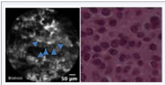
Figure 1 Morphological appearances of urothelial mucosa and urothelial cancer on pCLE and corresponding histology images. The nucleus of individual cells on pCLE were readily visualized as fluorescent white dots with connective tissues on a less fluorescent background (arrows). The morphological patterns of pCLE were like those of histopathological study.
The morphological patterns of pCLE were like those of histopathological study. Several authors reported the efficacy of pCLE for detection of bladder urothelial cancer by using fluorescein [6-9]. Because of limited visual field and depth, pCLE cannot be proposed for complete exploration of the bladder. pCLE can, however, be combined with certain procedures, such as NBI or PDD, to obtain histologic images of targeted areas. pCLE can also provide histologic information about the tumor area and edges. The natural fluorescence of tissues without a fluorescent dye is insufficient for pCLE [12] Fluorescein is approved by the Food and Drug Administration, and has been used in clinical practice for many years by gastroenterologists [12-14].The problem of using fluoresce in is that fluoresce cannot stain cytoplasm and nuclei of the cells. Li et al., reported the efficacy of acrifravine for CLE in patients who underwent upper gastrointestinal CLE [10]. Recently, Chan et al., reported that acrifravine could stain cell nuclei of breast cancer in their ex vivo study [15]. Acrifravine is harmful in the eyes or mucosa if inhaled, and causes skin irritation. Therefore, we tested ethacridine, one of the derivatives of acrifravine that is approved as an antiseptic drug in the urological, gynecological, and dermatological fields in Japan [16]. There were no data available on adverse effects of ethacridine; however, it is available for purchase without prescription at drug store in Japan. Generally, ethacridine seemed to be considered safe in Japan. Ethacridine is one of derivative of acridine orange, and acridine orange was densely to lysosomes and acidic vesicles, which are rich in tumor cells [17]. Kusuzaki et al., reported that acridine orange specifically binds to malignant tumors and immediately accumulates in tumor cells [11]. In this study, ethacridine also stained the nuclei of the urothelial cells similarly to acridine orange. Fluorescein is safe, but it is approved for ophthalmological uses only in Japan, and cannot stain the nucleus of the any cancer cell. pCLE-based fluorescein can visualize only architecture and cell size without nucleus. In this study, ethacridine could stain the cell nucleus. In this study, we used 0.01% of ethacridine for 60 seconds and cell nucleus was clearly observed. Further study is needed to optimize the staining conditions (e.g., concentration of ethacridine, staining time, etc.). Regardless, ethacridine has a potential to visualize the cancer cell nucleous by using pCLE, and it seemed to be easier to determine cancerous lesion compared to pCLE-based fluoresce in.
In this study, we compared the images of pCLE with histopathological findings under the same horizontal view. Therefore, we observed almost the same lesion by pCLE and microscopically from the same horizontal view. We confirmed accuracy of pCLE compared to histopathological examination by one-to-o e under TURBT procedure in vivo. This new technique might be useful detection of carcinoma in situ without unnecessary random biopsies. pCLE might be alternative biopsy method with less invasive as optical biopsy.
CONCLUSION
pCLE by using ethacridine can clearly detect the cancerous lesion. The next step is intravesical ethacridine instillation to determine the margin of cancerous lesion during transurethral resection. pCLE by using intravesical ethacridine instillation might be useful and less invasive.


