Teaching Neuroimaging: The Radiological Findings Point to the Genetic Cause in Two Brothers with Sensorineural Hearing Loss
- 1. Division of Neuropaediatrics, Hospital for Children and Adolescents, University Hospital Leipzig, Germany
- 2. Institute of Human Genetics, University of Leipzig, Medical Center, Germany
- 3. Clinic of Otolaryngology, Head and Neck Surgery, University Hospital Leipzig, Leipzig, Germany
- 4. Department of Pediatric Radiology, Hospital for Children and Adolescents, University Hospital Leipzig, Germany
Keywords
X-Linked Deafness; POU3F4 Gene; Corkscrew-Like Shaped Auditory Canal
Clinical Presentation
Two half-brothers of a healthy mother were diagnosed with bilateral sensorineural hearing loss. The maternal grandfather was deaf. Exome sequencing revealed a maternally inherited, hemizygous, likely pathogenic variant NM_000307.5:c. 1000A>G, p.(Lys334Glu) in the POU3F4 gene in both brothers. Both brothers received an MRI and CT scan to plan cochlear implantation. MRI showed that the cochlea had a very wide opening to the internal auditory canal on both sides with a corkscrew-like shape. This was confirmed on CT and indicates incomplete partition type III (X-linked deafness). Mutations in the POU3F4 gene lead to non-syndromic, X-linked deafness (MIM #304400). Absence of a cochlear modiolus with a dilated lateral internal auditory canal and a thickened stapes footplate as a pathognomonic radiological pattern of this disease were reported [1]. The radiological findings already point to the genetic cause in these patients.
Figure 1
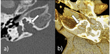
Figure 1: CT of the older brother at the age of 5 years. It shows bilateral cochlear incomplete partition type III. Both the right (a) and the left (b) cochlea lack the modiolus and the cribriform plate with preserved interscalar septa. This appearance has been referred to as “corkscrew” sign (arrows).


