Loading

JSM Dental Surgery

3D Relationship of Maxillary Posterior Teeth and Sinus Floor for Immediate Extraction Implant Placement

Research Article | Open Access | Volume 5 | Issue 1

  • 1. Department of Bioengineering, Università degli Studi di Napoli Federico, Italy
+ Show More - Show Less
Corresponding Authors
Francesco Amato, Department of Bioengineering, Università degli Studi di Napoli Federico, Italy
Abstract

The objective of the study was to analyze the anatomical relationship between the sinus floor and the root apexes of maxillary premolars and molars to evaluate the amount of basal bone available for immediate extraction implant placement.

Study Design: Maxillary arches Cone-Beam Computerized Tomographic (CBCT) scans taken at one dental clinic were analyzed. The sub-sinusal bone height (SBH) in the premolar region was measured, in the first and second molar area the inter-radicular septum bone height (ISBH) was also measured.

Results: Six hundred (600) CBCT scans were examined. In the first premolar region the findings were: 1.5% root apex inside the sinus; in 4.9% it approximated the sinus floor; in 17.5% distance varied between 1 and 3mm; 36.4% distance between 3 and 7mm; and 39.6% >7mm. In the second premolar region the findings were: 13.9% inside the sinus; 11.8% approximating the sinus floor; 30.8% distance 1-3mm; 28.3% distance 3-7 mm; 15.1% >7mm. The first molar area showed 26.2% ISBH 7 mm. The second molar area showed 14.3% ISBH 7 mm.

Conclusion: Pre-operatory tridimensional radiological diagnosis is essential to safely insert an implant in fresh extraction sockets of maxillary premolars and molars in order to avoid complications such as sinus floor perforation.

Keywords

• Subsinus basal bone

• Immediate placement

• Maxillary sinus

• Cone-beam CT

• Membrane perforation

CITATION

Amato F (2023) 3D Relationship of Maxillary Posterior Teeth and Sinus Floor for Immediate Extraction Implant Placement. JSM Dent Surg 5(1): 1036.

INTRODUCTION

Although the use of osseointegrated implants to replace missing teeth has become a predictable procedure with high implant survival rates [1,2], success rates in the posterior maxilla are lower due to the poor quality and inadequate height of bone [3]. Furthermore, after tooth loss, the periosteum of the maxillary sinus can exhibit increased osteoclastic activity, which can cause bone resorption 4 and a resumption of the physiological sinus- floor pneumatization process [5,6].The result is a progressive reduction of the bone height. Subsinus alveolar bone height (SBH) has been defined as the distance between the floor of the maxillary sinus and the alveolar crest [7]. A lack of SBH may be treated in different ways depending on the degree of atrophy. Options include sinus lift augmentation, vertical bone regeneration of the alveolar ridge, interpositional grafting, short implants, or use of alternative sites (tuberal, pterygoid, zygoma, or tilted) [8-10]. A valid alternative to prevent post-extraction sinus-floor pneumatization and avoid the need for grafting procedures can be immediate extraction implant placement.

Several authors have advocated that one of the main criteria for achieving good primary stability with implants immediately placed in extraction sites is to engage the implant in the bone apical to the root apex [11-13].When taking this approach, however, it is essential to be aware of the amount of basal bone and the relationship between the root apex and the anatomical landmarks apical to it: the anterior part of the sinus floor for the premolars and the posterior portion of the sinus floor for the molars.

SBH has been investigated by several authors [7,14-16], using panoramic radiographs of edentulous maxillae. Other authors have used three-dimensional diagnostic tools to gather anatomical information about this area [17-21].

The aim of this CBCT study was to further investigate the relationship between the sinus floor and the root apexes in the maxillary premolar and molar area, to the interradicular bone septum height (IBSH) in the molar area, and to determine the amount of bone apical to the root apexes available for implant insertion in the posterior region.

MATERIAL AND METHODS

All the cone-beam computerized tomographic (CBCT) scans of patients with fully or partially dentate maxillary arches that were taken at a private dental. The 3D radiographical examination were obtained using a Gendex CB-500 machine. The software system was periodically calibrated and controlled. The same operator, three times for each landmark, took all the measurements and repeated each operation two times to achieve a more accurate precision. A mean value of the two measurements was carried out. Depending on the obtained bone height, the findings were distributed in five Groups for the premolars and four Groups for the molars. A percentage value of the distribution of the teeth in the corresponding Group was carried out.

Patients inclusion criteria were the following: the patients scanned had to be 20 years or older, at least two maxillary premolars and/or molars had to be present on each side, no impacted teeth or other pathology could be present in the posterior maxillary alveolar bone. Smoking was not considered an exclusion criteria.

On each scan, the measures were carried out using each the dedicated software I-Cat Vision. For the premolars the distance between the root apex and the closest point of the sinus floor was recorded: Sub-sinus Bone Height (SBH). In the molars the distance between the roof of the forcation and the closest point of the sinus floor: inter-radicular septum bone height (ISBH) was measured.

Findings for the premolars were classified into five groups: The root apex was located inside the sinus (Group A); The root apex was in contact with the sinus floor, but not penetrating in the sinus (Group B); The SBH was < 3 mm (Group C); The SBH was more than 3 mm but less than 7 mm (Group D); The SBH was more than 7 mm (Group E). Figure 1 presents examples of each of the five categories.

Classification in five different categories for premolars based on sub-sinusal bone height (SBH).

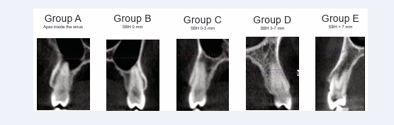
Figure 1: Classification in five different categories for premolars based on sub-sinusal bone height (SBH).

The findings for the inter-radicular septum bone height (ISBH) in the molar region were classified into three groups: ISBH < 4 mm (Group A); ISBH >4-7 mm Group B; ISBH > 7 mm Group C. Figure 2 presents examples of each of the three categories.

Classification in three different categories for molars, based on interradicular septum bone height (ISBH).

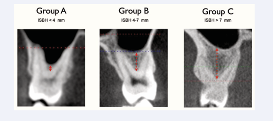
Figure 2: Classification in three different categories for molars, based on interradicular septum bone height (ISBH).

RESULTS

A total of 600 CBCT scans from 325 female and 275 male patients were analyzed. The patients mean age was 46. The scans included 977 first premolars. Fifteen of these belong to Group A (1.5%); 48 were in Group B (4.9 %); 171 in Group C (17.5%); 356 in Group D (36.4%), Group D); 387 in Group E (39.6%).

A total of 939 second premolars were analyzed. One hundred thirty-one of these belong to Group A (13.9%); 111 were in Group B (11.8%); 289 in Group C (30.8%); 266 in Group D (28.3%); 142 in Group E (15.1%).

Table 1 summarizes the findings for all premolars. In the molar area, a total of 1.231 first molars were analyzed, 323 (26.2%) had an ISBH of less than 4mm (Group A), 472 (38.3%) had an ISBH of 4 - 7 mm (Group B), 436 (35.4%) had an ISBH of more than 7 mm (Group C).

A total of 1.250 second molars were analyzed, 179 (14.3%) had an ISBH of less than 4mm (Group A), 578 (46.2%) had an ISBH of 4 - 7 mm (Group B), 493 (39.4%) had an ISBH of more than 7 mm (Group C). Table 2 summarizes the molar findings.

DISCUSSION

The anatomical relationship between the premolar and molar roots and the sinus floor in the posterior maxilla area is articulated and has a high individual variability [22]. Various factors influence the dimension of the sinus, including growth, function, and pneumatization. The alveolar dental process, breathing physiology, and functional changes all further contribute to the high differentiation of the maxillary sinus [23].

The position of the sinus floor is a limiting factor that influences surgical treatment and implant insertion in the posterior maxilla. The post-extraction bone remodeling and the consequent sinus pneumatization that often takes place after tooth loss can reduce the available bone crest to the level where bone augmentation procedures are necessary prior to implant placement [24].

When the vertical bone atrophy is related to the pneumatization of the maxillary sinus, elevation of the sinus floor has been suggested to enable implant placement [25]. Depending on the amount of residual bone, different bone-augmentation techniques have been proposed, including the lateral window approach described by Boyne and James (1980) and Tatum (1986) [26,27].The crestal approach, in which the Schneiderian membrane is lifted using an osteotome, was first described by Summers (1994) [28]. In both techniques, the simultaneous insertion of implants is recommended if the initial bone height is a minimum of 4 to 6 mm [29]. Otherwise, a delayed insertion is preferred [30].

While sinus augmentation has been demonstrated to be a predictable technique for increasing bone height in deficient posterior maxillary ridges prior to implant placement, the mean survival rates of implants placed in sinus-grafted areas are lower than those of implants placed in native bone after functional loading [31-33]. Some recent studies have compared the survival rates of standard length implants (more than 8mm) in lifted sinus versus the use of short implants (≤8mm). These studies have clearly demonstrated comparable survival rates between longer implants in grafted sinus and extra short implants, but higher biological complications when a sinus lift was performed, sinus membrane perforation can occur more frequently in the augmented sinus than for extra short implants [34,35]. In order to reduce the complications, the surgical time, the costs and the morbidity the use of extra short implants can represent a valid surgical treatment if a correct and scrupulous cases selection is performed [36,37].

A valid alternative to prevent post-extraction alveolar bone atrophy and thus avoid the need for grafting can be immediate extraction placement [38]. The immediate extraction placement technique has become a common procedure for replacing hopeless teeth. Its major advantage is to reduce the duration and invasiveness of the treatment [39-41], yielding higher patient-satisfaction levels than delayed implant placement [42]. Immediate implant placement in the esthetic zone has become a therapeutic first choice for clinicians [43], while in molar regions,some difficulties must be overcome. The predictability of the post-extraction implant surgery in such sites depends upon the close approximation of the bone walls to the implant surface as well as the bone density, quality, and availability [44]. Some have argued that in order to achieve good primary stability at the time of immediate extraction placement, the bone apical to the root apex or the interradicular bone in case of multi-rooted teeth should be engaged by the implant threads [11-14].

The crestal extension of the maxillary antrum and its relationship with premolar and molar roots can be a limiting factor for immediate extraction implant placement. The available basal bone between the roots apexes and floor of the sinus can only be precisely measured after a three-dimensional diagnostic examination [45]. The present study demonstrates that in the first premolar area in about 25% of the cases there is little (< 3mm) or no bone apical to the root apex to engage with the implant to achieve primary stability. In the second premolar area, this percentage increases to more than 60% of the cases. Thus, care should be taken when immediate extraction placement is performed not to extend the osteotomy beyond the root apex in order to avoid sinus perforation. As a consequence selecting an implant with a diameter coherent or larger than the actual mesio-distal size of the alveolus could be a valid choice to achieve immediate mechanical stability after tooth extraction.

In the molar area the inter-radicular septum has been indicated as the ideal site for implant insertion both for mechanical stability and for the prosthetic axis [46].Thus the dimension of the septum represent a key factor for successful immediate implant placement.

The results of this study indicate that more than 60% of the first and second molars had an ISBH of less than 7mm, as a consequence immediate extraction placement in the inter radicular bone septum of molars in most of the cases should be performed applying a sinus elevation with the crestal approach (osteotome technique) or using ultra-short implants in order to minimize the risks of sinus-membrane perforation.

In all the CBCT scans that were analyzed, the distance between root apexes and maxillary sinuses was least for all the second premolars and first molars. This finding was not uniform, however, because of the anatomical variability of sinus growth.

One of the notable anatomical characteristics was the symmetry of the anatomical conditions in either side of each patient’s mouth, both in the premolar and in the molar areas. This can probably be explained by the equivalent development of sinus pneumatization on both sides of the mouth and the fact that the premolar and molar roots on each side of any person’s mouth tend to be the same shape and length.

CONCLUSION

The results of this study show that in about 25% of the first premolar cases there is a close proximity between the root apexes and the inferior border of the sinus floor that would not allow for apical stabilization in the basal bone of an immediate extraction implant. The same condition was found in more than 55% of the cases in the second premolar region. A valid alternative to achieve immediate implant primary stability could be to use a wide diameter implant to engage the mesial and distal walls of the alveolar socket. In the molar area both the first and second molar showed in more than 60% of the cases an inter-radicular septum bone height inadequate to insert a standard length implant. Alternatively, when immediate extraction implant placement is being carried out, the use of short implants or the use of osteotomes, rather than burs, to deepen the apical part of the osteotomy and elevate the sinus floor may be indicated to minimize the risk of membrane perforation and allow for standard length implant placement.

REFERENCES

1. Fugazzotto PA, Gulbransen HJ, Wheeler SL, Lindsay JA. The use of IMZ osseointegrated implants in partially and completely edentulous patients: success and failure rates of 2.023 implant cylinders up to 60 months in function. Int J Oral Maxillofac Implants. 1993; 8: 617-621.

2. Leimola-Virtanen R, Peltola J, Oksala E, Helenius H, Happonen RP. ITI titanium plasma-sprayed screw implants in the treatment of edentulous mandibles: a follow-up study of 39 patients. Int J Oral Maxillofac Implants. 1995; 10: 373-378.

3. Khatiblou FA. Sinus floor augmentation and simultaneous implant placement, part 1: the 1-stage approach. J Oral Implantol. 2005; 31: 205-208.

4. Misch C. Density of bone: effect on treatment planning, surgical approach and healing. In: Misch C, ed. Contemporary Implant Dentistry. St Louis, Mo: Mosby; 1993: 469-485.

5. Wehrbein H, Diedrich P. Progressive pneumatization of the basal maxillary sinus after extraction and space closure [in German]. Fortschr Kieferorthop. 1992; 53: 77-83.

6. Ohba T, Langlais RP, Morimoto Y, Tanaka T, Hashimoto K. Maxillary sinus floor in edentulous and dentate patients. Indian J Dent Res. 2001; 12: 121-122

7. Kopecka D, Simunek A, Brazda T, Rota M, Slezak R, Capek L. Relationship between subsinus bone height and bone volume requirements for dental implants: A human radiographic study. Int J Oral Maxillofac Implants 2012; 27: 48-54.

8. Ulm CW, Solar P, Gsellmann B, Matejka M, Watzek G. The edentulous maxillary alveolar process in the region of the maxillary sinus-A study of physical dimensions. Int J Oral Maxillofac Surg. 1995; 24: 279-282.

9. Jensen OT. Combined sinus grafting and Le Fort I Procedures. In: Jensen OT (ed). The Sinus Bone Graft. Chicago: Quintessence.1999: 191-200.

10. Schleier P, Bierfreund G, Schultze-Mosgau S, Moldenhauer F, Küpper H, Freilich M. Simultaneous dental implant placement and endoscope guided internal sinus floor elevation: 2-year post-loading outcomes. Clin Oral Implants Res. 2008; 19: 1163-1170.

11. Maksoud MA. Immediate implants in fresh posterior extraction sockets: report of two cases. J Oral Implant. 2001; 27: 123-126.

12. Crespi R, Capparè P, Gherlone E. Fresh socket implants in periapical infected sites in human. J Periodontol. 2010; 81: 378-383.

13. Bhale M, Neely AL, Kolhatkar S. Immediate implant placement: clinical decisions, advantages and disvantages. J Prosthodont. 2008; 17: 576-581.

14. Penarrocha M, Uribe R, Balaguer J. Immediate implants after extraction. A review of the current situation. Med. Oral. 2004; 234 242.

15. Monsour PA, Dudhia R. Implant radiography and radiology. Aust Dent J. 2008; 53: S11-S25.

16. Ulm CW, Solar P, Gsellmann B, Matejka M, Watzek G. The edentulous maxillary alveolar process in the region of the maxillary sinus—A study of physical dimensions. Int J Oral Maxillofac Surg. 1995; 24: 279-282.

17. Hedes?iu M, Balog C, Preda DM, Baciut G, Fildan F, Pop A, et al. The accuracy of alveolar crest dimensions measurement for dental implants. In vitro study. Rev Med Chir Soc Med Nat Ias?i. 2008; 112: 224-228.

18. Klinge B, Petersson A, Maly P. Location of the mandibular canal: Comparison of macroscopic findings, conventional radiography, and computed tomography. Int J Oral Maxillofac Implants. 1989; 4: 327 332.

19. Eberhardt JA, Torebinejad M, Christiansen EL. A computed tomographic study of the distances between the maxillary sinus floor and the apices of the maxillary posterior teeth. Surg Oral Med Oral Pathol. 1992; 73: 345-356.

20. Sharan A, Madjar D. Correlation between maxillary sinus floor topography and related root position of posterio teeth using panoramic and cross-sectional computed tomography imaging. Oral Surg Oral Med oral Pathol Oral Endod. 2006; 102: 375-381.

21. Yoshimine S, Nishihara K, Nozoe E, Yoshimine M, Nakamura N. Topographic analysis of maxillary premolars and molars and maxillary sinus cone beam computed tomography. Implant Dent. 2012; 21: 528-535.

22. Jang JK, Kwak SW, Ha JH, Kim HC. Anatomical relationship of maxillary posterior teeth with the sinus floor and buccal cortex J Oral Rehabil 2017; 44: 617-625

23. Lana JP, Carneiro PM, Machado VC, Souza PE, Manzi FR, Horta MC. Anatomic variations and lesions of the maxillary sinus detected in cone beam computed tomography for dental implants. Clin Oral Implants Res. 2012; 23: 1398-1403.

24. Araújo MG, Lindhe J. Dimensional ridge alterations following tooth extraction. An experimental study in the dog. J Clin Periodontol. 2005; 32: 212-218.

25. Raghoebar GM, Onclin P, Boven GC, Vissink A, Meijer HJA Long-term effectiveness of maxillary sinus floor augmentation: A systematic review and meta-analysis J Clin Periodontol. 2019; 46: 307-318.

26. Tatum H Jr. Maxillary and sinus implant reconstruction. Dent Clin North Am. 1986; 30: 207-229

27. Boyne PJ, James RA. Grafting of the maxillary sinus floor with autogenous marrow and bone. J Oral Surg. 1980; 38: 613-616.

28. Summers RB. A new concept in maxillary implant surgery: the osteotome technique. Compendium. 1994; 15: 154-156.

29. Summers RB. The osteotome technique, part 3: less invasive methods of elevating the sinus floor. Compendium. 1994; 15: 698.

30. Smiler DG, Johnson P, Lozada J, Misch C, Rosenlicht JL, Tatum OH, et al. Sinus lift grafts and endosseous implants: treatment of the atrophic posterior maxilla. Dent Clin North Am. 1992; 36: 151-186.

31. Avila-Ortiz G, Wang HL, Galindo-Moreno P, Mish CE, Rudek I, Neiva R. Influence of lateral window dimensions on vital bone formation following maxillary sinus augmentation. Int J oral Maxillofac Implants. 2012; 27: 1240-1238.

32. Pjetursson BE, Tan WC, Zwahlen M, Lang NP. A systematic review of the success of sinus floor elevation and survival of implants in- serted in combination with sinus floor elevation. J Clin Periodontol. 2008; 35: 216-240.

33. Wallace SS, Froum SJ. Effect of maxillary sinus augmentation on the survival of endosseous dental implants. A systematic review. Ann Periodontol. 2003; 8: 328-343.

34. Thoma DS, Zeltner M, Husler J, Hammerle CH, Jung RE. EAO Supplement Working Group 4- EAO CC 2015 Short implants wersus sinus lifting with longer implants to restore the posterior maxilla: a systematic review. Clin Oral Implants Res. 2015; 26: 154-169.

35. Felice P, Pistilli R, Piattelli M, Soardi E, Corvino V, Esposito M. Posterior atrophic jaws rehabilitated with prostheses supported by 5 x 5mm implants with novel nanostructured calcium-incorporated titanium surface or by longer implants in augmented bone. Preliminary results from a randomized controlled trial. Eur J Oral Implantol. 2012; 5: 149-161.

36. Guljè FL, Raghoebar GM, Vissink A, Meijer HJ. Single crowns in the resorbed posterior maxilla supported by either 6-mm implants or by 11-mm implants combined with sinus floor elevation surgery: a 1-year randomised controlled trial. Eur J Oral Implantol. 2014; 7: 247-255.

37. Lemos CA, Ferro-Alves ML, Okamoto R, Mendonça MR, Pellizzer EP. Short dental implants versus standard dental implants placed in the posterior jaws: A systematic review and meta-analysis. J Dent. 2016; 47: 8-17.

38. Stephen T Chen 1, Thomas G Wilson Jr, Christoph, Hämmerle HF. Immediate or early placement of implants following tooth extraction: review of biologic basis, clinical procedures, and outcomes Int J Oral Maxillofac Implants. 2004; 19: 12-25

39. Barzilay I, Graser GN, Caton J, Shenkle G. Immediate implantation of pure titanium threaded implants into extraction sockets. J Dent Res. 1988; 67: 234.

40. Barzilay I, Graser G, Iranpour B, Natiella JR. Immediate implantation of a pure titanium implant into an extraction socket: Report of a pilot procedure. Int J Oral Maxillofac Implants. 1991; 6: 277-284.

41. Lazzara RJ. Immediate implant placement into extraction sites: Surgical and restorative advantages. Int J Periodontics Restorative Dent. 1989; 9: 332-343.

42. Quirynen M, Van Assche N, Botticelli D, Berglundh T. How does the timing of implant placement to extraction affect outcome? Int J Oral Maxillofac Implants. 2007; 22: 203-223.

43. Atieh MA, Ahmad AH, Payne AGT, Duncan WJ. Immediate loading with single implant crowns: A systematic review and meta-analysis. Int J Prosthodont. 2009; 22: 151-160.

44. Atieh MA, Payne AG, Duncan WJ, Cullinan MP. Immediate restoration/ loading of immediately placed single implants: is it an effective bimodal approach? Clin Oral Implants Res. 2009; 20: 645-659.

45. Park YB, Jeon HS, Shim JS, Lee KW, Moon HS. Analysis of the anatomy of the maxillary sinus septum using 3-dimensional computed tomography. J Oral Maxillofac Surg. 2011; 69: 1070-1078.

46. Amato F, Polara G. Immediate Implant Placement in Single-Tooth Molar Extraction Sockets: A 1- to 6-Year Retrospective Clinical Study. Int J Periodontics Restorative Dent. 2018; 38: 495-501

Amato F (2023) 3D Relationship of Maxillary Posterior Teeth and Sinus Floor for Immediate Extraction Implant Placement. JSM Dent Surg 5(1): 1036

Received : 25 Oct 2023
Accepted : 21 Nov 2023
Published : 24 Nov 2023
Journals
Annals of Otolaryngology and Rhinology
ISSN : 2379-948X
Launched : 2014
JSM Schizophrenia
Launched : 2016
Journal of Nausea
Launched : 2020
JSM Internal Medicine
Launched : 2016
JSM Hepatitis
Launched : 2016
JSM Oro Facial Surgeries
ISSN : 2578-3211
Launched : 2016
Journal of Human Nutrition and Food Science
ISSN : 2333-6706
Launched : 2013
JSM Regenerative Medicine and Bioengineering
ISSN : 2379-0490
Launched : 2013
JSM Spine
ISSN : 2578-3181
Launched : 2016
Archives of Palliative Care
ISSN : 2573-1165
Launched : 2016
JSM Nutritional Disorders
ISSN : 2578-3203
Launched : 2017
Annals of Neurodegenerative Disorders
ISSN : 2476-2032
Launched : 2016
Journal of Fever
ISSN : 2641-7782
Launched : 2017
JSM Bone Marrow Research
ISSN : 2578-3351
Launched : 2016
JSM Mathematics and Statistics
ISSN : 2578-3173
Launched : 2014
Journal of Autoimmunity and Research
ISSN : 2573-1173
Launched : 2014
JSM Arthritis
ISSN : 2475-9155
Launched : 2016
JSM Head and Neck Cancer-Cases and Reviews
ISSN : 2573-1610
Launched : 2016
JSM General Surgery Cases and Images
ISSN : 2573-1564
Launched : 2016
JSM Anatomy and Physiology
ISSN : 2573-1262
Launched : 2016
Annals of Emergency Surgery
ISSN : 2573-1017
Launched : 2016
Annals of Mens Health and Wellness
ISSN : 2641-7707
Launched : 2017
Journal of Preventive Medicine and Health Care
ISSN : 2576-0084
Launched : 2018
Journal of Chronic Diseases and Management
ISSN : 2573-1300
Launched : 2016
Annals of Vaccines and Immunization
ISSN : 2378-9379
Launched : 2014
JSM Heart Surgery Cases and Images
ISSN : 2578-3157
Launched : 2016
Annals of Reproductive Medicine and Treatment
ISSN : 2573-1092
Launched : 2016
JSM Brain Science
ISSN : 2573-1289
Launched : 2016
JSM Biomarkers
ISSN : 2578-3815
Launched : 2014
JSM Biology
ISSN : 2475-9392
Launched : 2016
Archives of Stem Cell and Research
ISSN : 2578-3580
Launched : 2014
Annals of Clinical and Medical Microbiology
ISSN : 2578-3629
Launched : 2014
JSM Pediatric Surgery
ISSN : 2578-3149
Launched : 2017
Journal of Memory Disorder and Rehabilitation
ISSN : 2578-319X
Launched : 2016
JSM Tropical Medicine and Research
ISSN : 2578-3165
Launched : 2016
JSM Head and Face Medicine
ISSN : 2578-3793
Launched : 2016
JSM Cardiothoracic Surgery
ISSN : 2573-1297
Launched : 2016
JSM Bone and Joint Diseases
ISSN : 2578-3351
Launched : 2017
JSM Bioavailability and Bioequivalence
ISSN : 2641-7812
Launched : 2017
JSM Atherosclerosis
ISSN : 2573-1270
Launched : 2016
Journal of Genitourinary Disorders
ISSN : 2641-7790
Launched : 2017
Journal of Fractures and Sprains
ISSN : 2578-3831
Launched : 2016
Journal of Autism and Epilepsy
ISSN : 2641-7774
Launched : 2016
Annals of Marine Biology and Research
ISSN : 2573-105X
Launched : 2014
JSM Health Education & Primary Health Care
ISSN : 2578-3777
Launched : 2016
JSM Communication Disorders
ISSN : 2578-3807
Launched : 2016
Annals of Musculoskeletal Disorders
ISSN : 2578-3599
Launched : 2016
Annals of Virology and Research
ISSN : 2573-1122
Launched : 2014
JSM Renal Medicine
ISSN : 2573-1637
Launched : 2016
Journal of Muscle Health
ISSN : 2578-3823
Launched : 2016
JSM Genetics and Genomics
ISSN : 2334-1823
Launched : 2013
JSM Anxiety and Depression
ISSN : 2475-9139
Launched : 2016
Clinical Journal of Heart Diseases
ISSN : 2641-7766
Launched : 2016
Annals of Medicinal Chemistry and Research
ISSN : 2378-9336
Launched : 2014
JSM Pain and Management
ISSN : 2578-3378
Launched : 2016
JSM Women's Health
ISSN : 2578-3696
Launched : 2016
Clinical Research in HIV or AIDS
ISSN : 2374-0094
Launched : 2013
Journal of Endocrinology, Diabetes and Obesity
ISSN : 2333-6692
Launched : 2013
Journal of Substance Abuse and Alcoholism
ISSN : 2373-9363
Launched : 2013
JSM Neurosurgery and Spine
ISSN : 2373-9479
Launched : 2013
Journal of Liver and Clinical Research
ISSN : 2379-0830
Launched : 2014
Journal of Drug Design and Research
ISSN : 2379-089X
Launched : 2014
JSM Clinical Oncology and Research
ISSN : 2373-938X
Launched : 2013
JSM Bioinformatics, Genomics and Proteomics
ISSN : 2576-1102
Launched : 2014
JSM Chemistry
ISSN : 2334-1831
Launched : 2013
Journal of Trauma and Care
ISSN : 2573-1246
Launched : 2014
JSM Surgical Oncology and Research
ISSN : 2578-3688
Launched : 2016
Annals of Food Processing and Preservation
ISSN : 2573-1033
Launched : 2016
Journal of Radiology and Radiation Therapy
ISSN : 2333-7095
Launched : 2013
JSM Physical Medicine and Rehabilitation
ISSN : 2578-3572
Launched : 2016
Annals of Clinical Pathology
ISSN : 2373-9282
Launched : 2013
Annals of Cardiovascular Diseases
ISSN : 2641-7731
Launched : 2016
Journal of Behavior
ISSN : 2576-0076
Launched : 2016
Annals of Clinical and Experimental Metabolism
ISSN : 2572-2492
Launched : 2016
Clinical Research in Infectious Diseases
ISSN : 2379-0636
Launched : 2013
JSM Microbiology
ISSN : 2333-6455
Launched : 2013
Journal of Urology and Research
ISSN : 2379-951X
Launched : 2014
Journal of Family Medicine and Community Health
ISSN : 2379-0547
Launched : 2013
Annals of Pregnancy and Care
ISSN : 2578-336X
Launched : 2017
JSM Cell and Developmental Biology
ISSN : 2379-061X
Launched : 2013
Annals of Aquaculture and Research
ISSN : 2379-0881
Launched : 2014
Clinical Research in Pulmonology
ISSN : 2333-6625
Launched : 2013
Journal of Immunology and Clinical Research
ISSN : 2333-6714
Launched : 2013
Annals of Forensic Research and Analysis
ISSN : 2378-9476
Launched : 2014
JSM Biochemistry and Molecular Biology
ISSN : 2333-7109
Launched : 2013
Annals of Breast Cancer Research
ISSN : 2641-7685
Launched : 2016
Annals of Gerontology and Geriatric Research
ISSN : 2378-9409
Launched : 2014
Journal of Sleep Medicine and Disorders
ISSN : 2379-0822
Launched : 2014
JSM Burns and Trauma
ISSN : 2475-9406
Launched : 2016
Chemical Engineering and Process Techniques
ISSN : 2333-6633
Launched : 2013
Annals of Clinical Cytology and Pathology
ISSN : 2475-9430
Launched : 2014
JSM Allergy and Asthma
ISSN : 2573-1254
Launched : 2016
Journal of Neurological Disorders and Stroke
ISSN : 2334-2307
Launched : 2013
Annals of Sports Medicine and Research
ISSN : 2379-0571
Launched : 2014
JSM Sexual Medicine
ISSN : 2578-3718
Launched : 2016
Annals of Vascular Medicine and Research
ISSN : 2378-9344
Launched : 2014
JSM Biotechnology and Biomedical Engineering
ISSN : 2333-7117
Launched : 2013
Journal of Hematology and Transfusion
ISSN : 2333-6684
Launched : 2013
JSM Environmental Science and Ecology
ISSN : 2333-7141
Launched : 2013
Journal of Cardiology and Clinical Research
ISSN : 2333-6676
Launched : 2013
JSM Nanotechnology and Nanomedicine
ISSN : 2334-1815
Launched : 2013
Journal of Ear, Nose and Throat Disorders
ISSN : 2475-9473
Launched : 2016
JSM Ophthalmology
ISSN : 2333-6447
Launched : 2013
Journal of Pharmacology and Clinical Toxicology
ISSN : 2333-7079
Launched : 2013
Annals of Psychiatry and Mental Health
ISSN : 2374-0124
Launched : 2013
Medical Journal of Obstetrics and Gynecology
ISSN : 2333-6439
Launched : 2013
Annals of Pediatrics and Child Health
ISSN : 2373-9312
Launched : 2013
JSM Clinical Pharmaceutics
ISSN : 2379-9498
Launched : 2014
JSM Foot and Ankle
ISSN : 2475-9112
Launched : 2016
JSM Alzheimer's Disease and Related Dementia
ISSN : 2378-9565
Launched : 2014
Journal of Addiction Medicine and Therapy
ISSN : 2333-665X
Launched : 2013
Journal of Veterinary Medicine and Research
ISSN : 2378-931X
Launched : 2013
Annals of Public Health and Research
ISSN : 2378-9328
Launched : 2014
Annals of Orthopedics and Rheumatology
ISSN : 2373-9290
Launched : 2013
Journal of Clinical Nephrology and Research
ISSN : 2379-0652
Launched : 2014
Annals of Community Medicine and Practice
ISSN : 2475-9465
Launched : 2014
Annals of Biometrics and Biostatistics
ISSN : 2374-0116
Launched : 2013
JSM Clinical Case Reports
ISSN : 2373-9819
Launched : 2013
Journal of Cancer Biology and Research
ISSN : 2373-9436
Launched : 2013
Journal of Surgery and Transplantation Science
ISSN : 2379-0911
Launched : 2013
Journal of Dermatology and Clinical Research
ISSN : 2373-9371
Launched : 2013
JSM Gastroenterology and Hepatology
ISSN : 2373-9487
Launched : 2013
Annals of Nursing and Practice
ISSN : 2379-9501
Launched : 2014
JSM Dentistry
ISSN : 2333-7133
Launched : 2013
Author Information X