Goldenhar Syndrome: Surgical Planning in a Severe Case with Cleft Lip and Palate, Coloboma and Macrostomia
- 1. Department of Plastic Surgery, University of Bologna, Italy
- 2. Department of Plastic Surgery, Bellaria Hospital, Italy
Abstract
Goldenhar syndrome is a rare congenital defect which may be characterized by a variety of anomalies each with a huge variability of degree. Its multimalformative aspect represents a challenging condition for the surgeon. Multidisciplinary team is often required. Surgical timing for correcting each defect should be planned from the beginning in order to avoid functional problems and to reduce the number of procedures. We present a severe case of Goldenhar syndrome to underline the importance of early surgical planning.
Citation
Villani R, Michelina VV, Fabbri E, Zarabini AG, Morselli PG (2017) Goldenhar Syndrome: Surgical Planning in a Severe Case with Cleft Lip and Palate, Coloboma and Macrostomia. JSM Head Face Med 2(1): 1003.
Keywords
• Goldenhar syndrome
• Cleft lip and palate
• Coloboma
• Macrostomia
INTRODUCTION
Goldenhar syndrome is a rare congenital defect, also known as oculo-auriculo-vertebral spectrum (OAVS), described in 1952 by Maurice Goldenhar on three patients with epibulbardermoids, preauricular appendages and mandibular hypoplasia. Since that time, many articles have been written characterizing the variety of anomalies associated with GS and underlining the variability of the degree of these abnormalities (Table 1) [1,2].
|
Table 1: GS deformities divided into groups according to the region they affect.
|
The occurrence of this defect differs among authors and is assessed from 1:3500 or 1:5600 to 1:45000 live births [3].
The etiopathogenesis of Goldenhar syndrome is still unclear. Some researchers believe that GS is a result of environmental factors appearing during fetal life. Early explanations centered on a stapedial artery dysfunction during embryological development, as this vessel supplies the first and second branchial or pharyngeal arches [4]. Nevertheless, the vasculogenic theory provides little explanation for the multiple and varied extra cranial findings. One alternative and more plausible hypothesis is that GS results from defective neural crest cells (NCC) development. In fact NCCs provide trophic influences to multiple organ groups, including those implicated in Goldenhar syndrome [5].
Due to the Goldenhar syndrome wide spectrum of anomalies, a significant confusion still exists regarding its nomenclature and classification. Cohen et al in 1989 recommended gathering all this entities under the umbrella term of OAVS. Several authors describe Goldenhar syndrome as a hemifacial microsomia with extra-craniofacial abnormalities and analyze its deformities with the OMENS-plus classification [6-8].
The treatment in Goldenhar syndrome patients is surgical, but it varies considerably depending on the deformity and its degree. Surgical planning is crucial and must consider the right timing to correct each anomaly.
We present a case of a Goldenhar syndrome affected newborn with right coloboma, right cleft lip and palate and macrostomia. We focus on the importance to decide the right timing to correct the different deformities in consideration of functional deficiencies and general health conditions of the patient.
PRESENTATION
The patient was a newborn boy who was a first child born by cesarean after a 38 weeks pregnancy. At birth the baby was vital, with APGAR score 1’ 10 and 5’ 10, with no respiratory problems, the weight was 2340 gr (<3° percentile), length was 43.5 cm (<3° percentile) and head circumference was 32 cm (4° percentile). General examination demonstrated the presence of right cleft lip and palate (Tessier n.4 cleft), lateral facial cleft extending from the right oral commissure (Tessier n.7 cleft) [9], multiple bilateral preauricular tags, right epibulbar dermoids and right upper eyelid coloboma. Rachis radiograph showed rib and vertebral anomalies. No kidney’s or heart’s defect was relieved. Cerebral echography demonstrated the presence of a corpus callosum lipoma. Based on the clinical and radiological features, diagnosis of Goldenhar syndrome was made (Figure 1).
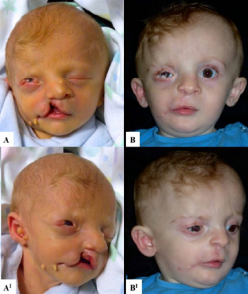
FA: preoperative; B: postoperative.
At 6-week age the patient underwent surgical correction of coloboma because of corneal decompensation signs, as corneal edema, which was not under control with topical medical treatment. The eyelid cleft had a 7 mm opening associated with 6 mm diameter epibulbar dermoid. Coloboma was corrected under general anesthesia by direct suture and superior cantholysis.
In consideration of the little child general clinic condition, after the approval from the anesthetist, the patient (at 10-month age) underwent a single surgical procedure for lip and palatoplasty, correction of macrostomia and nasal septum deformity, excission of preauricolar tags.
Under general anesthesia, with an orotracheal intubation, palatoplasty was performed after positioning a Digman gag. The borders of the cleft, the palate and the inferior area of the vomer were infiltrated with adrenaline 1:80000. Afterward, an incision was performed on the oral mucosa, along the cleft margins with appropriate dimension to be folded to create a satisfying nasal layer. Muscles were realigned meticulously on midline. Then two longitudinal bipedicled flaps were prepared with medial subperiosteal dissection. Palate closure was obtained by flaps medialisation. Particular attention was given to separate layer reconstruction (triple layer closure: nasal mucosa, muscle layer, oral mucosa).
During cheiloplasty cleft n.7 was corrected before cleft n.4 by separate layers reconstruction. Afterward, cleft n.4 was corrected by rotation of the flap which was medial to the cleft and advancement of the lateral flap, again with particular attention to layers reconstruction and Cupid’ bow junction.
During cheiloplasty, septal deviation correction was performed. The caudal end of the inferior border of the cartilaginous septum was separated from the premaxilla, and the cartilage in excess was removed by an inferior wedge-shaped chondrectomy. Multiple incisions on the concave aspect of the septal cartilage were performed to break the cartilage memory. Then the caudal septum was repositioned in the midline and sutured to the periosteum.
No after surgery complications presented and one year postoperative follow up was regular with no functional problems (Figure 2).
DISCUSSION
Goldenhar syndrome patients need early detailed evaluation due to their frequent several deformities. Many functions could be affected from the very beginning of life, such as the early neonatal respiratory deficiency, or the following suction, swallowing and vocal problems. Since the beginning also vision and hearing should be checked for impairment, and cardiac, kidney and brain ultrasound studies are recommended too in order to discard associated defects in this organs.
Treatment of eyelid defects depends on the extent of involvement and the risk of corneal decompensation. Initial therapy should be conservative, consisting of topical lubrificants or bandage contact lenses. Surgical repair is warranted if corneal decompensation is caused by dehydration and / or trichiasis [10].
One stage cheilo-palatoplasty before the end of the first year of life enables the quick implementation of the next stages of treatment, which facilitates both orthodontic treatment and speech therapy [11]. Moreover a minor number of general anesthesia procedures during infancy could reduce the possibility of poor neurodevelopment. There is in fact mounting and convincing preclinical evidence in rodents and nonhuman primates that anaesthetics in common clinical use are neurotoxic to the developing brain in vitro and cause long-term neurobehavioural abnormalities in vivo[12,13].
Primary rhinoseptoplasty with simultaneous cheiloplasty led to a reduction in morphological and functional impairment of nasal breathing, better symmetry, and anthropometric parameters more similar to those of the normal population [14].
In conclusion multimalformative patients represent a challenging condition for the surgeon. Multidisciplinary team is often required. Surgical timing to correct each defect must be planned from the beginning to avoid functional problems and to reduce the number of procedures.


