Oral Cavity Squamous Cell Carcinoma: Contemporary Evidence-Based Management and Multidisciplinary Care
- 1. Braman Comprehensive Cancer Center at Mount Sinai Medical Center, USA
- 2. Sociedad Quirugica S.C at American British Cowdray Medical Center, Mexico
Abstract
Head and neck cancers comprise a heterogeneous group of malignancies arising from the oral cavity, pharynx, larynx, sinonasal tract, salivary glands, and related upper aerodigestive sites. In the United States, cancers of the oral cavity and pharynx account for approximately 2.9% of all new cancer diagnoses, with an estimated 60,480 new cases and 13,150 deaths in 2026. Despite advances in diagnosis, surgical technique, radiation delivery, systemic therapy, and supportive care, many patients continue to present with locoregionally advanced disease, and survival remains strongly influenced by tumor subsite, stage, nodal burden, extranodal extension, margin status, depth of invasion, human papillomavirus status, tobacco and alcohol exposure, and access to multidisciplinary care.
Oral cavity squamous cell carcinoma remains primarily a surgically managed disease, with treatment guided by anatomic subsite, depth of invasion, clinical and pathologic nodal status, margin assessment, perineural and lymphovascular invasion, and adverse pathologic features. Contemporary management requires coordinated evaluation by head and neck surgical oncology, reconstructive surgery, radiation oncology, medical oncology, pathology, radiology, dentistry, speech and swallowing therapy, nutrition, rehabilitation, pain management, psychosocial oncology, and survivorship teams. Current guideline-based treatment integrates surgery, neck management, risk-adapted adjuvant radiation or chemoradiation, and—in selected locally advanced resectable head and neck squamous cell carcinoma—perioperative immunotherapy, supported by the phase III KEYNOTE-689 trial showing improved event-free survival with neoadjuvant and adjuvant pembrolizumab added to standard therapy.
This review summarizes the contemporary epidemiology, staging, diagnostic workup, surgical principles, neck management, reconstructive considerations, indications for adjuvant therapy, systemic treatment advances, surveillance, survivorship, and multidisciplinary management of oral cavity and related head and neck cancers, with emphasis on evidence-based decision-making and functional outcomes.
Keywords
• Oral Cavity Cancer
• Head and Neck Cancer
• Squamous Cell Carcinoma
• Depth of Invasion
• Neck Dissection
• Adjuvant Therapy
• Immunotherapy
• Multidisciplinary Care
Citation
Arrangoiz R, Cordera F (2026) Oral Cavity Squamous Cell Carcinoma: Contemporary Evidence-Based Management and Multidisciplinary Care. JSM Head Neck Cancer Cases Rev 6(1): 1013.
INTRODUCTION
In the year 2022 head and neck cancer accounted for 389,846 new cases of cancer worldwide (Age-Standardized Rate / ASR 4.0/100,000; 16th most common) and 188,438 deaths (ASR 1.9/100,000; 15th leading cause of cancer death) [1,2]. About 66% of incident cases and 75% of deaths occurred in Asia [1,2]. The highest regional rates are seen in Melanesia and South-Central Asia; Papua New Guinea ranks among the countries with the greatest age standardized incidence [1,2].
Globally, the burden is driven by tobacco (smoked and smokeless) and alcohol; in South and parts of Southeast Asia, areca (betel) quid often combined with smokeless tobacco, is a dominant exposure [3,4]. A 2024 International Agency for Research on Cancer (IARC) population attributable fraction analysis estimated that about one third (≈ 31%) of all oral cavity cancers worldwide were due to smokeless tobacco or areca-nut use, with the largest absolute numbers in South-Central Asia [3].
In contrast to the oropharynx, HPV accounts for a small minority of oral cavity cancers, a 2025 Journal of the National Cancer Institute (JNCI) analysis estimated that 3.9% of U.S. oral cavity cancers (2016 to 2020) were attributable to HPV16 [5].
In 2026, about 73,240 head and neck cancers (oral cavity, pharynx, and larynx) will be diagnosed in the United States, representing roughly 3.6% of all new cancers, with 16,900 deaths projected [6]. Within this group, oral cavity and pharynx cancers account for 60,540 new cases (43,200 men; 17,340 women) and 12,900 deaths, with incidence nearly three times higher in men (Table 1) [6]. Oropharyngeal cancer (OCP) incidence has risen by 0.7% per year (between 2012 to 2022), driven by HPV-positive oropharyngeal tumors, while non-oropharyngeal oral sites have declined. Mortality for OCP has increased ~ 0.6% to 0.8% per year (between 2009 to 2023), mainly for base of the tongue and tonsil. At diagnosis, most patients with head and neck cancer have regional or distant disease (≈ 54% regional, 13% distant) [6].
Table 1: Estimated New Cases and Mortality for the Year 2026 in the United States [6].
|
|
Estimated New Cases |
Estimated Deaths |
|
60, 540 (M: 43,200; F: |
12,900 (M: 9,200; F: |
|
Pharynx |
17,340) |
3,700) |
|
20,300 (M: 14,300; F: 6000) |
3,300 (M: 2,200; F: 1100) |
|
15,900 (M: 9,200; F: 6,700) |
3,400 (M: 2,100; F: 1,300) |
|
21,800 (M: 18,000; F: 3,800) |
4,700 (M: 3,700; F: 1000) |
|
2,540 (M: 1,500; F: 1,040) |
1,550(M: 1,200; F: 300) |
|
cavity |
|
|
Patients with head and neck cancers often develop second primary tumors (SPTs), because the entire upper aerodigestive mucosa has been exposed to carcinogens (“field cancerization”) [7,8]. Contemporary population studies still place the annual SPT rate at 3% to 7%, with a 36% cumulative risk by 20 years, SPTs account for a substantial share of late deaths in this population [7,8]. Most SPTs occur in the upper aerodigestive tract or the lung (50% to 75%) [9,10], multiple series report roughly half in the head and neck / esophageal region and 20% to 25% in lung; pooled data suggest a majority (60%) occur in head and neck overall [1-11]. Survivors of HPV positive oropharyngeal cancer have a lower SPT risk and longer time to SPT than HPV-negative patients, though vigilance remains necessary [12,13]. Continuing to smoke after diagnosis increases the risks of SPTs and death; smoking cessation after HNSCC improves cancer-specific and overall survival, so building cessation support into survivorship care is very important [14].
SURGICAL ANATOMY OF THE ORAL CAVITY
The oral cavity is a complex anatomical region composed of multiple subsites lined by stratified squamous epithelium and supported by muscular, glandular, and neurovascular structures. It extends from the vermilion border of the lips anteriorly to the oropharyngeal isthmus posteriorly. Precise definition of the oral cavity subsites is essential for accurate tumor classification, staging, and surgical planning. Its boundaries are defined superiorly by a line extending from the vermilion border of the lips posteriorly to the junction of the hard and soft palates, inferiorly from the vermilion border to the circumvallate papillae of the oral tongue, and laterally by the oral mucosa extending posteriorly to the anterior tonsillar pillars.
These boundaries delineate the oral cavity from the oropharynx and are critical in distinguishing tumors that differ substantially in etiology, biology, staging systems, and treatment algorithms [15].
Within these limits, the oral cavity is subdivided into several distinct subsites: the lips, buccal mucosa, upper and lower alveolar ridges and gingiva, retromolar trigone, floor of mouth, hard palate, and the anterior two thirds of the tongue (oral tongue). Each subsite possesses unique anatomic relationships that influence tumor spread and operative strategy. Alveolar ridge and gingival tumors frequently invade adjacent maxillary or mandibular bone early, often necessitating marginal or segmental mandibulectomy or maxillectomy. Retromolar trigone lesions are particularly aggressive, with early extension into the mandible, masticator space, pterygomandibular raphe (is a tendinous band of fascia that connects the oral cavity to the pharynx), and oropharynx, frequently requiring wide composite resections. Tumors of the floor of mouth and oral tongue are characterized by thin mucosa, minimal anatomic barriers, and proximity to critical neurovascular structures, predisposing them to early muscular invasion and regional nodal metastasis; in these subsites, depth of invasion is a key determinant of T classification and neck management [15,16].
Lips, Buccal Mucosa, and Retromolar Trigone
The oral mucosa is demarcated from the external skin at the vermilion border of the lips, a clinically important landmark in tumor spread and staging. The buccal mucosa overlies and is firmly adherent to the buccinator muscle, which plays a key role in mastication and maintaining food between the teeth. The parotid (Stensen’s) duct opens into the buccal mucosa opposite the maxillary second molar at the parotid papilla, making this area relevant for both salivary pathology and tumor extension. Posteriorly, the retromolar trigone overlies the pterygomandibular raphe, a fibrous band connecting the buccinator and superior pharyngeal constrictor muscles. Due to this anatomical relationship, malignancies in this region can easily spread into both the oral cavity and oropharynx. Additionally, the proximity of the lingual nerve and inferior alveolar nerve, located medial to the mandibular ramus, facilitates early perineural invasion, a recognized pathway for tumor dissemination. The buccal mucosa receives its blood supply from branches of the maxillary artery (buccal branch), while sensory innervation arises from the maxillary (V2) and mandibular (V3) divisions of the trigeminal nerve [17,18].
Floor of the Mouth
The floor of the mouth is a horseshoe-shaped structure supported by the mylohyoid muscles, which form a muscular diaphragm separating the oral cavity from the neck. In the anterior midline, the submandibular ducts (Wharton’s ducts) open at the sublingual caruncles on either side of the lingual frenulum.
The sublingual glands and ducts lie superior to the mylohyoid muscle, creating a potential space for tumor spread. The mylohyoid muscles attach to the mylohyoid line of the mandible and meet in a median raphe. Variations or deficiencies in this raphe may allow herniation of the sublingual gland into the neck, which is clinically relevant in distinguishing plunging ranulas from other neck masses. The close relationship between mucosa, salivary glands, and muscle facilitates early invasion of malignancies into deeper cervical structures [17,18].
Hard Palate
The hard palate is a horizontal bony plate that forms a subsection of the palate of the mouth. It forms the anterior two-thirds of the roof of the oral cavity. The hard palate is comprised of two facial bones: the palatine process of the maxilla and the paired palatine bones. The hard palate contains several landmarks, including the incisive foramen and the greater and lesser palatine foramina. They serve as a passageway for the neurovascular structures that supply the structures of the oral cavity. The mucosa of the hard palate is tightly bound to the periosteum of the maxilla, limiting mobility but allowing direct tumor invasion into bone. The lateral submucosa contains the greater palatine neurovascular bundle, while the posterior half houses numerous minor salivary glands, predominantly mucous secreting. The greater palatine artery, a branch of the maxillary artery, emerges from the greater palatine foramen near the second maxillary molar and courses anteriorly toward the incisive canal, where it anastomoses with the nasopalatine artery. Preservation of this artery is critical in surgical procedures such as the maxillary swing approach. Sensory innervation is provided by the greater and lesser palatine nerves (branches of V2), which may serve as routes for perineural tumor spread to the pterygopalatine fossa and skull base [17,18].
Tongue
The embryologic origins of the tongue first appear at 4 weeks’ gestation. The first branchial arch is responsible for the development of the tongue derivatives. It gives rise to two lateral lingual swellings and one median swelling (known as the tuberculum impar). The two lateral lingual swellings grow over the tuberculum impar and merge, forming the anterior two thirds of the tongue. Portions of the second, third, and fourth branchial arches give rise to the base of the tongue. The intrinsic musculature of the tongue derives from occipital somite’s which give rise to myoblasts. The tongue is divided into an oral (anterior two-thirds) and pharyngeal (posterior one-third) part by the sulcus terminalis, which originates at the foramen cecum. The dorsal surface contains specialized mucosa with filiform, fungiform, foliate, and circumvallate papillae, whereas the ventral surface is smooth and continuous with the floor of the mouth. The tongue contains intrinsic muscles (superior/inferior longitudinal, transverse, and vertical muscles), which lack fascial planes and allow rapid multidirectional tumor spread.
In contrast, involvement of extrinsic muscles (genioglossus, hyoglossus, styloglossus, palatoglossus) indicates advanced disease and deeper invasion. Arterial supply is derived from branches of the lingual artery (dorsal lingual, deep lingual, and sublingual arteries), while venous drainage occurs via the lingual veins into the facial and retromandibular veins. Motor innervation is provided by the hypoglossal nerve (CN XII), except for the palatoglossus, which is innervated by the pharyngeal plexus (via the vagus nerve). Sensory innervation of the anterior two-thirds is via the lingual nerve (V3), with taste sensation carried by the chorda tympani branch of the facial nerve (CN VII) [10-20].
Alveolar Process and Dentition
The alveolar processes of the maxilla and mandible contain the dental sockets (alveoli) that house the teeth. These structures are covered by firmly attached gingival (buccal) mucosa, which is continuous with the mucosa of the lips, floor of the mouth, and hard palate. This mucosa is tightly bound to the underlying periosteum, forming a barrier that can, however, be breached by invasive malignancies, allowing spread into adjacent bone. The inferior alveolar nerve, a branch of the mandibular division of the trigeminal nerve (CN V3), provides sensory innervation to the lower teeth and gingiva. It enters the mandibular foramen on the medial surface of the mandibular ramus and travels within the mandibular canal to supply the dentition, terminating as the mental nerve. Due to its intraosseous course, this nerve is particularly vulnerable to perineural invasion, a significant pathway for tumor spread in oral cancers. Arterial supply to the lower teeth and gingiva is provided by the inferior alveolar artery, a branch of the maxillary artery. This vessel constitutes the primary endosteal blood supply to the mandible. In addition, the periosteal vessels supply the cortical bone. Disruption of these vascular systems, whether by tumor invasion, trauma, or radiotherapy, can predispose to complications such as osteonecrosis [10-18].
The vascular supply of the oral cavity is derived predominantly from branches of the external carotid artery. The lingual artery supplies the oral tongue and floor of mouth, the facial artery contributes to the lips, buccal mucosa, and submandibular region, and branches of the maxillary artery supply the hard palate, alveolar ridges, and retromolar trigone. This rich arterial network has important implications for oncologic resection, flap design, and hemostasis during surgery. Venous drainage parallels arterial anatomy and occurs primarily through the lingual, facial, and pterygoid venous systems, ultimately draining into the internal jugular vein. Awareness of these venous pathways is critical during deep tongue and floor-of mouth resections, where venous bleeding may be brisk and difficult to control [16].
Lymphatic drainage of the oral cavity is extensive and highly interconnected, accounting for the high incidence of regional metastasis even in early-stage disease.
The primary nodal basins at risk include level IA (submental), level IB (submandibular), and level II (upper deep jugular) lymph nodes. Lesions of the lips, anterior floor of mouth, and anterior oral tongue commonly drain to level IA and IB, whereas more posterior oral tongue and floor-of-mouth tumors preferentially drain to level IB and II. Cross-midline lymphatic drainage is common, particularly for midline or near-midline tumors of the oral tongue and floor of mouth, underscoring the need for careful consideration of bilateral neck treatment. This predictable yet overlapping lymphatic anatomy forms the anatomic basis for elective neck dissection or sentinel lymph node biopsy in clinically node-negative oral cavity squamous cell carcinoma [21].
The boundaries of the oral cavity are:
• Superior border - from the vermillion border to the junction of the hard and soft palates.
• Inferior border - from the vermillion border to the circumvallate papillae of the oral tongue.
• The lateral border - is the mucosa of the mouth up to the anterior tonsillar pillars.
In his classic 1972 study, Lindberg systematically evaluated the anatomic distribution of cervical lymph node metastases in patients with previously untreated squamous cell carcinoma of the upper respiratory and digestive tracts. The analysis was based on careful clinical examination of the neck and correlation with the primary tumor site, allowing Lindberg to characterize not only which nodal levels were involved, but also how frequently and in what sequence metastases occurred. The results convincingly demonstrated that cervical nodal spread follows orderly, predictable patterns determined by primary tumor subsite [21].
For oral cavity carcinomas, Lindberg found that metastatic involvement was overwhelmingly concentrated in the upper cervical nodal basins. The highest incidence of metastases occurred in the submandibular and submental regions (corresponding to modern level IB and IA), followed closely by the upper deep jugular nodes (level II). In contrast, involvement of lower jugular nodes (levels III and IV) was uncommon in early disease and typically occurred only in the setting of advanced primary tumors or extensive nodal burden. True “skip metastases” to the lower neck without involvement of upper nodal levels were rare, supporting a stepwise pattern of lymphatic spread [21].
The study also demonstrated subsite-dependent differences in nodal metastatic risk within the oral cavity. Tumors of the oral tongue and floor of mouth exhibited the highest rates of cervical metastasis, reflecting the dense and bilateral lymphatic networks of these regions.
In comparison, cancers of the lip, hard palate, and maxillary alveolus showed a lower overall incidence of nodal disease. These findings provided early anatomic explanations for the variable prognosis observed among oral cavity subsites and highlighted the need for tailored neck management strategies [21].
A particularly important result was Lindberg’s observation of contralateral and bilateral nodal metastases, especially in tumors located near or crossing the midline. Oral tongue and floor-of-mouth lesions demonstrated a substantial risk of bilateral nodal involvement, even when the primary tumor appeared clinically lateralized. This finding was critical in challenging the assumption that unilateral neck treatment was sufficient in all cases and laid the foundation for modern recommendations favoring bilateral neck dissection or irradiation in selected patients with midline or deeply infiltrative oral cavity tumors [21].
Lindberg further correlated primary tumor extent with nodal disease burden. Larger and more locally advanced tumors were associated with a higher incidence of nodal metastases and involvement of multiple nodal levels. Although depth of invasion was not formally measured at the time, the results strongly suggested that deeper and more aggressive tumors possessed greater metastatic potential, an observation that anticipated the later incorporation of depth of invasion into contemporary staging systems [21,22].
The results of Lindberg’s study established several enduring principles: (1) cervical lymph node metastases from oral cavity squamous cell carcinoma are predictable and subsite-specific, (2) nodal spread typically progresses in a sequential fashion from upper to lower neck levels, (3) oral tongue and floor-of-mouth cancers carry the highest risk of regional metastasis, and (4) bilateral nodal involvement is common in midline oral cavity tumors. These findings provided the conceptual and anatomic basis for elective neck treatment and ultimately for the development of selective neck dissection, making Lindberg’s work a cornerstone of modern head and neck surgical oncology [21].
The oral cavity is constantly exposed to carcinogens found in inhaled or ingested substances, making it the most frequent site for malignant epithelial tumors in the head and neck region [4-15]. Carcinogens in the oral cavity commonly include tobacco, alcohol, and betel nuts. While human papilloma virus (HPV) is strongly linked to oropharyngeal cancers, its connection to oral cancer is less certain. Primary oral cavity tumors can develop from the surface epithelium, minor salivary glands, submucosal soft tissue, or dento-alveolar tissues. Over 90% of oral cavity cancers are squamous cell carcinomas, which will be the main focus of this review.
EPIDEMIOLOGY OF ORAL CAVITY SQUAMOUS CELL CARCINOMA
Oral cavity squamous cell carcinoma (OCSCC), defined as malignancy arising from the oral tongue, floor of mouth, buccal mucosa, gingiva, alveolar ridge, retromolar trigone, and hard palate, represents a distinct clinicopathologic entity within head and neck oncology [10-15]. When restricted to these subsites, OCSCC accounts for approximately 220,000 to 240,000 new cases and 110,000 to 120,000 deaths worldwide each year, making it one of the most common malignancies of the upper aerodigestive tract globally, but substantially less frequent than estimates that combine oral cavity with pharyngeal cancers [23,24].
Sex Distribution
OCSCC demonstrates a pronounced male predominance, historically comprising 66% to 95% of all cases, depending on geographic region and subsite [4-25]. Contemporary data indicate a male-to-female ratio of approximately 3:1, although this ratio varies by anatomic location and has narrowed over time, particularly in Western countries [26]. Tumors of the floor of mouth, retromolar trigone, and alveolar ridge show the greatest male predominance, whereas oral tongue squamous cell carcinoma exhibits a smaller gender gap and, in some series, a rising incidence among women [27].
Age Distribution
The incidence of OCSCC increases with age, with most patients presenting between 50 and 70 years. Risk rises sharply after the fifth decade of life, reflecting cumulative exposure to tobacco, alcohol, and other carcinogens [28]. Nevertheless, oral tongue SCC is increasingly diagnosed in younger patients (< 45 years) who frequently lack traditional risk factors, suggesting alternative etiologic pathways distinct from classic carcinogen-driven disease [4].
Geographic Variation
Marked geographic heterogeneity exists in the incidence of OCSCC. The highest rates are observed in South and Southeast Asia, where oral cavity cancer ranks among the most common malignancies in men [23-29]. This distribution closely parallels the prevalence of betel quid and areca nut chewing, frequently combined with tobacco and slaked lime. These exposures are strongly associated with cancers of the buccal mucosa, gingiva, and retromolar trigone, as well as with oral potentially malignant disorders [30-32]. In contrast, in North America and Europe, OCSCC more commonly involves the oral tongue and floor of mouth, reflecting different exposure patterns and oral habits [31].
Epidemiology in the United States
In the United States, oral cavity cancer accounts for approximately 35,000 to 38,000 new cases annually, representing roughly 60% of non-oropharyngeal head and neck squamous cell carcinomas [31]. Incidence remains higher in men, particularly in urban populations, where combined exposure to tobacco and alcohol predominates. Among women, particularly in rural regions, an increased risk of OCSCC has been associated with smokeless tobacco (snuff) use [4-31]. Importantly, unlike oropharyngeal squamous cell carcinoma, HPV plays a minimal etiologic role in true oral cavity SCC, and HPV-driven epidemiologic trends do not apply to this disease entity [5].
Anatomic Subsite Distribution
In Western populations, the oral tongue is the most common subsite, accounting for 35% to 45% of cases, followed by the floor of mouth (20% to 25%), gingiva/ alveolar ridge (15% to 20%), buccal mucosa (10% to 15%), and hard palate and retromolar trigone (< 10%) [33]. In contrast, in regions with prevalent betel nut use, buccal mucosa and gingiva predominate [4-31].
Mortality and Survival
Despite advances in surgical technique and adjuvant therapy, overall 5-year survival for OCSCC remains approximately 50% to 60%, with prognosis strongly stage-dependent [31]. Early-stage disease (T1 to T2, N0) is associated with survival exceeding 70% to 80%, whereas advanced-stage disease with cervical nodal metastases carries a 5-year survival closer to 30% to 40% [31].
ETIOLOGY AND RISK FACTORS FOR ORAL CAVITY SQUAMOUS CELL CARCINOMA
OCSCC is a multifactorial malignancy resulting from complex interactions between environmental exposures, lifestyle factors, and host susceptibility. Despite advances in molecular oncology, the majority of OSCC cases worldwide remain attributable to preventable risk factors, particularly tobacco and alcohol use, with geographic variation reflecting cultural practices and socioeconomic conditions [4-34].
Tobacco Exposure
Tobacco use is the most important etiologic factor for OCSCC and head and neck squamous cell carcinoma overall [35]. Large population-based studies and pooled analyses demonstrate that approximately 80% to 85% of head and neck cancers are attributable to tobacco exposure, either alone or in combination with alcohol [4-39]. A clear dose response relationship has been consistently shown across cigarette smoking, cigar smoking, pipe smoking, chewing tobacco, and snuff use [34-39].
Contrary to common patient perceptions, cigar smoking is not safer than cigarette smoking. Epidemiologic studies demonstrate that cigar smoking confers a comparable risk of oral cavity cancer, with differences primarily in tumor subsite distribution rather than overall carcinogenicity. Cigar smokers have a higher incidence of tumors involving the lips, floor of mouth, and anterior tongue due to prolonged mucosal exposure and higher concentrations of carcinogens in cigar smoke [35-41].
Smokeless tobacco products are strongly associated with OCSCC, particularly cancers of the gingiva, buccal mucosa, and alveolar ridge. This association is especially pronounced in South Asia, China, Taiwan, and parts of Southeast Asia, where betel quid chewing, often combined with tobacco and areca nut, is highly prevalent [30-42]. Areca nut has been classified as a Group 1 carcinogen by the International Agency for Research on Cancer (IARC) [43]. Chronic use leads to oral potentially malignant disorders, including leukoplakia, erythroplakia, and oral submucous fibrosis, which carry a significant risk of malignant transformation [30-45].
Continued tobacco use after diagnosis is associated with a significantly increased risk of second primary tumors and decreased overall survival compared with former or never smokers [46-48]. Secondhand smoke exposure has also been associated with an increased risk of head and neck cancer, although the magnitude of risk is lower than that associated with direct tobacco use [49].
Alcohol Consumption
Alcohol consumption is an independent risk factor for OCSCC, although it is a weaker carcinogen than tobacco when considered alone [4-51]. Ethanol metabolism produces acetaldehyde, a recognized carcinogen that increases epithelial permeability and promotes DNA damage [52].
The combined use of tobacco and alcohol has a synergistic, multiplicative effect on OCSCC risk. Individuals who smoke and consume alcohol heavily have a 30- to 36-fold increased risk of developing oral cavity cancer compared with abstainers [37-39]. This interaction represents one of the strongest epidemiologic associations in head and neck oncology.
Oral Hygiene, Dental Status, and Local Factors
Poor oral hygiene, chronic mucosal trauma, and edentulism have been associated with an increased risk of OCSCC, particularly in populations with limited access to dental care [53-55]. Chronic inflammation and altered oral microbiota are proposed mechanisms, although residual confounding by tobacco and alcohol remains a limitation in some studies [54].
The role of alcohol-containing mouthwashes remains controversial. Some observational studies suggest a possible association between long-term use of high-alcohol mouth rinses and OSCC, but causality has not been established, and findings remain inconsistent [55,56].
Dietary and Nutritional Factors
Dietary intake appears to influence OSCC risk.
Epidemiologic studies indicate that diets rich in fruits and vegetables, particularly those containing vitamin A, β-carotene, and α-tocopherol, are associated with a reduced risk of oral cavity cancer [57-60]. However, randomized chemoprevention trials have not consistently confirmed a protective effect, and routine supplementation is not recommended [61-64].
Consumption of mate (yerba mate), a hot herbal beverage widely consumed in South America, has been associated with an increased risk of oral cavity and upper aerodigestive tract cancers [65,66]. Thermal injury from high-temperature consumption is believed to play a major etiologic role [67].
Ultraviolet Radiation
Ultraviolet radiation is a well-established risk factor for squamous cell carcinoma of the lip, particularly the lower lip [68-70]. Approximately one-third of patients with lip cancer report outdoor occupations involving chronic sun exposure [70]. Fair skin, cumulative UV exposure, and lack of lip protection further increase risk [70].
Cannabis Use
An association between marijuana use and head and neck cancer has been suggested in some studies; however, results are inconsistent and often confounded by concurrent tobacco use [71,72]. At present, the independent contribution of cannabis to OSCC risk remains uncertain.
Genetic Susceptibility and Inherited Syndromes
Inherited disorders affecting DNA repair and telomere maintenance confer a markedly increased risk of OSCC. Fanconi anemia, caused by defects in DNA repair genes, is associated with early-onset OSCC, often in the absence of traditional risk factors [73-75]. Dyskeratosis congenita, a telomere biology disorder, is characterized by mucocutaneous abnormalities and bone marrow failure and is associated with a significantly elevated risk of OSCC, particularly involving the tongue and floor of mouth [36-76].
MOLECULAR AND CELLULAR MECHANISMS OF CARCINOGENESIS IN ORAL CAVITY SQUAMOUS CELL CARCINOMA
Oral cavity squamous cell carcinoma (OCSCC) arises through a multistep, multilevel carcinogenic process characterized by the accumulation of genetic alterations, epigenetic dysregulation, and progressive disruption of key cellular signaling pathways. These changes occur within a background of chronic carcinogen exposure and inflammation and are best understood through the framework of clonal evolution and field cancerization, concepts that have direct implications for surgical margins, recurrence, and second primary tumors.
Carcinogen exposure and early molecular injury
Tobacco smoke remains the dominant etiologic factor in OCSCC and contains numerous mutagenic compounds capable of inducing DNA adducts, oxidative damage, and permanent genomic alterations. These effects generate a high somatic mutation burden and promote the development of genetically altered epithelial fields that may appear histologically normal yet harbor oncogenic changes, a phenomenon known as field cancerization [77-79]. Alcohol acts synergistically with tobacco by increasing mucosal permeability and enhancing carcinogen exposure [51]. Modern sequencing studies have demonstrated that driver mutations can be detected in normal-appearing oral mucosa adjacent to tumors, supporting the concept that malignant transformation often begins long before invasive cancer is clinically evident [80].
Early genetic events: tumor suppressor loss and cell cycle deregulation
Among the earliest and most frequent genetic events in OCSCC are alterations involving chromosomes 3p and 9p, particularly loss of heterozygosity (LOH) at 9p21 affecting the CDKN2A (p16INK4A) locus [81,82]. These alterations disrupt cell-cycle control through unchecked CDK4/6 activity and RB pathway inactivation. Multiple longitudinal studies of oral potentially malignant disorders (OPMDs) have shown that LOH at 3p and / or 9p strongly predicts malignant transformation, independent of histologic grade [81-83].
Mutations of TP53, typically occurring early in tobacco related OCSCC, further compromise genomic integrity and apoptosis, facilitating clonal expansion and progression [77-84]. TP53 dysfunction is strongly associated with chromosomal instability and accumulation of additional oncogenic events, underscoring its central role as a “gatekeeper” in oral carcinogenesis.
Growth factor signaling and proliferative drive
Aberrant activation of the epidermal growth factor receptor (EGFR) pathway is a hallmark of head and neck squamous cell carcinoma, including OCSCC. Tobacco smoke extracts have been shown to directly activate EGFR signaling in vitro, leading to downstream induction of cyclin D1 (CCND1) and accelerated G1–S cell-cycle progression [85,86]. EGFR activation also stimulates cyclooxygenase-2 (COX-2) and prostaglandin E2 (PGE2) production, creating a positive feedback loop that sustains proliferation, angiogenesis, and invasion [87]. These pathways remain clinically relevant, as EGFR signaling continues to represent a therapeutic target in advanced disease.
Disruption of squamous differentiation programs
Genomic profiling studies have identified frequent alterations in the NOTCH signaling pathway in OCSCC, highlighting the importance of impaired epithelial differentiation in carcinogenesis [88]. NOTCH normally functions as a tumor suppressor in stratified squamous epithelium by promoting terminal differentiation; its disruption favors maintenance of a progenitor-like state and malignant progression. These findings emphasize that OCSCC is driven not only by proliferative signaling but also by loss of normal differentiation controls.
Chromosomal instability and aneuploidy
As OCSCC progresses, tumors commonly acquire widespread copy-number alterations and aneuploidy, reflecting underlying chromosomal instability [77-89]. Increasing evidence suggests that aneuploidy may act as a driver, rather than merely a consequence, of malignant transformation by enabling rapid phenotypic adaptation and selection of aggressive clones [90]. DNA ploidy abnormalities detected in OPMDs have been associated with a higher risk of progression to invasive carcinoma, reinforcing their potential role as prognostic biomarkers in clinical practice [91].
Telomere dysfunction and telomerase reactivation
Early telomere shortening followed by telomerase reactivation is another key event in oral carcinogenesis. Recent studies have identified TERT promoter mutations as relatively enriched in oral cavity tumors compared with other head and neck subsites, suggesting a role in immortalization and tumor maintenance [92,93]. While not yet incorporated into routine clinical decision making, TERT alterations represent an emerging area of translational interest.
HPV-associated pathways: an alternative mechanism
Unlike oropharyngeal SCC, HPV-driven carcinogenesis is less common in the oral cavity. When present, transcriptionally active HPV induces malignant transformation through viral oncoproteins E6 and E7, which inactivate p53 and retinoblastoma protein, respectively [77-94]. HPV-associated tumors typically demonstrate fewer chromosomal deletions at 3p, 9p, and 17p, supporting an alternative molecular pathway distinct from tobacco-related disease.
Epigenetic methylation alterations and promoter hyper
Epigenetic dysregulation plays a critical role in OCSCC progression. Promoter hypermethylation leads to transcriptional silencing of multiple tumor suppressor genes, including p16, DAPK, E-cadherin, and RAR-β [95-97]. These changes contribute to loss of cell-cycle control, impaired apoptosis, and increased invasiveness. Importantly, methylation signatures are being explored as noninvasive biomarkers using saliva or oral rinse samples for early detection and surveillance [98].
Tumor microenvironment and inflammatory signaling
Beyond epithelial-intrinsic alterations, OCSCC progression is shaped by a permissive tumor microenvironment characterized by chronic inflammation, immune evasion, and stromal remodeling. COX-2/PGE2 signaling promotes angiogenesis and immunosuppression, while certain TP53 alterations have been linked to an immune-cold phenotype with reduced responsiveness to immunotherapy [99,100]. Emerging data also implicate oral microbiome dysbiosis in modulating inflammation and carcinogenic signaling, adding another layer of complexity to disease pathogenesis [101,102].
DIAGNOSIS OF SQUAMOUS CELL CARCINOMA OF THE ORAL CAVIT
Early diagnosis and prompt referral to a specialist with expertise in head and neck oncologic surgery are critical, because stage at presentation remains the dominant prognostic factor in oral cavity squamous cell carcinoma (OCSCC) and other head and neck malignancies [4]. Multiple population-based and institutional series have shown that diagnostic and treatment delays are associated with more advanced stage at diagnosis and worse overall survival, particularly when the interval from first symptoms to diagnosis or from diagnosis to treatment initiation exceeds 4 to 6 weeks [103-106]. Early detection permits less extensive surgery, reduced need for adjuvant therapy, lower treatment-related morbidity, and improved disease-specific survival.
A detailed history must systematically document established risk factors for head and neck squamous cell carcinoma, including lifetime tobacco exposure (type, amount, pack-years), alcohol consumption (quantity, pattern of use), betel quid or areca nut chewing, prior head and neck irradiation, and occupational exposures [4-10]. Additional host factors such as poor oral hygiene, chronic trauma from ill-fitting dentures, immunosuppression, and family history of head and neck cancer should also be explored [107,108]. Although human papillomavirus (HPV) plays a central role in oropharyngeal carcinogenesis, its contribution to classic oral cavity SCC is more modest; nevertheless, high-risk sexual behavior and prior anogenital HPV-associated disease should be noted.
Any adult with symptoms referable to the upper aerodigestive tract lasting longer than two weeks, or with an unexplained neck mass, should undergo a thorough head and neck evaluation with a high index of suspicion for malignancy [4-109]. This is particularly important in older patients, smokers, and heavy alcohol users. The history should characterize symptom onset, duration, and progression, as well as prior evaluations or treatments that may have contributed to diagnostic delay.
Because of field cancerization, patients with head and neck squamous cell carcinoma have an increased risk of synchronous and metachronous second primary tumors in the upper aerodigestive tract, lung, and esophagus. Contemporary series report a synchronous second primary rate of approximately 3% to 7% at initial workup [4-110]. Consequently, a careful evaluation of the entire upper aerodigestive tract is required at diagnosis. In many centers this includes flexible nasopharyngolaryngoscopy in clinic, with selective panendoscopy (esophagoscopy and bronchoscopy) reserved for patients with specific symptoms, high-risk behaviors, or suspicious imaging rather than as a routine “blind” procedure in all patients [111-113].
Oral cavity cancers typically produce symptoms related to the upper aerodigestive tract, including changes in swallowing, speech, hearing, and breathing. During the interview, particular attention should be paid to epiphora, dysphagia, odynophagia, globus sensation, dysphonia or hoarseness, difficulty articulating words, epistaxis, ear pain, hemoptysis, otic fullness, halitosis, sialorrhea or xerostomia, nasal obstruction, sensations of malocclusion, oral cavity pain, trismus, and any unexplained weight loss or fatigue [4-112]. A comprehensive physical examination is mandatory in every patient, with special emphasis on the head and neck examination (inspection and palpation of all oral cavity subsites, oropharynx, nasopharynx when accessible, and neck), otoscopic examination, indirect or flexible laryngoscopy, and when indicated nasopharyngolaryngoscopy. A focused neurologic examination with particular attention to cranial nerves V and VII–XII is essential to detect perineural spread or skull base involvement [4-112].
Table 2 summarizes common symptoms and signs of head and neck cancer, including those related to oral cavity lesions and those suggestive of more extensive regional or skull base disease (e.g., middle ear effusion, atrophy of masticatory muscles, or loss of corneal reflex) [4-112]. The initial clinical assessment often provides insight into both severity and chronicity of disease.
Table 2: Symptoms and Signs of Head and Neck Cancer
|
Epiphora |
Oral cavity pain |
|
Sialorrhea |
Non-healing ulcerated lesion |
|
Xerophthalmia |
Halitosis |
|
Xerostomia |
Ill-fitting dentures |
|
Dysphagia |
Nasal Obstruction |
|
Odynophagia |
Middle ear effusion |
|
Globus sensation |
Mal occlusion |
|
Dysphonia |
Atrophy of the masseter muscle |
|
Difficulty articulating words |
Atrophy of the temporalis muscle |
|
Epistaxis |
Decrease tongue mobility |
|
Hemoptysis |
Absence of corneal reflex |
|
Otalgia |
|
The most frequent presenting complaint in patients with oral cavity tumors is a nonhealing sore or ulcer in the mouth or on the lips. An indurated ulcer with raised, rolled edges, particularly when associated with spontaneous or contact bleeding, is highly suspicious for malignancy and should be biopsied without delay [4-10]. Oral mucosal SCC may also present as an exophytic verrucous or papillary mass, a firm infiltrative plaque, or as erythroplakia or mixed erythroleukoplakia [15]. Lesions of the floor of the mouth often manifest as ulcerated or red patches or as papillary proliferations; these have a particularly high rate of occult nodal metastases. Gingival cancers may present as ulceration, proliferative masses, or apparent periodontal disease that fails to respond to conventional dental therapy [15]. Tongue cancers frequently appear as infiltrating ulcers or indurated masses; decreased tongue mobility, deviation on protrusion, or pain radiating to the ear suggests deep muscular invasion or involvement of the hypoglossal, lingual, or glossopharyngeal nerves [10].
Symptoms such as nasal obstruction or recurrent epistaxis in a patient with an apparent “oral cavity” lesion should raise concern for extension into the nasal cavity, maxillary antrum, or nasopharynx, especially with tumors arising from the hard palate or upper alveolus. Malocclusion or progressive trismus often indicates extension into the masticator space, pterygoid musculature, or temporomandibular joint [4]. A unilateral middle ear effusion in an adult may reflect obstruction of the eustachian tube by a nasopharyngeal or skull base lesion. Loss of the corneal reflex suggests involvement of the ophthalmic division (V1) of the trigeminal nerve, whereas atrophy of the masseter or temporalis muscles implies involvement of the mandibular division (V3).
These findings mandate careful radiologic assessment of potential perineural spread to the skull base [4]. Approximately one third of patients with oral cavity cancer present with a palpable neck mass, sometimes in the absence of obvious intraoral symptoms [112-114]. Cervical nodal metastases may be the initial manifestation of an otherwise occult primary of the oral tongue, floor of mouth, or other upper aerodigestive tract site. On inspection, cancer of the oral mucosa may appear as an indurated ulcer with raised edges or as an exophytic lesion (Figures 1 and 2). SCC of the floor of the mouth may present as a red or ulcerated plaque or as a papillary mass (Figure 3). Tumors of the hard palate often present as exophytic papillary or nodular lesions and may extend superiorly to involve the nasal cavity or maxillary sinus.
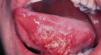
Figure 1: Ulcerated Lesion of the Tongue
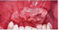
Figure 2: Exophytic Lesion of the Floor of the Mouth

Figure 3: Exophytic Papillary Carcinoma of the Buccal Mucosa
The differential diagnosis of oral cavity SCC is broad and includes other malignant epithelial and mesenchymal tumors, such as minor salivary gland neoplasms (e.g., mucoepidermoid carcinoma; Figure 4), adenoid cystic carcinoma, sarcomas, melanoma, and lymphoma [15]. A variety of benign or inflammatory conditions can mimic SCC, including pyogenic granuloma, chronic traumatic ulcers, aphthous ulcers, lichen planus, specific infections such as tuberculosis and syphilis, and a range of benign epithelial tumors. Benign squamous papillomas and keratoacanthomas (Figure 5) may present as exophytic or infiltrative lesions; although many are indolent, those with infiltrative growth patterns may cause local tissue destruction and occasionally harbor dysplasia or early invasive carcinoma, underscoring the need for histologic confirmation of atypical or persistent lesions.
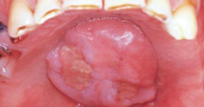
Figure 4: Mucoepidermoid carcinoma of the Palate
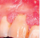
Figure 5: Papilloma
Histopathologic confirmation is mandatory. Biopsy of the primary tumor can often be performed in the clinic under local anesthesia using punch instruments or biopsy forceps (Figure 6), particularly for accessible lesions of the oral tongue, buccal mucosa, or lower alveolus. For larger or posteriorly located lesions, or in patients with a strong gag reflex, biopsy under sedation or general anesthesia may be preferable [4-15]. The specimen should be obtained from the indurated margin of the lesion, avoiding necrotic or heavily keratinized areas, and should be of sufficient depth to permit accurate assessment of invasion and, when possible, depth of invasion (DOI), which now forms part of the T category in the AJCC staging system [22]. Repeat biopsy should be considered if the initial sample is superficial or non-diagnostic in the setting of persistent clinical suspicion.
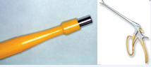
Figure 6: Punch Biopsy and Forceps to Perform the Biopsy
Fine-needle aspiration (FNA) cytology, ideally ultrasound-guided, is the first-line diagnostic modality for evaluating cervical lymphadenopathy in this context [4-118]. It is minimally invasive, rapid, and cost-effective, and can reliably distinguish metastatic SCC from other pathologies. Modern series and meta-analyses report sensitivities generally in the 80% to 95% range and specificities exceeding 90%, with overall diagnostic accuracy frequently above 85% to 90% for cervical lymph node metastases [4-119]. Nevertheless, limitations remain, particularly for low-grade lymphomas, necrotic nodes, cystic nodal metastases, and small-volume disease, so a negative FNA does not exclude malignancy when clinical suspicion is high. In such cases, repeat ultrasound-guided FNA, possibly using on-site cytologic evaluation, may be appropriate.
Core needle biopsy of cervical lymph nodes is increasingly used in some centers when lymphoma is suspected or when prior FNAs are repeatedly non diagnostic; however, in the setting of a suspected mucosal head and neck SCC with an unknown primary, most guidelines continue to recommend FNA as the initial investigation [112]. Open excisional nodal biopsy should be avoided as an early diagnostic step, because it can compromise subsequent neck dissection, increase the risk of regional recurrence, and may negatively impact survival [4-112]. Open biopsy is reserved for cases in which a diagnosis cannot be established after adequate imaging and at least two non-diagnostic FNAs (with or without core biopsies) and ideally should be performed by the surgeon who will provide definitive treatment, with preparedness to proceed to formal neck dissection if metastatic SCC is confirmed [4].
Cross-sectional imaging plays a central role in the staging and surgical planning of oral cavity cancer. Contrast-enhanced computed tomography (CT) is widely available and remains the workhorse modality for evaluating primary tumor extent, bone involvement (maxilla and mandible), and cervical nodal metastases [4-10]. CT provides excellent spatial resolution and reliably differentiates fat, muscle, and cortical bone. For bone invasion, both CT and MRI demonstrate high, though variable, diagnostic performance; reported sensitivities for CT range from approximately 50% to 90%, with specificities around 60% to 100%, while MRI sensitivities range from 58% to over 90% with specificities around 73% to 100% [114-124]. Some studies suggest that MRI may be more sensitive for early medullary invasion, whereas CT may better delineate cortical erosion (Figure 7); others show broadly comparable performance, and recent work suggests that combining CT and MRI or using advanced techniques such as subtraction CT may further improve accuracy in selected cases [114-125].
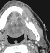
Figure 7: CT of the Head and Neck Showing Bone Erosion
Magnetic resonance imaging offers superior soft-tissue contrast and is particularly useful for assessing tongue musculature, floor-of-mouth invasion, masticator space and parapharyngeal extension, perineural spread, and intracranial or skull base involvement [120-124]. High-field MRI and optimized sequences allow reasonably accurate estimation of DOI, although several series indicate a tendency toward DOI overestimation compared with histopathology [126-128]. Systematic reviews indicate that MRI, CT, and intraoral ultrasound each have strengths and limitations in DOI assessment; intraoral high-frequency ultrasound appears particularly accurate for DOI in early (T1–T2) tongue lesions, whereas MRI or CT may be more informative for deeper or more posterior tumors [126-130].
In recent years, intraoral ultrasound and refined CT/MRI techniques have emerged as valuable adjuncts for preoperative DOI measurement and nodal risk stratification. High-frequency intraoral ultrasound has demonstrated strong correlation between sonographic and pathological DOI in early tongue SCC and may outperform MRI for small, superficial lesions [129,130]. Standard preoperative CT and PET/CT have also been shown to provide reasonably accurate DOI estimates for deeper tumors, potentially aiding preoperative T-staging and neck management decisions, though specificity remains limited, and histopathology remains the gold standard [128]. Fluorodeoxyglucose positron emission tomography combined with CT (FDG PET/CT) is not routinely required for all early-stage oral cavity cancers but has an established role in selected scenarios [120].
PET/CT can identify distant metastases, detect synchronous second primary tumors, refine nodal staging, and assist radiotherapy planning, particularly in stage III to IV disease, recurrent or residual tumors, and cases with equivocal findings on conventional imaging [112-132]. Prospective and retrospective series demonstrate that PET/CT frequently provides additional diagnostic information beyond CT or MRI alone and can alter management in a meaningful proportion of patients. However, PET/CT is associated with a significant rate of false positives, and current guidelines from NCCN and EHNS–ESMO–ESTRO recommend its selective use, tailored to stage, primary site, symptoms, and risk factors, rather than as a routine test in all patients with early T1 to T2, N0 oral cavity disease [107,108].
In summary, the diagnostic workup of oral cavity SCC requires coordinated integration of clinical assessment, histopathologic confirmation, and appropriate imaging. Early recognition of suspicious oral lesions, systematic documentation of risk factors, meticulous examination of the entire upper aerodigestive tract, judicious use of FNA and imaging, and timely referral to a multidisciplinary head and neck oncology team are essential steps to reduce diagnostic delay, allow appropriate staging, and improve oncologic and functional outcomes [4-9].
PATHOLOGY AND HISTOLOGIC GRADE OF ORAL CAVITY TUMORS
More than 90% of malignant tumors arising in the oral cavity and throughout the head and neck region are squamous cell carcinomas (SCCs), originating from the stratified squamous epithelium lining the upper aerodigestive tract [4-134]. The World Health Organization (WHO) classification recognizes several distinct histologic variants of SCC based on architectural growth pattern, cytologic features, and stromal interactions, which may have diagnostic and, in selected cases, prognostic relevance.
Histologic Variants of Oral Cavity Squamous Cell Carcinoma
According to the WHO Classification of Head and Neck Tumors, squamous cell carcinomas are subdivided into the following histologic variants [4-134]:
• Conventional (keratinizing and non-keratinizing)
• Verrucous carcinoma
• Basaloid squamous cell carcinoma
• Papillary squamous cell carcinoma
• Spindle cell (sarcomatoid) carcinoma
• Acantholytic squamous cell carcinoma
• Adenosquamous carcinoma
• Carcinoma cuniculatum
With the exception of carcinoma cuniculatum, which arises almost exclusively in the oral cavity-most commonly the plantar-like mucosa of the gingiva, alveolar ridge, or floor of mouth-all other variants may occur throughout the head and neck region [135].
Histologic variants account for approximately 10% to 15% of all head and neck SCCs, with verrucous, papillary/exophytic, spindle cell (sarcomatoid), basaloid, and adenosquamous carcinomas representing the most frequently encountered subtypes in the oral cavity [135-138]. Each variant demonstrates a characteristic histomorphologic appearance that may raise specific differential diagnostic considerations, particularly in limited biopsy specimens.
Historically, SCC variants were considered to have outcomes similar to conventional SCC when matched for stage. However, emerging data suggest that certain variants-particularly basaloid, adenosquamous, and spindle cell carcinomas, may be associated with more aggressive local behavior, higher rates of nodal metastasis, or worse disease-specific survival, especially when arising in the oral cavity rather than the oropharynx [135-140]. In contrast, verrucous carcinoma and carcinoma cuniculatum are typically low-grade malignancies characterized by pushing borders, minimal metastatic potential, and excellent prognosis when adequately resected [141-143].
Despite these differences, tumor stage, depth of invasion (DOI), nodal status, and adverse pathologic features remain the dominant determinants of prognosis, and management is generally guided by these factors rather than histologic subtype alone [136-144].
Histologic Grading Systems
One of the earliest attempts at histologic grading of SCC was proposed by Broders, who introduced a semi-quantitative system based on the proportion of differentiated (keratinizing) versus undifferentiated tumor cells [142]. This grading system remains widely referenced and classifies SCC into four grades based on the extent of keratinization:
• Well-differentiated: > 75% keratinization
• Moderately differentiated: keratinization 50%
• Poorly differentiated: 25% to 50% keratinization
• Undifferentiated (anaplastic): < 25% keratinization
Although histologic grade reflects tumor differentiation, multiple contemporary studies have demonstrated that histologic grade alone is an inconsistent predictor of clinical behavior and survival in oral cavity SCC [135-144]. As a result, grading has largely been supplanted by more biologically relevant pathologic parameters.
Adverse Histopathologic Features and Prognostic Indicators
Several histopathologic features have been shown to correlate more reliably with aggressive tumor behavior and poor outcomes than histologic grade alone. These include:
• Perineural invasion (PNI)
• Lymphovascular invasion (LVI)
• Extracapsular (extranodal) extension (ENE)
• Depth of invasion (DOI)
• Positive or close surgical margins
Among these, depth of invasion has emerged as one of the most powerful predictors of occult nodal metastasis and survival and is now incorporated into the AJCC 8th edition staging system for oral cavity SCC [22-145]. The presence of ENE and PNI is strongly associated with increased locoregional recurrence and decreased disease specific survival and frequently influences decisions regarding adjuvant therapy [144-146].
Immunohistochemistry and Diagnostic Ancillary Studies
Immunohistochemical (IHC) studies play a critical role in the evaluation of poorly differentiated or undifferentiated tumors, particularly when routine hematoxylin and eosin (H&E) staining is insufficient to establish lineage. Squamous cell carcinomas consistently express epithelial markers, including cytokeratins, and are typically immunoreactive for:
• AE1/AE3 (pan-cytokeratin cocktail)
• Pan-cytokeratin (CK-pan)
• CK5/CK6
• p63 and p40
Among these, p40 is considered the most specific marker for squamous differentiation and is particularly useful in distinguishing SCC from poorly differentiated adenocarcinoma, sarcoma, or melanoma [137]. IHC may also assist in distinguishing spindle cell (sarcomatoid) carcinoma from true mesenchymal sarcomas and in confirming adenosquamous differentiation in biphasic tumors by demonstrating epithelial lineage [147].
ORAL CAVITY POTENTIALLY MALIGNANT DISORDERS AND PROGRESSION TO INVASIVE SQUAMOUS CELL CARCINOMA
Oral cavity squamous cell carcinoma (OCSCC) develops through a well-recognized multistep process characterized by progressive histopathologic and molecular alterations, evolving from epithelial hyperplasia and dysplasia to carcinoma in situ and ultimately invasive malignancy. Clinically apparent oral cavity potentially malignant disorders (OCPMDs) represent key intermediates along this continuum and may either harbor occult carcinoma at presentation or undergo malignant transformation over time [4-9].
Leukoplakia and erythroplakia are the most frequently encountered OCPMDs. These lesions may present as small focal patches or extensive verrucous plaques, with surface changes ranging from homogeneous white plaques to mixed red-white or ulcerated lesions. Leukoplakia (Figure 8) is defined clinically as a white plaque of questionable risk after exclusion of other known diseases and is thought to arise from chronic mucosal exposure to carcinogens such as tobacco, alcohol, and betel nut. Histopathologically, leukoplakia is most often associated with hyperkeratosis and epithelial hyperplasia; in the absence of epithelial dysplasia, the risk of malignant transformation remains relatively low, generally reported at less than 5% in large longitudinal series [4-150]. However, the presence and grade of dysplasia markedly alter this risk profile.
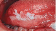
Figure 8: Leukoplakia
In contrast, erythroplakia (Figure 9) presents as a red, friable mucosal lesion, often sharply demarcated from adjacent normal tissue, and is far more ominous biologically. Histologic examination reveals moderate-to severe epithelial dysplasia, carcinoma in situ, or frankly invasive carcinoma in a substantial proportion of cases. Contemporary series consistently demonstrate invasive carcinoma in approximately 35% to 40% of erythroplakias at the time of biopsy, underscoring the need for prompt tissue diagnosis and definitive management [4-45].
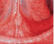
Figure 9: Erythroplakia
According to the World Health Organization (WHO) classification (Table 3), oral epithelial precursor lesions are stratified based on architectural and cytologic abnormalities. Squamous cell hyperplasia reflects an increased number of epithelial cells without cytologic atypia and may involve acanthosis of the spinous layer and/or basal cell hyperplasia within the progenitor compartment. Epithelial dysplasia is defined by disordered maturation, loss of stratification, and cytologic atypia, and is graded as mild (squamous intraepithelial neoplasia [SIN] 1), moderate (SIN 2), or severe dysplasia/carcinoma in situ (SIN 3) [135-151].
Table 3: World health organization (WHO) classification of precursor lesions of the oral cavity [135].
|
Lesion Type |
Description |
Additional Terms |
|
Squamous cell hyperplasia |
Increase in the number of cells |
Spinous layer (acanthosis) Basal/parabasal cells (progenitor compartment) Basal cell hyperplasia |
|
Dysplasia |
Cellular atypia, loss of normal cellular maturation, loss of epithelial stratification |
Mild dysplasia -(squamous intraepithelial neoplasia 1) Moderate dysplasia -(squamous intraepithelial neoplasia 2) Severe dysplasia or carcinoma in situ -(squamous intraepithelial neoplastic 3) |
The risk of progression to invasive carcinoma correlates strongly with dysplasia severity. While mild dysplasia carries a relatively low transformation rate, severe dysplasia and carcinoma in situ are associated with a substantial risk of concurrent occult invasion, reported in up to 20% to 25% of cases in surgical series [45].
These data reinforce the clinical imperative for early biopsy, accurate histopathologic grading, and definitive treatment of high-grade precursor lesions to prevent progression to advanced OCSCC.
FIELD CANCERIZATION (FIELD DEFECT) IN ORAL CAVITY SQUAMOUS CELL CARCINOMA
The concept of field cancerization is central to understanding the natural history, recurrence patterns, and long-term risk of second primary tumors in oral cavity squamous cell carcinoma (OCSCC). First described by Slaughter and colleagues in 1953, field cancerization refers to diffuse epithelial injury resulting from chronic exposure to carcinogens, leading to a large area of genetically altered mucosa predisposed to malignant transformation rather than a single localized event [78].
Biological Basis
At the molecular level, field cancerization is characterized by clonal expansion of premalignant epithelial cells that harbor early genetic and epigenetic alterations. Common early events include loss of heterozygosity (LOH) at chromosomal regions 3p, 9p (CDKN2A/p16), and 17p (TP53), which may be detected in histologically normal mucosa adjacent to invasive tumors [78-82].
These altered clones can persist after surgical resection and subsequently acquire additional mutations, progressing to dysplasia, carcinoma in situ, or invasive carcinoma [79]. Next-generation sequencing studies have demonstrated that multiple spatially distinct lesions within the oral cavity may share identical driver mutations, confirming a common clonal origin and supporting the biological basis of field cancerization [152]. Conversely, genetically distinct tumors may also arise independently within the same carcinogen-exposed mucosal field, explaining the coexistence of both clonally related “second field tumors” and true second primary cancers [153].
Clinical Manifestations
Clinically, field cancerization manifests as widespread mucosal abnormalities, including leukoplakia, erythroplakia, and varying grades of epithelial dysplasia extending beyond the margins of the primary tumor [45].
Multifocal and synchronous lesions within the oral cavity
Patients with OCSCC have a 20% to 40% lifetime risk of developing a second primary malignancy, most commonly within the oral cavity, oropharynx, larynx, esophagus, or lungs [46-154]. Persistent tobacco and alcohol exposure significantly increase this risk by maintaining and expanding the altered epithelial field [39].
Surgical and Oncologic Implications
For head and neck surgeons, field cancerization has important practical implications. Histologically negative surgical margins do not necessarily indicate biologically normal tissue, as genetically altered mucosa may extend beyond the resection boundary and contribute to local recurrence [155]. This explains why local failure can occur despite adequate margin width on routine histopathologic assessment. Furthermore, distinguishing between local recurrence and second primary tumors is often challenging but clinically relevant, as second primaries reflect ongoing field damage rather than technical failure of surgery [156]. As a result, comprehensive long-term surveillance of the entire upper aerodigestive tract is mandatory, with a low threshold for biopsy of new or evolving mucosal lesions [108].
Emerging Research and Future Directions
Recent research has focused on refining the clinical application of the field cancerization concept. Molecular margin analysis using LOH profiling or mutation mapping has been explored to better define oncologically relevant margins beyond conventional histology [155-157]. Noninvasive approaches, including salivary diagnostics and oral brush biopsies, are being investigated to detect molecular alterations within at-risk fields [158].
Chemoprevention strategies targeting the premalignant field, such as retinoids, COX-2 inhibitors, and metabolic agents, have shown mixed results and are not currently standard of care [159,160]. More recently, attention has shifted toward the role of chronic inflammation and immune evasion within the field, suggesting that immunomodulatory approaches may eventually play a role in preventing progression of field defects [161].
TNM CLASSIFICATION OF HEAD AND NECK TUMORS: CURRENT STANDARDS AND ANTICIPATED EVOLUTION TOWARD THE 2026 ERA
The Tumor–Node–Metastasis (TNM) classification system remains the fundamental framework for staging head and neck malignancies, underpinning prognostic assessment, treatment selection, and clinical trial stratification worldwide.
The currently adopted system for head and neck cancer staging is the American Joint Committee on Cancer (AJCC) 8th Edition, harmonized with the Union for International Cancer Control (UICC) TNM classification. This edition marked a decisive transition from purely anatomic staging toward a more biologically informed model, particularly for squamous cell carcinoma (SCC) [22-33].
Current Standard (AJCC 8th Edition)
Among the most impactful changes introduced in the AJCC 8th Edition is the incorporation of depth of invasion (DOI) into T classification for oral cavity squamous cell carcinoma. DOI has consistently demonstrated superior prognostic relevance compared with greatest surface dimension, showing strong associations with local control, occult nodal metastasis, and disease-specific survival. Its integration into T staging corrected longstanding deficiencies in risk stratification and has had immediate clinical implications, particularly in guiding elective neck dissection and adjuvant treatment decisions [145-162].
Nodal staging was similarly refined through the formal inclusion of extranodal extension (ENE) as a defining feature of advanced nodal disease. ENE is now recognized as a dominant adverse prognostic factor, independently associated with increased locoregional recurrence, distant failure, and reduced overall survival [163-165]. The addition of ENE acknowledges the biologic aggressiveness of nodal disease beyond size and laterality alone and has reinforced its central role in postoperative risk stratification and multimodality treatment planning [33-162].
A further paradigm shift was the creation of a distinct TNM system for HPV-associated oropharyngeal squamous cell carcinoma, reflecting its markedly favorable prognosis compared with HPV-negative disease. This modification prevented systematic overstaging of HPV-mediated tumors and provided a staging framework that more accurately correlates with survival outcomes. Importantly, this change has facilitated the rational design of treatment de-escalation trials aimed at reducing long-term toxicity while preserving oncologic efficacy [33-166].
Anticipated Evolution Toward the 2026 TNM (AJCC 9th Edition)
Although the AJCC 9th Edition has not yet been formally released, multiple validation studies and staging task force publications strongly suggest that future TNM iterations will continue to move toward a hybrid anatomic–biologic staging model [33-166]. Anticipated refinements include more granular characterization of DOI and ENE (rather than binary thresholds), improved handling of borderline and heterogeneous nodal disease, and further alignment of clinical and pathologic staging to minimize stage migration [166].
There is also increasing recognition that traditional TNM variables alone are insufficient to fully capture tumor behavior. Molecular alterations, immune microenvironment features, and radiologic biomarkers have demonstrated prognostic significance independent of TNM stage [152-168]. As such, the post-2026 staging paradigm is expected to explore structured integration of validated biomarkers, particularly for HPV-negative disease, where outcomes remain heterogeneous despite similar anatomic stage [152-169].
Another anticipated area of refinement involves the relationship between TNM staging and functional outcomes and patient-reported quality of life, especially as survivorship increases [170]. The growing emphasis on de-escalation strategies and treatment personalization underscores the need for staging systems that predict not only survival but also treatment-related morbidity.
Prognostic and Research Implications
The evolution of TNM staging in head and neck cancer reflects a broader shift in oncology toward biologically informed, patient-centered care. The AJCC 8th Edition significantly improved prognostic discrimination and clinical applicability; however, ongoing research highlights the limitations of anatomy-based staging in an era of precision medicine. Future TNM updates will likely serve as a foundational scaffold upon which molecular, immunologic, and functional data are layered, rather than a standalone determinant of outcome [33-167].
For clinical research, these developments carry major implications. Trial design, eligibility criteria, and risk stratification increasingly depend on nuanced staging definitions. As TNM evolves beyond static anatomic descriptors, prospective validation of novel staging modifiers will be essential to ensure reproducibility, global applicability, and clinical relevance.
In summary, while the AJCC 8th Edition remains the current standard for head and neck cancer staging, the forthcoming 2026-era TNM system is poised to further integrate biologic insight, refine prognostic accuracy, and support personalized treatment strategies (Tables 4,5,6,7). Continued collaboration between pathologists, surgeons, radiation oncologists, and translational scientists will be critical to ensure that staging systems evolve in parallel with advances in head and neck cancer care.
Table 4: TNM Classification Primary Tumor (T) [22].
|
Category |
Definition |
|
Tis |
Carcinoma in situ |
|
T1 |
Tumor ≤ 2 cm in greatest dimension and DOI ≤ 5 mm |
|
T2 |
Tumor ≤ 2 cm with DOI > 5 mm and ≤ 10 mm OR tumor > 2 cm but ≤ 4 cm with DOI ≤ 10 mm |
|
T3 |
Tumor > 4 cm OR any tumor with DOI > 10 mm |
|
T4a |
Moderately advanced local disease: invasion through cortical bone of mandible or maxilla, involvement of maxillary sinus, or invasion of skin of the face |
|
T4b |
Very advanced local disease: invasion of masticator space, pterygoid plates, skull base, or encasement of internal carotid artery |
Table 5: TNM Classification of Oral Cavity Cancer - Lymph Nodes (N) [22].
|
Category |
Definition |
|
N0 |
No regional lymph node metastasis |
|
N1 |
Single ipsilateral lymph node ≤ 3 cm without ENE |
|
N2a |
Single ipsilateral lymph node ≤ 3 cm with ENE |
|
N2b |
Multiple ipsilateral lymph nodes, none > 6 cm, without ENE |
|
N2c |
Bilateral or contralateral lymph nodes, none > 6 cm, without ENE |
|
N3a |
Lymph node > 6 cm without ENE |
|
N3b |
Any lymph node(s) with extranodal extension (ENE+), or single node > 3 cm with ENE |
Table 6: TNM Classification of Oral Cavity Cancer - Distance metastasis (M) [22].
|
Category |
Definition |
|
M0 |
No distant metastasis |
|
M1 |
Distant metastasis present |
Table 7: Anatomical staging and prognostic groups [22].
|
Stage |
TNM Definition |
|
Stage 0 |
Tis N0 M0 |
|
Stage I |
T1 N0 M0 |
|
Stage II |
T2 N0 M0 |
|
Stage III |
T3 N0 M0 or T1–T3 N1 M0 |
|
Stage IVA |
T4a N0–N2 M0 or T1–T3 N2 M0 |
|
Stage IVB |
Any T N3 M0 or T4b any N M0 |
|
Stage IVC |
Any T any N M1 |
Notes: DOI >10 mm automatically upstages to T3, even if surface diameter is small, ENE+ nodal disease = N3b, resulting in Stage IVB, Stage grouping reflects anatomic extent, not treatment response or biology
Prognosis in head and neck cancer is strongly correlated with disease stage at the time of diagnosis (Table 8). Patients presenting with early-stage disease (stage I) demonstrate favorable outcomes, with reported 5-year disease-specific survival rates exceeding 80% in most contemporary series. In contrast, survival declines substantially for patients with locally advanced disease (stage III–IV), with 5-year survival rates frequently falling below 40%, reflecting the combined impact of larger primary tumors, regional nodal involvement, and higher rates of distant failure. The presence of cervical lymph node metastases remains one of the most powerful adverse prognostic factors, reducing survival by approximately 50% even in patients with otherwise small primary tumors. Despite advances in diagnostic imaging and multidisciplinary management, the majority of patients with head and neck cancers continue to present with advanced-stage disease (stage III or IV), underscoring the persistent challenges of early detection and timely diagnosis [22-169].
Table 8: Prognostic Risk Groups in Oral Cavity Cancer [22].
|
Risk Group |
Typical AJCC Stage |
Key Defining Features |
Prognostic Implications |
|
Low Risk |
Stage I–II |
DOI ≤10 mm, N0, ENE− |
Excellent disease-specific survival; surgery alone often sufficient |
|
Intermediate Risk |
Stage III, selected IVA |
DOI >10 mm, single node ≤3 cm, ENE− |
Increased locoregional recurrence risk; consider adjuvant RT based on pathology |
|
High Risk |
Stage IVA–IVB |
ENE+, multiple nodes, T4 disease |
High risk of locoregional and distant failure; adjuvant chemoradiation indicated |
|
Very High Risk |
Stage IVC |
Distant metastasis |
Poor prognosis; systemic therapy ± palliative intent |
PATTERNS OF RELAPSE
Despite advances in surgical technique, radiotherapy delivery, and systemic therapy, patterns of failure in oral cavity squamous cell carcinoma (OCSCC) continue to be dominated by locoregional relapse. Contemporary series consistently demonstrate that local and regional recurrences account for approximately 70% to 85% of treatment failures, even after apparently adequate primary therapy with curative intent. Local recurrence at or adjacent to the primary tumor bed remains the most common mode of failure, reflecting the biologic phenomenon of field cancerization, subclinical tumor spread, and the intrinsic aggressiveness of a subset of tumors characterized by adverse pathologic features such as depth of invasion, perineural invasion, lymphovascular invasion, and positive or close margins. Several modern institutional and population-based analyses confirm that most local recurrences occur within the first 24 months after treatment, underscoring the importance of intensive early surveillance [108-171].
Regional failure, primarily within the cervical lymphatic basin, represents the second most common site of relapse and is closely associated with initial nodal burden, extracapsular extension (ENE), and inadequate pathologic nodal clearance. Recent data from multi-institutional cohorts suggest that even in patients undergoing comprehensive neck dissection and adjuvant radiotherapy, regional recurrence rates remain between 10% and 20% in node-positive disease, particularly in pN2 to pN3 tumors and those with ENE [169-173]. Importantly, salvage outcomes for isolated regional recurrence remain modest, with 5-year disease-specific survival rarely exceeding 30% to 40%, emphasizing the prognostic weight of regional control.
In contrast, distant metastases are less frequent at initial diagnosis but increase substantially with advancing stage and recurrent disease.
The lungs represent the most common site of distant spread, accounting for approximately 70% to 80% of distant metastases, followed by bone and liver. Contemporary series report distant metastatic rates of 8% to 15% overall, rising to 20% to 30% in patients with advanced T-stage, bulky nodal disease, or ENE, particularly in the setting of locoregional recurrence [152-175]. Autopsy and end-of-life studies continue to demonstrate distant metastases in 10% to 30% of patients at the time of death, suggesting that systemic dissemination may be underrecognized during life, especially in patients who succumb early to locoregional progression.
These evolving patterns of failure have reinforced the role of functional imaging, particularly 18F-FDG PET/CT, in the evaluation of suspected recurrence or progression. PET/CT has demonstrated superior sensitivity for detecting occult distant metastases and synchronous locoregional disease compared with conventional imaging, particularly in the post-treatment setting where anatomic distortion limits the accuracy of CT and MRI. Multiple prospective and retrospective studies have shown that PET/CT alters clinical management in 20% to 30% of patients evaluated for recurrent head and neck cancer, frequently by identifying previously unsuspected distant disease and thereby avoiding non-beneficial salvage surgery or reirradiation [170].
Collectively, modern evidence confirms that locoregional failure remains the dominant pattern of relapse in OCSCC, while distant metastases emerge as a significant competing risk in advanced and recurrent disease. These data support aggressive optimization of primary local and regional therapy, risk-adapted adjuvant treatment, and the judicious use of PET/CT in recurrence assessment to guide appropriate salvage strategies and avoid futile interventions.
EVIDENCE-BASED MANAGEMENT OF ORAL CAVITY SQUAMOUS CELL CARCINOMA
Management of oral cavity squamous cell carcinoma (OCSCC) is best delivered through a multidisciplinary pathway because cure is tightly linked to achieving local control while preserving critical functions (speech, swallowing, mastication) and appearance, and because the typical patient population frequently has significant comorbidity and complex social determinants that influence treatment feasibility and adherence.
Contemporary national guidance emphasizes that treatment options for oral cavity tumors should be reviewed within a multidisciplinary team (MDT) and finalized through consensus with the patient and carers, with explicit communication of treatment intent (curative vs palliative) early in the pathway [4-176].
A MDT for OCSCC would typically include a head and neck surgical oncologist, radiation oncologist, medical oncologist, head and neck radiologist, pathologist (including anatomic pathology expertise in margin assessment), dentist/oral medicine specialist, prosthodontist/ maxillofacial prosthodontist, reconstructive surgeon (microvascular and regional flap reconstruction), speech-language pathologist, dietitian/nutritionist, physical therapist (including jaw/neck range-of-motion and shoulder rehabilitation), oncology nursing, social work, and psychology/psychiatry for distress screening and psychosocial intervention [4-10].
Neurorehabilitation and occupational therapy support are often integrated for return-to-work and functional independence planning, especially after extensive resections and neck dissections. Evidence from institutional cohort analyses suggests that care delivered after formalization of a dedicated multidisciplinary tumor board is associated with improved survival, supporting MDT care as more than logistical convenience and aligning it with best-practice quality [177].
MDT value is not limited to survival endpoints. Head and neck tumor boards frequently refine staging, reconcile imaging–pathology discordances, and modify treatment plans, and are commonly structured to embed ancillary services (speech language pathology, physical therapy, nutrition, psychology, social work) directly into the treatment plan to minimize avoidable morbidity and delays [177].
Primary tumor management and surgical oncologic principles
Across guideline systems, surgery is consistently positioned as the mainstay for most resectable oral cavity tumors, reflecting both oncologic effectiveness and the practical advantage of obtaining definitive pathologic staging (margin status, depth of invasion, perineural invasion, lymphovascular invasion, nodal yield, extranodal extension) that directly drives adjuvant therapy selection. The official guidance endorsed by specialty associations in the states that surgery remains the mainstay for oral cavity tumors and recommends tumor resection with a clinical clearance of 1 cm where vital structures permit, underscoring that the “clinical” margin goal is often larger than the final histologic margin due to specimen shrinkage and anatomic constraints [10-176].
A key publication-quality nuance is that “surgery vs radiation therapy” equivalence is more defensible for selected early head and neck subsites than for oral cavity mucosa as a whole. A global scoping review of head and neck treatment guidelines maps that, while surgery and radiotherapy remain the principal modalities for early stage disease internationally, recommendations vary by subsite and resource context, and organ-preservation radiotherapy is particularly emphasized in larynx/ oropharynx rather than oral cavity [178].
In practice for OCSCC, many professional summaries of guideline-concordant care describe single-modality management for early-stage lesions with a preference toward surgery when feasible, reserving definitive radiotherapy for selected cases based on patient factors, access, and anticipated functional trade-offs [179,180].
Margin adequacy remains one of the highest-leverage determinants of local control and adjuvant therapy escalation in OCSCC, yet the field continues to debate the optimal histologic “clear” distance. Contemporary evidence emphasizes that the widely applied 5 mm cutoff for a clear margin has limited evidential basis as a universal discriminator across subsites and margin planes, and that risk likely behaves on a continuum rather than at a single threshold [136-182]. This has stimulated interest in improved intraoperative margin assessment, including whole-specimen imaging (ultrasound, fluorescence, MRI) and refinements in frozen-section workflows; however, current systematic reviews conclude that available techniques have heterogeneous accuracy and often insufficient sensitivity for reliably identifying “close” (< 5 mm) margins across the full specimen surface, particularly at deep planes [183].
Locally advanced primary tumors often require composite resections (e.g., floor of mouth and tongue resections with segmental mandibulectomy when bone is involved, or extensive soft-tissue resection requiring flap reconstruction) and planned airway and feeding strategies. Many guidelines highlight that many anterior lesions are accessible transorally, whereas larger or more posterior tumors may require access procedures such as lingual release or lip-split with mandibulotomy to allow controlled resection and reconstruction [4-176].
Adjuvant therapy selection is fundamentally risk-adapted. Guidelines specifically note that adjuvant radiochemotherapy improves control rates in the presence of positive margins or advanced neck disease, reflecting the evidence base for postoperative concurrent cisplatin based chemoradiotherapy in high-risk head and neck squamous cell carcinoma [4-176].
The EORTC 22931 randomized trial reported lower 5-year cumulative local/regional relapse with concurrent cisplatin plus radiotherapy versus radiotherapy alone (with higher grade ≥3 acute toxicity in the combined arm) [146]. The RTOG 9501 demonstrated improved local-regional control and disease-free survival with postoperative concurrent cisplatin but at the cost of substantially increased adverse effects; long-term follow up suggests that benefit is concentrated in subsets with microscopically involved margins and/or extranodal spread [184].
The timing and continuity of postoperative radiotherapy also matters. A focused oral cavity cohort study demonstrated that longer overall postoperative radiotherapy treatment time was strongly associated with worse locoregional control and survival, supporting a publication-grade emphasis on preventing avoidable delays and maintaining treatment package discipline when adjuvant radiotherapy is indicated.
Management of the clinically negative and positive neck
The neck is not an optional consideration in OCSCC: cervical metastases substantially worsen prognosis, and the clinically node-negative (cN0) neck still harbors meaningful rates of occult disease [185]. The clinical practice guideline for neck management in squamous cell carcinoma of the oral cavity and oropharynx highlights that 10% to 40% of patients without clinical nodal disease will have occult metastases and frames effective neck management as a central component of high-quality oncologic care [186].
For patients undergoing curative-intent surgery for the primary tumor, ASCO recommends an ipsilateral elective neck dissection (END) for cT2 to cT4, cN0 oral cavity cancers, and importantly, also recommends END for cT1, cN0 disease, with the caveat that selected highly reliable patients may be managed with close surveillance incorporating specialized neck ultrasonography [186].
For a cN0 neck undergoing END, ASCO specifies that the dissection should include levels Ia, Ib, II, and III, and that an adequate dissection should yield at least 18 lymph nodes, aligning surgical quality with measurable oncologic benchmarks [186].
For a clinically node-positive (cN+) neck, ASCO recommends an ipsilateral therapeutic selective neck dissection that includes levels Ia, Ib, IIa, IIb, III, and IV, again with a minimum node yield expectation (≥ 18), and suggests considering level V dissection in multistation disease [186]. These recommendations also provide a framework for standardization, moving away from ambiguous or historical “named” dissections toward explicit reporting of levels removed, which is consistent with the consensus nomenclature updates from the American Head and Neck Society and collaborating societies [187].
Randomized evidence supports proactive management of the cN0 neck. In a landmark randomized trial comparing END to therapeutic (delayed) neck dissection in early stage node-negative oral cancer, END improved 3-year overall survival (80.0% vs 67.5%) and disease-free survival (69.5% vs 45.9%), establishing END as a survival improving intervention rather than merely a staging exercise [188].
This trial evidence synergizes an evidence based approach to treating the neck as part of the initial definitive management for most oral cavity primaries, and with other similar guidelines “elective neck treatment should be offered for all oral cavity tumors,” reflecting the historically high stakes of regional failure in this disease [17-176]. Contralateral neck management is driven by anatomic risk. ASCO recommends contralateral neck dissection for a contralateral cN+ neck, and indicates that elective contralateral dissection may be offered for oral tongue and/or floor-of-mouth primaries that are T3/T4 or approach midline, capturing the clinical observation that midline proximity and posterior subsites increase bilateral drainage risk [186]. When surgery is not feasible, neck radiotherapy becomes the elective modality. ASCO specifies that elective radiotherapy to an undissected neck (50 to 56 Gy in 25 to 30 fractions) may be efficacious and should be delivered when an END cannot be performed, reinforcing the principle that nodal basins “at risk” should still receive definitive elective treatment within the selected modality [186].
The extent of elective neck dissection, particularly whether to routinely include level IV, has been controversial, especially for oral tongue primaries. A meta-analysis focused on skip metastasis to level IV in cN0 OCSCC reported a fixed-effects level IV involvement estimate of 2.53% and skip metastasis of 0.50%, concluding that selective neck dissection limited to levels I to III is adequate for most cN0 OCSCC patients and that routine level IV dissection is not justified on available data [189]. These findings complement standardization efforts that define supraomohyoid/selective dissections as I to III and encourage explicit level reporting when surgeons extend dissections based on subsite-specific concerns.
Depth of invasion and tumor thickness in risk stratification
A modern, publication-ready discussion of “tumor thickness” must distinguish conventional tumor thickness (which can be influenced by exophytic growth) from depth of invasion (DOI), which more directly captures biologic infiltration and metastatic propensity (Table 9 and 10). The 8th edition staging system introduced a major change for oral cavity tumors by incorporating DOI into T-category assignment, reflecting accumulating evidence that deeper invasion is associated with higher risk of nodal metastasis and worse disease-specific outcomes [22-185].
Table 9: Illustrative risk stratification by DOI in early oral tongue SCC [191].
|
DOI Category |
Nodal metastasis |
Local recurrence |
|
1-5mm |
23% |
15% |
|
6-10mm |
34% |
20% |
|
>10mm |
53% |
40% |
Table 10: Thickness of oral cancer predicts survival and failure [192].
|
Tumor Thickness |
5 y Overall Survival |
Treatment Failure |
|
< 2 mm |
97 % |
2 % |
|
2 – 8 mm |
83% |
|
|
> 8 mm |
65% |
45% |
Consistent with this shift, guidelines emphasize that DOI is now a pathologic feature that should be routinely reported because it influences staging and can alter treatment planning, including neck management [108 178]. Data from early oral tongue SCC cohorts illustrate how nodal metastasis and local recurrence risk rise with DOI strata aligned to AJCC cut points [169-190].
These values summarize one early oral tongue SCC dataset and should be contextualized as subsite- and cohort-dependent, but they demonstrate the steep risk gradient that motivates elective neck treatment even for “small” primaries [190].
A key question for publication is not whether DOI predicts nodal disease (it does), but where to place the operative threshold for END or for SLNB-guided strategies. Practice varies, and thresholds in the literature span a wide range (Figure 10). A recent open-access cohort specifically evaluated DOI 4 mm as a decision threshold in “early” oral cancers; nodal metastasis was substantially higher when DOI ≥ 4 mm (25.4%) versus DOI < 4 mm (4.7%), supporting DOI ≥ 4 mm as a pragmatic cutoff for END in that population [193].
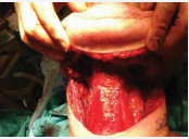
Figure 10: Bilateral Supraomohyoid Neck Dissection for a Tongue Cancer > 4 mm in Thickness
In contrast, analyses of guideline interpretation note that current NCCN guidance has recommended considering END when DOI exceeds 3 mm, while acknowledging that the evidence supporting a single cutoff is imperfect and often derived from occult metastasis rates rather than survival outcomes [108-194].
Historically, tumor thickness was also used as a surrogate for biologic aggressiveness. A classic early-stage oral cavity study reported a significant prognosis shift around a 4 mm thickness cutoff, with favorable local control and survival below this level [195]. Later oral tongue series similarly observed that regional failures after primary surgery were concentrated among tumors ≥ 4 mm thickness, reinforcing that invasive depth metrics are clinically meaningful even in pT1 to pT2 N0 cohorts [196].
The most defensible publication stance is therefore twofold: (1) DOI-(and/or meticulously measured invasion depth) should be treated as a core prognostic variable and reported systematically in pathology, and (2) DOI based thresholds should be presented as risk stratification tools that guide neck management within overall clinical context (subsite, laterality/midline proximity, imaging, patient reliability for surveillance), rather than as rigid rules [185].
Sentinel lymph node biopsy in early-stage disease
Sentinel lymph node biopsy (SLNB) seeks to stage the cN0 neck accurately while reducing morbidity relative to routine END, particularly in early-stage disease. In the oral cavity, SLNB has matured from feasibility studies to multicenter prospective data and randomized comparison.
A major randomized, multicenter noninferiority trial compared SLNB-navigated neck dissection to standard elective neck dissection in early-stage OCSCC and demonstrated noninferior 3-year overall survival (87.9% vs 86.6%) and noninferior 3-year disease-free survival (78.7% vs 81.3%), with significantly better neck functionality scores in the SLNB group, supporting SLNB-guided surgery as an accepted de-escalation strategy in appropriately selected settings with established expertise [197].
Prior multicenter evidence also supports diagnostic performance, while emphasizing that performance metrics vary by technique and subsite. The Dutch multicenter trial in cN0, T1 to T2 oral cancer reported sensitivity 80% and negative predictive value (NPV) 88%, with high rates of neck control; these values are directionally consistent with the concept that SLNB can concentrate definitive neck surgery on patients most likely to benefit [198]. In a large multi-institutional U.S. trial framework, gamma probe directed SLNB in early oral SCC was reported as feasible with an NPV of 96% for nodal metastasis [199]. However, SLNB is not uniformly “plug-and-play” across oral cavity subsites.
Modern reviews highlight the “shine-through” phenomenon, particularly relevant to floor-of-mouth tumors, where tracer activity at the injection site obscures nearby sentinel nodes (often in level I), yielding lower sensitivity and accuracy than in other subsites. One review quantified this subsite effect, reporting lower SLNB sensitivity for floor-of-mouth tumors than for other oral cavity locations, anchoring the common recommendation that floor-of-mouth primaries require special caution or alternative strategies depending on local expertise and imaging adjuncts [200].
Taken together, a publication-quality position is that SLNB is an evidence-supported alternative to routine END for selected early OCSCC, particularly T1 to T2 cN0 disease, when performed in centers with mature protocols, nuclear medicine support, and validated pathology workflows; it is best presented as a technique-sensitive strategy with known subsite limitations rather than as a universal replacement for END [197].
Surgical access to oral cavity tumors must balance oncologic adequacy with preservation of function and minimization of morbidity [4]. The choice of approach depends on tumor size, depth of invasion, subsite, proximity to the mandible, anticipated need for composite resection, and reconstructive requirements. Contemporary management emphasizes achieving wide en bloc resection with histologically negative margins while preserving speech, swallowing, and aesthetic integrity whenever possible [33-108].
Transoral (Peroral) Approach
Small, anteriorly located T1 to T2 lesions of the oral tongue, floor of mouth, buccal mucosa, or gingiva can often be managed via a transoral approach. This technique avoids external incisions and minimizes morbidity. With appropriate exposure using mouth gags and retractors, and in selected cases transoral laser microsurgery, adequate oncologic margins can be achieved in carefully selected tumors [201].
Multiple contemporary series demonstrate equivalent oncologic outcomes for early-stage oral cavity cancers treated transorally compared with open approaches, provided that negative margins are obtained, and appropriate management of the neck is performed [169]. Margin status remains one of the strongest predictors of local control and survival; positive or close margins significantly increase recurrence risk and worsen disease specific survival [143,144].
Lower Cheek Flap (Lip-Splitting Approach)
For larger or more posterior tumors, particularly those requiring mandibular resection or simultaneous neck dissection, the lower cheek flap provides excellent exposure. This approach involves a midline lip-splitting incision that may extend laterally into the neck, facilitating access to both the primary tumor and cervical lymphatics [17]. It remains one of the most versatile approaches for oral cavity tumors except for those primarily involving the upper gingiva and hard palate. The lower cheek flap allows for marginal or segmental mandibulectomy and facilitates microvascular reconstruction, particularly with fibula free flaps [202].
Modern refinements in incision design and meticulous closure techniques have reduced the risk of lip incompetence and aesthetic deformity, although lower lip scarring and transient sensory disturbance may occur [17]. When orbicularis oris continuity is restored and reconstruction is performed in experienced centers, long-term speech and swallowing outcomes are generally favorable [203].
Visor Flap
The visor flap offers exposure to anterior oral cavity lesions while avoiding lip splitting. It is particularly useful for select anterior floor-of-mouth or alveolar ridge tumors [17]. However, bilateral mental nerve sacrifice is typically required, resulting in permanent chin anesthesia. This sensory loss may contribute to lower lip incompetence, drooling, and articulation difficulty. Because of these functional consequences and limited posterior exposure, its use has declined in favor of approaches preserving neural integrity [17-204].
Upper Cheek Flap (Weber–Ferguson Approach)
Extensive tumors of the upper gingiva, hard palate, and maxilla may require an upper cheek flap such as the Weber Ferguson incision or its modifications. This approach provides wide exposure for partial or total maxillectomy and facilitates midfacial reconstruction [205]. Advances in microvascular reconstruction, including scapular and fibular osteocutaneous flaps and virtual surgical planning, have significantly improved aesthetic and functional outcomes following these resections [206].
Mandibulotomy for Access to Posterior Tumors
For large posterior oral cavity tumors approaching the base of the tongue without mandibular invasion, mandibulotomy provides necessary exposure while preserving oncologic principles. The three principal techniques, lateral, median (midline), and paramedian, have distinct complication profiles.
Lateral Mandibulotomy
Lateral mandibulotomy, typically performed posterior to the canine or premolar region, is associated with several disadvantages. Asymmetric muscular forces across the osteotomy can stress healing and may necessitate intermaxillary fixation. Transection of the inferior alveolar nerve produces permanent sensory loss to the lower lip and chin distal to the osteotomy [207-209].
Additionally, disruption of endosteal blood supply may predispose to devascularization of the distal mandibular segment. When postoperative radiation therapy is administered, the osteotomy often lies within the high-dose field, increasing the risk of delayed union or osteoradionecrosis [17-208]. Reported mandibulotomy complication rates range from 10% to 25%, with higher rates in irradiated patients [17].
Median (Midline) Mandibulotomy
Midline mandibulotomy preserves bilateral inferior alveolar nerve function and endosteal blood supply but usually requires extraction of a central incisor to prevent root exposure [210]. Division of the genioglossus and geniohyoid muscles may delay recovery of swallowing and tongue mobility. Although union rates are acceptable in experienced hands, functional considerations limit routine use [17-210].
Paramedian Mandibulotomy
Paramedian mandibulotomy is widely preferred in high-volume centers. The osteotomy is placed between the lateral incisor and canine, preserving inferior alveolar and mental nerves and maintaining genioglossus/geniohyoid attachments [4-209]. Division is generally limited to the mylohyoid muscle, minimizing long-term swallowing dysfunction. Comparative series report lower rates of sensory deficit and osteoradionecrosis compared with lateral approaches [208, 209]. Proper fixation techniques further reduce nonunion risk [207,208].
Mandibular Invasion and Patterns of Spread
Understanding mandibular invasion pathways is essential when determining whether marginal or segmental mandibulectomy is indicated. Contrast-enhanced CT and MRI improve detection of cortical and medullary invasion, although sensitivity remains imperfect [121-211].
McGregor et al., first described differences in mandibular invasion according to dentition status [179]. In edentulous mandibles, invasion commonly occurs through cortical thinning along the occlusal surface. In dentulous mandibles, tumor spread may follow periodontal ligament spaces, and intact teeth may partially delay direct cortical invasion. Alveolar ridge resorption in edentulous patients reduces vertical bone height, facilitating earlier medullary involvement [179,180].
Contemporary pathologic analyses confirm that true medullary invasion is associated with higher local recurrence and worse disease-specific survival [212]. A systematic review and meta-analysis demonstrated that marginal mandibulectomy yields oncologic outcomes comparable to segmental resection when invasion is limited to superficial cortex and negative margins are achieved [212,213].
Segmental mandibulectomy is indicated for extensive cortical destruction or medullary involvement [204-213].
Contemporary Reconstruction and Functional Outcomes
Microvascular free tissue transfer, particularly fibula osteocutaneous flaps, allows restoration of mandibular continuity and dental rehabilitation with osseointegrated implants, significantly improving quality of life [202-214]. Virtual surgical planning and patient-specific cutting guides have improved reconstructive precision and reduced operative time without increasing complication rates [206]. Functional outcomes are increasingly recognized as key endpoints. Speech intelligibility, swallowing efficiency, and aesthetic satisfaction correlate strongly with preservation of neuromuscular structures and accurate reconstruction [203]. Early speech and swallowing therapy and coordinated prosthodontic rehabilitation significantly enhance long-term functional recovery [17].
Surgical access to oral cavity tumors must be individualized according to tumor characteristics, planned resection, reconstructive requirements, and anticipated adjuvant therapy. Paramedian mandibulotomy and contemporary reconstructive techniques have reduced morbidity while maintaining oncologic rigor. A detailed understanding of mandibular invasion patterns, particularly differences between dentulous and edentulous mandibles, remains critical in selecting between marginal and segmental resections. Integration of modern imaging, reconstructive innovation, and functional outcome assessment continues to refine surgical management and improve survival and quality-of-life outcomes in OCSCC.
RADIATION THERAPY IN ORAL CAVITY SQUAMOUS CELL CARCINOMA (OCSCC)
Radiation therapy (RT) for cancers of the oral cavity is delivered most commonly as external beam radiotherapy (EBRT), now typically intensity-modulated RT (IMRT) with image guidance, either as definitive treatment or as an adjuvant modality after surgery. Interstitial brachytherapy (low-dose rate, pulsed-dose rate, or high-dose rate) retains an important, highly selected role as monotherapy for small, well-lateralized early lesions (classically mobile tongue/floor of mouth), or as a boost to intensify dose to the primary bed while limiting exposure of uninvolved mucosa and salivary glands.
Contemporary consensus guidance emphasizes that brachytherapy’s major limitation in oral cavity disease is not target dose intensification per se, but rather the inability of implantation alone to simultaneously treat elective regional lymphatics, particularly in tumors with meaningful risk of occult nodal metastasis, making combined-modality approaches (EBRT to primary + nodal basins with brachytherapy boost to the primary) appropriate in selected cases when organ/function preservation is prioritized and expertise is available [215,216].
Definitive RT: technique, targets, dose, and overall treatment time
For definitive (nonoperative) RT, EBRT typically encompasses the primary tumor and at-risk regional lymphatics, including clinically negative nodal levels when the estimated risk of occult metastasis justifies elective nodal irradiation (ENI). IMRT-based planning with simultaneous integrated boost (SIB) is standard in many centers, enabling differential dose painting (higher dose to gross disease, intermediate dose to high-risk subclinical regions, and lower dose to elective nodal volumes) while improving dose conformity and sparing organs at risk (parotid/submandibular glands, mandible, oral cavity “avoidance” mucosa, pharyngeal constrictors when relevant). When superficial, very small mucosal primaries are treated non-surgically, carefully selected cases may be approached with limited-field techniques (electrons or intraoral cone therapy) or brachytherapy alone; however, this strategy is best restricted to lesions with a clearly low risk of nodal involvement and with rigorous imaging/ clinical follow-up [215,216].
Definitive RT is usually delivered once daily over 6 to 7 weeks to a biologically tumoricidal dose (commonly 66 to 70 Gy to gross disease in 2.0 to 2.12 Gy fractions, with ENI typically 50 to 54 Gy, institution- and risk-adapted) [217-219]. A consistent finding across multiple datasets in head and neck cancer is that prolongation of overall treatment time (OTT), from interruptions, replanning delays, intercurrent illness, or inadequate supportive care, correlates with inferior locoregional control, plausibly due to accelerated tumor repopulation; consequently, avoidable breaks should be minimized and interruptions compensated when feasible [220-222].
Toxicity profile and mitigation
Although RT avoids surgical resection of oral cavity tissues, treatment-related morbidity can be clinically significant and long-lasting. Common and/or high impact toxicities include xerostomia, dysgeusia, mucosal fibrosis and soft-tissue contracture with trismus, dental deterioration, osteoradionecrosis of the mandible, dysphagia (often multifactorial), and hypothyroidism after lower neck irradiation. Modern IMRT reduces, but does not eliminate, many of these risks, and outcomes are strongly influenced by pretreatment dental optimization, meticulous mandibular and salivary gland sparing when oncologically safe, early nutritional and swallowing intervention, and coordinated survivorship surveillance [223].
Postoperative RT (PORT): indications, dose, and timing
PORT is recommended when pathologic features predict a high likelihood of locoregional recurrence despite complete macroscopic resection. One of the initial studies supporting the use of postoperative radiotherapy (PORT) in head and neck cancer is the RTOG 73-03 trial [224].
This randomized study included patients with locally advanced head and neck squamous cell carcinomas involving the supraglottic larynx, hypopharynx, oral cavity, and oropharynx. Patients were assigned to receive either preoperative radiotherapy (50 Gy) followed by surgery or surgery followed by postoperative radiotherapy (60 Gy). With long-term follow-up of approximately 10 years, the study demonstrated significantly improved locoregional control in the postoperative radiotherapy group (65%) compared with the preoperative radiotherapy group (48%) (p = 0.04). A trend toward improved overall survival was also observed (38% vs 33%), although this did not reach statistical significance (p = 0.10). Importantly, rates of surgical and radiation-related complications were comparable between the two treatment arms [224].
A prospective randomized trial from MD Anderson Cancer Center evaluated optimal postoperative radiation dose in 240 patients with resected stage III to IV head and neck squamous cell carcinomas, including tumors of the oral cavity, oropharynx, hypopharynx, and larynx [225]. Patients who received doses <54 Gy experienced significantly higher locoregional failure rates compared with those treated with ≥57.6 Gy (p = 0.02). A clear dose response relationship was not observed beyond 57.6 Gy, except in patients with extracapsular nodal extension (ENE), for whom improved control required doses of at least 63 Gy [225].
Additionally, the presence of “risk clusters”, defined as two or more adverse features, such as oral cavity primary, close or positive margins, perineural invasion, >2 positive lymph nodes, nodal size >3 cm, treatment delay >6 weeks, or poor performance status, was associated with increased risk of failure and similarly supported escalation to approximately 63 Gy [225].
Moderate to severe complications occurred in 7.1% of patients and were more frequent with doses exceeding 63 Gy, suggesting that further dose escalation does not improve the therapeutic ratio and may increase toxicity. Overall, postoperative radiotherapy, typically delivered to 60 to 66 Gy over 6 to 7 weeks, significantly reduces locoregional recurrence in patients with adverse pathologic features, decreasing failure rates from approximately 50% to 15% to 20% in high-risk populations [165-228]. The indications for postoperative radiotherapy are now well established and are summarized in Table 11.
Table 11: Indications for postoperative radiotherapy [165, 184, 226-228].
|
Close or positive margins |
|
An affected lymph node > 3 cm |
|
Multiple lymph nodes involved |
|
Extra capsular extension (ECE) |
|
Patients who had an open biopsy of a suspicious neck node and did not undergo neck dissection at the time |
|
Perineural invasion, lymphovascular space invasion, invasion of cartilage, bone or deep soft tissues |
|
Recommendation of the surgeon due to intraoperative findings |
The most consistently validated “high-risk” indications include microscopically positive margins and extranodal extension (ENE). Two landmark randomized trials established that, in resected stage III/IV head and neck squamous cell carcinoma, adding concurrent cisplatin to PORT improves locoregional control and disease-free survival, with the clearest benefit in patients with positive margins and/or ENE; long-term follow-up confirms durable disease-control gains in these subsets, balanced against increased acute toxicity and potential late non-cancer mortality in some analyses [146-230].
Additional “intermediate-risk” factors frequently used to justify PORT (without mandatory chemotherapy) include multiple positive lymph nodes, pT3 to T4 primary disease, lymphovascular invasion (LVI), perineural invasion (PNI), close margins (institution-specific definitions), and other adverse features indicating aggressive biology or inadequate surgical clearance. These factors were incorporated into the eligibility criteria and risk stratification of the landmark postoperative trials, particularly EORTC 22931 and RTOG 9501 (Table 12 and 13), although the most consistent benefit from adding concurrent cisplatin was limited to positive margins and/or ENE [146-229]. Current NCCN guidance similarly lists multiple positive nodes, PNI, vascular invasion, lymphatic invasion, and pT3 to T4 disease as adverse pathologic features generally supporting postoperative RT [231].
Table 12: Comparison of RTOG 9501 and EORTC 22931 [146, 184, 229].
|
Feature |
RTOG 9501 |
EORTC 22931 |
|
Study design |
Phase III randomized trial |
Phase III randomized trial |
|
Population |
Resected stage III to IV HNSCC |
Resected stage III to IV HNSCC |
|
Sample size |
459 patients |
334 patients |
|
Radiation dose |
60 to 66 Gy |
60 to 66 Gy |
|
Chemotherapy |
Cisplatin 100 mg/m² (days 1, 22, 43) |
Cisplatin 100 mg/m² (days 1, 22, 43) |
|
High-risk inclusion criteria |
Positive margins, extracapsular extension (ENE), ≥2 positive lymph nodes |
Positive margins, ENE, plus broader criteria: T3–T4, PNI, LVI, nodal disease |
|
Primary endpoint |
Locoregional control |
Progression-free survival |
|
Locoregional control |
Improved with CRT (82% vs 72%, p=0.01) |
Improved with CRT |
|
Disease-free survival |
Improved (p=0.04) |
Improved |
|
Overall survival |
No significant difference (p=0.19) |
Significant improvement (53% vs 40%, p=0.02) |
|
Follow-up |
Median ~45.9 months |
Median ~60 months |
|
Toxicity (Grade ≥3) |
Higher with CRT (77% vs 34%) |
Higher with CRT |
|
Key interpretation |
Benefit driven mainly by ENE and positive margins |
Broader inclusion → greater OS benefit observed |
Table 13: Results from the EORTC 22931 and RTOG 9501 Studies [146, 184, 229].
|
EORTC 22931 RTOG 9501 |
||||||
|
End Point |
RT |
RT + CT |
P Value |
RT |
RT + CT |
P Value |
|
5 y Disease Free Survival |
36% |
47% |
0.04 |
61% |
78% |
0.004 |
|
LR Control |
69% |
82% |
0.007 |
72% |
82% |
0.01 |
|
OS |
40% |
53% |
0.002 |
57% |
63% |
0.19 |
For oral cavity primaries specifically, increasing depth of invasion (DOI) correlates with occult nodal risk and worse outcomes and is commonly integrated into multidisciplinary recommendations regarding elective neck management and adjuvant therapy; however, DOI alone should be contextualized with the full pathologic risk profile rather than used as a single binary trigger for PORT [191-232].
Finally, quality metrics emphasize timeliness: multiple analyses demonstrate that initiating PORT beyond 6 weeks after surgery is associated with worse outcomes, supporting coordinated perioperative planning (dental clearance pathways, early radiation consultation, expedited simulation) to minimize avoidable delay [233, 234].
The adjuvant treatment for patients with oral cavity cancers is summarized in table 14.
Table 14: Summary of Adjuvant Treatment of Oral Cavity Cancers
|
Risk Based on Factors for Treatment Failure |
Management |
|
Low Risk |
No adjuvant radiation |
|
Intermediate Risk (- Margin, - ECE) |
Radiation therapy (60 Gy) |
|
High Risk (+ Margin and/or + ECE) |
Chemo radiation (60 to 66 Gy) with Cisplatin |
The randomized phase II trial RTOG 0234 evaluated the feasibility and efficacy of adding concurrent systemic therapy to postoperative radiation plus cetuximab in patients with high-risk resected head and neck squamous cell carcinoma (HNSCC) [235]. A total of 238 patients with stage III to IV disease and adverse pathologic features, including positive surgical margins, extracapsular nodal extension (ENE), or ≥2 involved lymph nodes, were enrolled following gross total resection. Participants were randomized to receive postoperative radiation therapy (60 Gy) with weekly cetuximab combined with either weekly cisplatin (30 mg/m²) or docetaxel (15 mg/m²). With a median follow-up of 4.4 years, the 2-year overall survival (OS) was 69% in the cisplatin arm versus 79% in the docetaxel arm, while 2-year disease-free survival (DFS) was 57% and 66%, respectively [235].
When benchmarked against the chemoradiotherapy arm of RTOG 9501 [229], both regimens demonstrated improved DFS, with hazard ratios of 0.76 (P = 0.05) for the cisplatin arm and 0.69 (P = 0.01) for the docetaxel arm, corresponding to absolute 2-year DFS gains of 2.5% and 11.1%, respectively. Importantly, treatment delivery was feasible, and toxicity was consistent with expectations for combined modality therapy, without unexpected safety signals.
The docetaxel-containing regimen showed a signal for improved survival outcomes compared with historical controls, providing the rationale for further investigation. This has led to the ongoing phase II/III trial RTOG 1216, which is comparing postoperative radiation with concurrent cisplatin versus docetaxel-cetuximab versus cisplatin-atezolizumab in high-risk HNSCC [236].
Despite the promising findings from RTOG 0234, high dose cisplatin-based chemoradiotherapy remains the standard of care for patients with positive margins or ENE, based on level I evidence from trials such as EORTC 22931 and RTOG 9501. The incorporation of cetuximab into the postoperative setting has not supplanted cisplatin due to lack of phase III superiority data. However, taxane-based regimens (particularly docetaxel) continue to be actively investigated, especially in patients who are cisplatin ineligible or as part of novel combination strategies with immunotherapy.
AREST Trial (ASCO 2026): Adjuvant Radiotherapy for Early-Stage Oral Cavity Squamous Cell Carcinoma
The role of postoperative radiotherapy (PORT) for patients with early-stage oral cavity squamous cell carcinoma (OSCC) harboring intermediate-risk pathological features has remained controversial for decades. Current recommendations are largely based on retrospective evidence, with established indications for PORT traditionally limited to patients with positive surgical margins, extranodal extension (ENE), or multiple metastatic lymph nodes [108-237]. The phase III AREST trial, presented at the 2026 American Society of Clinical Oncology (ASCO) Annual Meeting, provides the first prospective randomized evidence addressing the benefit of PORT in patients with intermediate-risk early-stage OSCC [238].
AREST was a multicenter, open-label, phase III randomized controlled trial that enrolled 392 patients with completely resected pT1 to pT2, pN0 oral cavity squamous cell carcinoma [238]. Adequate surgery was defined as complete tumor resection with histologically negative margins (≥ 5 mm) and ipsilateral level I to III neck dissection yielding at least 16 lymph nodes [238]. Eligible patients demonstrated one or more intermediate-risk pathological features, including depth of invasion (DOI) between 5 and 10 mm, perineural invasion (PNI), lymphovascular invasion (LVI), or poor tumor differentiation [238]. Patients were randomized to receive either postoperative radiotherapy or observation following surgery.
The primary endpoint was locoregional recurrence free survival (LRFS). Adjuvant radiotherapy significantly reduced the risk of locoregional recurrence compared with observation [238]. The greatest benefit was observed among patients with oral tongue squamous cell carcinoma [238]. Disease-free survival also favored the PORT arm, although no statistically significant improvement in overall survival has been demonstrated with the current duration of follow-up [238]. As expected, acute and late treatment related toxicities were more common in patients receiving postoperative radiotherapy [238].
The AREST trial represents an important advance in the management of early-stage OSCC because it provides the first level I evidence supporting postoperative radiotherapy in carefully selected patients with intermediate-risk pathological features [238]. These findings suggest that PORT should be strongly considered for patients with pT1 to pT2, N0 disease who demonstrate adverse pathological features such as DOI of 5 to 10 mm, PNI, LVI, or poor differentiation despite otherwise favorable pathological findings. Nevertheless, because an overall survival benefit has not yet been demonstrated, treatment decisions should remain individualized, balancing improved locoregional control against the potential acute and long-term functional morbidity associated with radiation therapy [108-238].
Longer follow-up and publication of the final peer reviewed manuscript will be important to determine whether these findings lead to modifications in future treatment guidelines.
Neoadjuvant Therapy for Locally Advanced Resectable Head and Neck Squamous Cell Carcinoma (HNSCC)
Neoadjuvant therapy for locally advanced resectable HNSCC has undergone a major paradigm shift following the June 2025 FDA approval of perioperative Pembrolizumab based on the landmark KEYNOTE-689 trial, representing the first major advance in this setting in more than two decades [239,240]. Historically, upfront surgery followed by risk-adapted adjuvant therapy remained the standard of care for resectable oral cavity and other non-nasopharyngeal head and neck cancers. However, the integration of immunotherapy into the perioperative setting has significantly altered treatment algorithms, particularly for patients with PD-L1–positive tumors.
The phase III KEYNOTE-689 trial randomized 714 patients with resectable stage III to IVA HNSCC to receive two cycles of neoadjuvant pembrolizumab (200 mg every 3 weeks) followed by surgery and adjuvant pembrolizumab concurrent with postoperative radiotherapy ± cisplatin, versus standard therapy alone [239]. At a median follow-up of 38.3 months, perioperative pembrolizumab significantly improved event-free survival (EFS) across all analyzed populations. In patients with PD-L1 combined positive score (CPS) ≥10, the 36-month EFS was 59.8% versus 45.9% (HR 0.66; 95% CI 0.49–0.88; P=0.004). Among patients with CPS ≥1, 36-month EFS was 58.2% versus 44.9% (HR 0.70; 95% CI 0.55–0.89; P=0.003), while in the overall study population the 36-month EFS was 57.6% versus 46.4% (HR 0.73; 95% CI 0.58–0.92; P=0.008) [239]. Importantly, neoadjuvant pembrolizumab did not impair surgical feasibility, with surgical completion rates of approximately 88% in both arms, and grade ≥3 immune-related adverse events occurred in 10% of patients receiving pembrolizumab [239]. Overall survival data remain immature, although early trends favor the pembrolizumab arm.
Based on these findings, the National Comprehensive Cancer Network Guidelines (v1.2026) now incorporate neoadjuvant pembrolizumab into the management of multiple resectable head and neck cancer subsites [231]. For oral cavity squamous cell carcinoma (stage III to IVA) with PD-L1 CPS ≥1, neoadjuvant pembrolizumab is listed as an “Other Recommended” option prior to surgery, followed by postoperative pembrolizumab with radiotherapy ± cisplatin depending on pathologic risk factors [231]. In p16 negative oropharyngeal, hypopharyngeal, and laryngeal cancers, neoadjuvant pembrolizumab is categorized as “Useful in Certain Circumstances” [231].
Notably, patients with N3 disease are excluded from these recommendations [231]. Adjuvant therapy after neoadjuvant pembrolizumab is determined according to postoperative pathologic risk stratification [231].
Patients without adverse pathologic features may receive radiotherapy with pembrolizumab followed by maintenance pembrolizumab. Patients with extranodal extension and/or positive margins are recommended to receive concurrent cisplatin-based chemoradiotherapy with pembrolizumab followed by adjuvant pembrolizumab [231]. Other intermediate-risk features may warrant postoperative radiotherapy with or without systemic therapy.
Before the immunotherapy era, induction chemotherapy represented the primary neoadjuvant approach for locally advanced HNSCC. The preferred induction regimen remains TPF (docetaxel, cisplatin, and 5-fluorouracil), supported by meta-analyses demonstrating superiority over PF (cisplatin/5-FU), with an overall survival hazard ratio for death of 0.72 [231-241]. Nevertheless, multiple randomized phase III trials, including DeCIDE and PARADIGM, failed to demonstrate a consistent survival advantage for adding induction chemotherapy before concurrent chemoradiation [241,242]. As a result, induction chemotherapy carries a category 3 designation in several head and neck subsites within the NCCN guidelines because of significant panel disagreement [231]. Despite this, induction chemotherapy may still play a role in selected patients with bulky symptomatic tumors, impending airway compromise, or laryngeal preservation strategies [241,242].
More recently, neoadjuvant chemoimmunotherapy combinations have demonstrated highly encouraging pathologic response rates in phase II studies [243 248]. Across multiple trials combining PD-1/PD-L1 inhibitors with platinum-based chemotherapy, pathologic complete response (pCR) rates of 27% to 56% and major pathologic response (MPR) rates of 49% to 82% have been reported [241,242]. A randomized phase II trial evaluating toripalimab combined with nab-paclitaxel/ cisplatin demonstrated significantly higher pCR (57.8% vs 34.9%; P=0.03) and MPR rates (82.2% vs 53.5%; P=0.004) compared with chemotherapy alone [248].
Additionally, innovative approaches incorporating low-dose radiotherapy with chemoimmunotherapy have produced pCR rates exceeding 60% in early-phase studies [247]. Although these strategies remain investigational and are not yet incorporated into NCCN guidelines, they represent one of the most rapidly evolving areas in head and neck oncology.
Another important recent development is the NIVOPOSTOP trial, which evaluated adjuvant Nivolumab combined with postoperative chemoradiotherapy in high-risk resected HNSCC [249]. Unlike KEYNOTE-689, NIVOPOSTOP did not include a neoadjuvant component.
The trial demonstrated improved 3-year disease free survival (63.1% vs 52.5%; HR 0.76), with benefits observed irrespective of PD-L1 status [249]. Interestingly, KEYNOTE-689 appeared to predominantly reduce distant recurrence, whereas NIVOPOSTOP improved locoregional control, suggesting potentially complementary biologic effects between perioperative and adjuvant immunotherapy strategies [249].
Collectively, these studies establish perioperative immunotherapy as a new therapeutic cornerstone in locally advanced resectable HNSCC. While perioperative pembrolizumab now represents a new standard of care for selected patients with PD-L1–positive resectable disease, ongoing trials are evaluating increasingly sophisticated neoadjuvant strategies incorporating checkpoint inhibitors, vaccines, bispecific antibodies, targeted agents, adaptive response-guided therapy, and chemoimmunotherapy combinations. These evolving approaches may further improve pathologic response rates, reduce recurrence, and ultimately redefine multidisciplinary management of head and neck squamous cell carcinoma. The most important message from ASCO 2026 is that the field is transitioning from simply adding immunotherapy to optimizing who receives it, when it is administered, and how it is combined with targeted therapies and biomarkers. While no new phase III trial matched the impact of KEYNOTE-689, advances in perioperative immunotherapy implementation, VEGF–PD-1 combinations, ctDNA-guided surveillance, and emerging ADCs collectively indicate that the next major paradigm shift in HNSCC will likely be biomarker-driven personalized therapy rather than a single new drug. Integration of Circulating Tumor DNA (ctDNA) in the Management of Head and Neck Squamous Cell Carcinoma Circulating tumor DNA (ctDNA) is emerging as a promising biomarker for the management of head and neck squamous cell carcinoma (HNSCC). Numerous studies have demonstrated that postoperative detection of ctDNA identifies minimal residual disease (MRD) and predicts recurrence months before radiographic or clinical evidence, making ctDNA one of the most sensitive prognostic biomarkers currently under investigation [250-252]. The greatest clinical utility has been demonstrated in HPV-positive oropharyngeal squamous cell carcinoma, where circulating HPV DNA (ctHPV DNA) has shown excellent sensitivity and specificity for surveillance, with persistent positivity strongly predicting disease recurrence [253,254]. In contrast, tumor-informed ctDNA assays for HPV-negative HNSCC, including oral cavity SCC, remain investigational but consistently correlate with recurrence risk, distant metastasis, and worse survival. Current research is focused on integrating ctDNA into risk-adapted treatment strategies. Ongoing trials are evaluating whether postoperative ctDNA positivity should guide treatment intensification (e.g., adjuvant chemoradiotherapy or immunotherapy), while ctDNA negative patients may safely undergo treatment de escalation. However, no prospective randomized trial has yet demonstrated improved survival from ctDNA guided management. Consequently, NCCN and other major guidelines do not currently recommend routine ctDNA testing outside clinical trials, although its incorporation into future personalized treatment algorithms appears likely.
POST-TREATMENT SURVEILLANCE IN HEAD AND NECK CANCER
Post-treatment surveillance in patients with head and neck squamous cell carcinoma (HNSCC) is guided by the risk of locoregional recurrence, distant metastasis, second primary malignancies, and treatment-related sequelae. The primary objectives of follow-up are early detection of potentially salvageable disease, identification of second primaries, and management of functional and treatment related toxicities.
Surveillance should include a thorough history and physical examination with particular emphasis on a comprehensive head and neck examination. Endoscopic evaluation with mirror and/or fiberoptic nasopharyngolaryngoscopy should be performed as clinically indicated [255,256].
Current guidelines recommend structured follow-up intervals based on the period of highest risk for recurrence. Patients should be evaluated every 1 to 3 months during the first year following treatment, every 2 to 6 months during the second year, every 4 to 8 months during years 3 through 5, and annually thereafter indicated [255,256]. Given that the majority of recurrences occur within the first 2 to 3 years, many high-volume centers adopt a more standardized approach with follow-up visits every 3 months during the first several years, extending to every 3 to 6 months through year 5. After 5 years, annual follow-up is appropriate, although continued surveillance may be individualized based on patient risk factors and comorbidities [231].
Post-treatment imaging plays an important role in establishing a new baseline and assessing treatment response. Cross-sectional imaging of the primary site and neck, often incorporating FDG-PET/CT, is recommended within 3 to 6 months after completion of therapy, particularly in patients treated with radiation therapy or chemoradiation [170-231].
Beyond this initial assessment, routine surveillance imaging is not universally recommended and should generally be guided by new or concerning signs and symptoms. In select high-risk patients with advanced-stage disease, periodic imaging may be considered, although this practice remains controversial and is not uniformly endorsed across guidelines [255].
Patients with a history of tobacco use remain at elevated risk for second primary malignancies, particularly lung cancer. Accordingly, chest imaging should be performed as clinically indicated and in alignment with established screening recommendations from the U.S. Preventive Services Task Force, including the use of annual low dose computed tomography in eligible individuals [257]. Continued surveillance for second primaries in the upper aerodigestive tract is also critical, given the concept of field cancerization.
Endocrine dysfunction, particularly hypothyroidism, is a well-recognized late effect of radiation therapy to the neck. The National Comprehensive Cancer Network recommends routine monitoring of thyroid function with thyroid-stimulating hormone (TSH) levels every 6 to 12 months indefinitely in patients who have received neck irradiation [231]. Early identification and treatment of hypothyroidism are essential to minimize long-term morbidity.
Contemporary survivorship care extends beyond oncologic surveillance and should incorporate multidisciplinary management of functional outcomes and quality of life. This includes assessment and intervention for speech and swallowing dysfunction, nutritional status, dental health, and psychosocial well-being. The American Head and Neck Society emphasizes the importance of coordinated survivorship care models that address both the oncologic and rehabilitative needs of this patient population [256-258]. Finally, risk factor modification remains a cornerstone of post-treatment care. Smoking cessation and alcohol counseling should be strongly encouraged, as continued exposure is associated with increased risks of recurrence, second primary tumors, and decreased overall survival [231-255].
RECURRENT ORAL CAVITY SQUAMOUS CELL CARCINOMA (OCSCC)
Recurrent oral cavity squamous cell carcinoma (OCSCC), including lesions of the oral tongue, floor of mouth, buccal mucosa, retromolar trigone, gingiva, and hard palate, requires an individualized, multidisciplinary approach guided by the site and extent of recurrence, prior treatment modalities, functional status, and resectability. Contemporary evidence and guideline based recommendations emphasize salvage surgery as the cornerstone of potentially curative therapy when feasible, while integrating systemic therapy and re-irradiation strategies in selected patients [231].
Salvage surgery remains the preferred treatment for resectable locoregional recurrences, particularly in patients who previously received definitive radiation therapy. Multiple modern series demonstrate that salvage resection offers the best chance for durable disease control, with 5-year overall survival (OS) rates ranging from approximately 25% to 40%, depending on margin status, disease-free interval, and nodal involvement [259].
Achieving negative margins is critical, as positive margins are consistently associated with significantly worse oncologic outcomes. In patients initially treated with surgery alone, repeat surgical resection, adjuvant radiation, or combined modality therapy may be considered based on recurrence characteristics and prior risk factors. For those who have not previously received radiation, adjuvant radiotherapy or chemoradiotherapy following salvage surgery improves locoregional control, particularly in the presence of high-risk features such as extracapsular nodal extension or close/positive margins [231-260].
For unresectable recurrence or patients who are not surgical candidates, systemic therapy plays a central role. While traditional cytotoxic chemotherapy alone has limited impact on long-term survival, the integration of immune checkpoint inhibitors has significantly changed the therapeutic landscape. The KEYNOTE-048 trial demonstrated that pembrolizumab (alone or in combination with platinum-based chemotherapy) improves overall survival compared with the EXTREME regimen in patients with recurrent or metastatic head and neck squamous cell carcinoma, particularly in tumors expressing PD-L1 (CPS ≥1 or ≥20) [261,262]. These findings have established immunotherapy as a first-line standard in this setting. In this international randomized study, 882 patients with untreated recurrent or metastatic HNSCC not amenable to curative local therapy were assigned to receive pembrolizumab monotherapy, pembrolizumab combined with platinum and 5-fluorouracil chemotherapy, or the EXTREME regimen consisting of cetuximab with platinum-based chemotherapy and 5-fluorouracil.
Patients were stratified according to programmed death ligand-1 (PD-L1) expression using the combined positive score (CPS). The study demonstrated that pembrolizumab monotherapy significantly improved overall survival compared with the EXTREME regimen in patients with PD-L1–positive tumors. Among patients with CPS ≥20, median overall survival was 14.9 months with pembrolizumab alone compared with 10.7 months with cetuximab-based chemotherapy. In patients with CPS ≥1, median overall survival was 12.3 months versus 10.3 months, respectively. Pembrolizumab combined with chemotherapy also improved survival outcomes across the overall study population, regardless of PD-L1 expression, with a median overall survival of 13.0 months compared with 10.7 months in the EXTREME arm.
Importantly, pembrolizumab monotherapy was associated with substantially lower rates of grade 3 or higher adverse events compared with cetuximab-based chemotherapy, supporting its role as an effective and less toxic treatment option for appropriately selected patients. Although response rates with pembrolizumab alone were lower than with combination chemotherapy, responses tended to be more durable. The results of KEYNOTE-048 established pembrolizumab monotherapy for patients with PD-L1–expressing recurrent or metastatic HNSCC and pembrolizumab plus platinum-based chemotherapy for patients requiring a more rapid response or with symptomatic disease as first-line standards of care. This trial represents one of the most important advances in systemic therapy for head and neck cancer in the immunotherapy era [261,262].
Re-irradiation, with or without concurrent systemic therapy, may be considered in carefully selected patients with good performance status and limited-volume recurrence. Advances in highly conformal techniques such as IMRT and proton therapy have improved toxicity profiles, though treatment remains associated with significant risks (e.g., osteoradionecrosis, carotid blowout).
Contemporary series report 2-year OS rates of approximately 20% to 40% in selected cohorts [231 263]. Emerging strategies include combined modality approaches such as chemotherapy with re-irradiation, integration of immunotherapy with radiation, and investigational approaches such as hyperthermia-assisted radiotherapy, which may enhance radiosensitivity in previously irradiated tissues. These approaches remain under active investigation, and enrollment in clinical trials is strongly encouraged, particularly given the historically poor outcomes associated with salvage therapy after prior multimodality treatment [264,265].
Overall, prognosis after recurrence remains guarded, with outcomes strongly influenced by the feasibility of complete surgical resection, prior treatment exposure, and disease biology. Early detection of recurrence and referral to high-volume multidisciplinary centers are critical to optimizing outcomes.
CONCLUSION
Cancer of the oral cavity comprises a diverse group of rare tumors that are often aggressive in their biologic behavior that require a multidisciplinary team approach to their management that includes a surgical oncologist, medical oncologist, radiation oncologist, dentist, oral maxillary surgeon, prosthodontist, rehabilitation therapists, rehabilitation speech therapist, as well as of emotional support by psychologists or social workers. Early referral to a center that has the expertise in the management of these complex tumors has been shown to improve outcomes and is highly encouraged.
REFERENCES
- Maan ADI, van de Ven SEM, Keereweer S, Cornelissen R, Siersema PD, Koch AD. Screening for second primary tumors in the aerodigestive tract in non-Asian populations with head and neck cancer - systematic review and meta-analysis. ESMO Gastrointest Oncol. 2025; 8: 100167.
- Ferlay J, Colombet M, Soerjomataram I, Parkin DM, Piñeros M, Znaor A, et al. Cancer statistics for the year 2020: An overview. Int J Cancer. 2021.
- Rumgay H, Nethan ST, Shah R, Vignat J, Ayo-Yusuf O, Chaturvedi P, et al. Global burden of oral cancer in 2022 attributable to smokeless tobacco and areca nut consumption: a population attributable fraction analysis. Lancet Oncol. 2024; 25: 1413-1423.
- Rodrigo Arrangoiz, Fernando Cordera, Manuel Munoz-Juarez, Eduardo Moreno Paquetin, Enrique Luque-de-Leon. Oral Cavity Cancer: Literature Review and Current Management, in In Understand Cancer: Research and Treatment. iConcept Press. 2015.
- Kava CM, Chaturvedi AK, Senkomago V, Mix JM, Markowitz LE, Kreimer AR, et al. The role of human papillomavirus 16 in oral cavity and laryngeal cancers in the United States. J Natl Cancer Inst. 2025; 117: 1492-1497.
- Society AC. Cancer Facts & Figures 2026. American Cancer Society: Atlanta, GA. 2026.
- Adjei Boakye E, Buchanan P, Hinyard L, Osazuwa-Peters N, Schootman M, Piccirillo JF. Incidence and Risk of Second Primary Malignant Neoplasm after a First Head and Neck Squamous Cell Carcinoma. JAMA Otolaryngol Head Neck Surg. 2018; 144: 727-737.
- Wang SW, Chan LP, Wang LF, Wu CW, Lin SH, Huang TY, et al. Secondary primary malignancy in patients with head and neck squamous cell carcinoma: 27-year experience from the perspective of diagnostic tools. PLoS One. 2022; 17: e0263773.
- Ridge JA. Cancer Management: A Multidisciplinary Approach Medical, Surgical, and Radiation Oncology. Head and Neck Tumors, ed. L.D.W. Damiel G. Haller, Kevin A, CAmphausen, William J, Hoskins. 2013, Cancer Network Home of the J Oncology: Cancer Network.
- Rodrigo Arrangoiz, Fernando Cordera, David Caba, Eduardo Moreno, Enrique Luque-de Leon, Manuel Munoz-Juarez. Oral tongue cancer: Literature review and current management. Cancer Rep Rev. 2018.
- Priante AV, Carvalho AL, Kowalski LP. Second primary tumor in patients with upper aerodigestive tract cancer. Braz J Otorhinolaryngol. 2010; 76: 251-256.
- Canavan JF. Second Primary Head and Neck Malignancies in Patients with Prior Human Papilloma Virus (HPV) Associated Oropharyngeal Squamous Cell Carcinoma (OPSCC) Treated With Radiation. Int J Radiation Oncology, Biology, Physics. 2021; 111: e410-e411.
- Andersen L, Jakobsen KK, Carlander AF, Garset-Zamani M, Friborg J, Kiss K, et al. The Incidence, Survival, and HPV Impact of Second Primary Cancer following Primary Oropharyngeal Squamous Cell Carcinoma: A 20-Year Retrospective and Population-Based Study. Viruses. 2022; 15: 34.
- Lee JJW, Kunaratnam V, Kim CJH, Pienkowski M, Hueniken K, Sahovaler A, et al. Cigarette smoking cessation, duration of smoking abstinence, and head and neck squamous cell carcinoma prognosis. Cancer. 2023; 129: 867-877.
- Jatin P Shah, Snehal G Patel, Bhuvanesh Singh, Richard Wong. Head and Neck Surgery and Oncology. 5th ed. Philadelphia, PA. 2020.
- Frank H Netter. Atlas of Human Anatomy. Gold-standard anatomic illustrations for oral cavity, vascular, and neural anatomy. 8th ed. ed. Philadelphia, PA: Elsevier. 2022.
- Jatin P Shah, Snehal G Patel, Bhuvanesh Singh. Jatin Shah’s Head and Neck Surgery and Oncology. 4th Edition ed. Philadelphia: Elsevier. 2012.
- Dalley AF II, Agur AMR. Moore’s clinically oriented anatomy (9th ed.). 9th ed. Wolters Kluwer. 2023.
- Sadler T. Head and Neck. Sun B. Langman’s Medical Embryology. 9TH ed, ed. L.W. Wilkins. Philadelphia. 2004.
- Oliver H Beahrs, MMEJ.Neck. Surgical Anatomy, the Embryologic and Anatomic Basis of Modern Surgery, ed. JE Skandalakis. Athens, Greece: Paschalidis Medical Publications. 2004; 1.
- Lindberg R. Distribution of cervical lymph node metastases from squamous cell carcinoma of the upper respiratory and digestive tracts. Cancer. 1972; 29: 1446-1449.
- Edge S, Byrd DR, Comptom CC.AJCC Cancer Staging Manual. 8th. ed. Lip and oral cavity cancer. New York, NY: Springer. 2018; 79-94.
- Sung H, Filho AM, Laversanne M, Ferlay J, Siegel RL, Soerjomataram I, et al. Global cancer statistics 2024: GLOBOCAN estimates of incidence and mortality worldwide for 34 cancers in 186 countries. CA Cancer J Clin. 2026; 76: e70090.
- (IARC). I.A.f.R.o.C. GLOBOCAN 2024. 2024.
- Warnakulasuriya S. Global epidemiology of oral and oropharyngeal cancer. Oral Oncol. 2009; 45: 309-316.
- Ng JH, Iyer NG, Tan MH, Edgren G. Changing epidemiology of oral squamous cell carcinoma of the tongue: A global study. Head Neck. 2017; 39: 297-304.
- Neville BW, Day TA. Oral cancer and precancerous lesions. CA Cancer J Clin. 2002; 52: 195-215.
- Patel SC, Carpenter WR, Tyree S, Couch ME, Weissler M, Hackman T, et al. Increasing incidence of oral tongue squamous cell carcinoma in young white women, age 18 to 44 years. J Clin Oncol. 2011; 29: 1488-1494.
- Sung H, Ferlay J, Siegel RL, Laversanne M, Soerjomataram I, Jemal A, et al. Global Cancer Statistics 2020: GLOBOCAN Estimates of Incidence and Mortality Worldwide for 36 Cancers in 185 Countries. CA Cancer J Clin. 2021; 71: 209-249.
- Warnakulasuriya S, Trivedy C, Peters TJ. Areca nut use: an independent risk factor for oral cancer. BMJ. 2002; 324: 799-800.
- Siegel RL, Kratzer TB, Giaquinto AN, Sung H, Jemal A. Cancer statistics, 2025. CA Cancer J Clin. 2025; 75: 10-45.
- Winn DM. Smokeless tobacco and cancer: the epidemiologic evidence. CA Cancer J Clin. 1988; 38: 236-243.
- Lydiatt WM, Patel SG, O’Sullivan B, Brandwein MS, Ridge JA, Migliacci JC, et al. Head and Neck cancers-major changes in the American Joint Committee on cancer eighth edition cancer staging manual. CA Cancer J Clin. 2017; 67: 122-137.
- Shield KD, Ferlay J, Jemal A, Sankaranarayanan R, Chaturvedi AK, Bray F, et al. The global incidence of lip, oral cavity, and pharyngeal cancers by subsite in 2012. CA Cancer J Clin. 2017; 67: 51-64.
- Shapiro JA, Jacobs EJ, Thun MJ. Cigar smoking in men and risk of death from tobacco-related cancers. J Natl Cancer Inst. 2000; 92: 333-337.
- Alter BP, Giri N, Savage SA, Rosenberg PS. Cancer in dyskeratosis congenita. Blood. 2009; 113: 6549-6557.
- Blot WJ, McLaughlin JK, Winn DM, Austin DF, Greenberg RS, Preston-Martin S, et al. Smoking and drinking in relation to oral and pharyngeal cancer. Cancer Res. 1988; 48: 3282-3287.
- Hashibe M, Brennan P, Benhamou S, Castellsague X, Chen C, Curado MP, et al. Alcohol drinking in never users of tobacco, cigarette smoking in never drinkers, and the risk of head and neck cancer: pooled analysis in the International Head and Neck Cancer Epidemiology Consortium. J Natl Cancer Inst. 2007; 99: 777-789.
- Hashibe M, Brennan P, Chuang SC, Boccia S, Castellsague X, Chen C, et al. Interaction between tobacco and alcohol use and the risk of head and neck cancer: pooled analysis in the International Head and Neck Cancer Epidemiology Consortium. Cancer Epidemiol Biomarkers Prev. 2009; 18: 541-550.
- Boffetta P, Hecht S, Gray N, Gupta P, Straif K. Smokeless tobacco and cancer. Lancet Oncol. 2008; 9: 667-675.
- Boffetta P, Nyberg F, Agudo A, Benhamou E, Jockel KH, Kreuzer M, et al. Risk of lung cancer from exposure to environmental tobacco smoke from cigars, cigarillos and pipes. Int J Cancer. 1999; 83: 805-806.
- Warnakulasuriya S, Fennell N, Diz P, Seoane J, Rapidis A; LDV Lifelong Learning Programme. An appraisal of oral cancer and pre-cancer screening programmes in Europe: a systematic review. J Oral Pathol Med. 2015; 44: 559-570.
- IARC, IAfRoC. IARC Monographs on the Evaluation of Carcinogenic Risks to Humans. International Agency for Research on Cancer: Lyon, France. 2012.
- Villa A, Woo SB. Leukoplakia-A Diagnostic and Management Algorithm. J Oral Maxillofac Surg. 2017; 75: 723-734.
- Warnakulasuriya S, Lodi G. Oral potentially malignant disorders: Proceedings from an expert symposium. Oral Dis. 2021; 27: 1859-1861.
- Day GL, Blot WJ. Second primary tumors in patients with oral cancer. Cancer. 1992; 70: 14-19.
- Do KA, Johnson MM, Doherty DA, Lee JJ, Wu XF, Dong Q, et al. Second primary tumors in patients with upper aerodigestive tract cancers: joint effects of smoking and alcohol (United States). Cancer Causes Control. 2003; 14: 131-138.
- Browman GP, Mohide EA, Willan A, Hodson I, Wong G, Grimard L, et al. Association between smoking during radiotherapy and prognosis in head and neck cancer: a follow-up study. Head Neck. 2002; 24: 1031-1037.
- Lee YC, Boffetta P, Sturgis EM, Wei Q, Zhang ZF, Muscat J, et al. Involuntary smoking and head and neck cancer risk: pooled analysis in the International Head and Neck Cancer Epidemiology Consortium. Cancer Epidemiol Biomarkers Prev. 2008; 17: 1974-1981.
- Bagnardi V, Rota M, Botteri E, Tramacere I, Islami F, Fedirko V, et al. Alcohol consumption and site-specific cancer risk: a comprehensive dose-response meta-analysis. Br J Cancer. 2015; 112: 580-593.
- Pelucchi C, Tramacere I, Boffetta P, Negri E, La Vecchia C. Alcohol consumption and cancer risk. Nutr Cancer. 2011; 63: 983-990.
- Seitz HK, Stickel F. Molecular mechanisms of alcohol-mediated carcinogenesis. Nat Rev Cancer. 2007; 7: 599-612.
- Marshall JR, Graham S, Haughey BP, Shedd D, O’Shea R, Brasure J, et al. Smoking, alcohol, dentition and diet in the epidemiology of oral cancer. Eur J Cancer B Oral Oncol. 1992; 28B: 9-15.
- Tezal M, Sullivan MA, Hyland A, Marshall JR, Stoler D, Reid ME, et al. Chronic periodontitis and the incidence of head and neck squamous cell carcinoma. Cancer Epidemiol Biomarkers Prev. 2009; 18: 2406-2412.
- Guha N, Boffetta P, Wünsch Filho V, Eluf Neto J, Shangina O, Zaridze D, et al. Oral health and risk of squamous cell carcinoma of the head and neck and esophagus: results of two multicentric case-control studies. Am J Epidemiol. 2007; 166: 1159-1173.
- Cole P, Rodu B, Mathisen A. Alcohol-containing mouthwash and oropharyngeal cancer: a review of the epidemiology. J Am Dent Assoc. 2003; 134: 1079-1087.
- Pavia M, Pileggi C, Nobile CG, Angelillo IF. Association between fruit and vegetable consumption and oral cancer: a meta-analysis of observational studies. Am J Clin Nutr. 2006; 83: 1126-1134.
- Garavello W, Lucenteforte E, Bosetti C, La Vecchia C. The role of foods and nutrients on oral and pharyngeal cancer risk. Minerva Stomatol. 2009; 58: 25-34.
- Garavello W, Lucenteforte E, Bosetti C, Talamini R, Levi F, Tavani A, et al. Diet diversity and the risk of laryngeal cancer: a case-control study from Italy and Switzerland. Oral Oncol. 2009; 45: 85-89.
- Lucenteforte E, Garavello W, Bosetti C, La Vecchia C. Dietary factors and oral and pharyngeal cancer risk. Oral Oncol. 2009; 45: 461-467.
- Lippman SM, Benner SE, Hong WK. Retinoids in chemoprevention of head and neck carcinogenesis. Prev Med. 1993; 22: 693-700.
- Lippman SM, Benner SE, Hong WK. Chemoprevention. Strategies for the control of cancer. Cancer. 1993; 72: 984-990.
- Lippman SM, Benner SE, Hong WK. Cancer chemoprevention. J Clin Oncol. 1994; 12: 851-873.
- Lippman SM, Glisson BS, Kavanagh JJ, Lotan R, Hong WK, Paredes-Espinoza M, et al. Retinoic acid and interferon combination studies in human cancer. Eur J Cancer. 1993; 29A: S9-S13.
- De Stefani E, Deneo-Pellegrini H, Mendilaharsu M, Ronco A. Diet and risk of cancer of the upper aerodigestive tract--I. Foods. Oral Oncol. 1999; 35: 17-21.
- De Stefani E, Ronco A, Mendilaharsu M, Deneo-Pellegrini H. Diet and risk of cancer of the upper aerodigestive tract--II. Nutrients. Oral Oncol. 1999; 35: 22-26.
- Loomis D, Guyton KZ, Grosse Y, Lauby-Secretan B, El Ghissassi F, Bouvard V, et al. Carcinogenicity of drinking coffee, mate, and very hot beverages. Lancet Oncol. 2016; 17: 877-878.
- Green A, MacLennan R. Freckles, moles, melanoma and the ozone layer. Med J Aust. 1990; 152: 388.
- Marks R, Rennie G, Selwood TS. Malignant transformation of solar keratoses to squamous cell carcinoma. Lancet. 1988; 1: 795-797.
- Rodrigo Arrangoiz, Joel Dorantes, Fernando Cordera, Manuel Muñoz Juarez, Eduardo Moreno Paquentin, Enrique Luque de León. Melanoma Review: Epidemiology, Risk Factors, Diagnosis and Staging. J Cancer Treatment and Res. 2016; 4: 1-15.
- Hashibe M, Straif K, Tashkin DP, Morgenstern H, Greenland S, Zhang ZF. Epidemiologic review of marijuana use and cancer risk. Alcohol. 2005; 35: 265-275.
- Browman GP, Wong G, Hodson I, Sathya J, Russell R, McAlpine L, et al. Influence of cigarette smoking on the efficacy of radiation therapy in head and neck cancer. N Engl J Med. 1993; 328: 159-163.
- Kutler DI, Auerbach AD, Satagopan J, Giampietro PF, Batish SD, Huvos AG, et al. High incidence of head and neck squamous cell carcinoma in patients with Fanconi anemia. Arch Otolaryngol Head Neck Surg. 2003; 129: 106-112.
- Rosenberg PS, Alter BP, Ebell W. Cancer risks in Fanconi anemia: findings from the German Fanconi Anemia Registry. Haematologica. 2008; 93: 511-517.
- Alter BP. Fanconi anemia and the development of leukemia. Best Pract Res Clin Haematol. 2014; 27: 214-221.
- Dokal I. Dyskeratosis congenita. Hematology Am Soc Hematol Educ Program. 2011; 2011: 480-486.
- Cancer Genome Atlas Network. Comprehensive genomic characterization of head and neck squamous cell carcinomas. Nature. 2015; 517: 576-582.
- SLAUGHTER DP, SOUTHWICK HW, SMEJKAL W. Field cancerization in oral stratified squamous epithelium; clinical implications of multicentric origin. Cancer. 1953; 6: 963-968.
- Braakhuis BJ, Tabor MP, Kummer JA, Leemans CR, Brakenhoff RH. A genetic explanation of Slaughter’s concept of field cancerization: evidence and clinical implications. Cancer Res. 2003; 63: 1727-1730.
- Jensen JM, Sjöstedt SMS, Carmona JL, Ahlborn LB, Vieira FG, Nielsen FC, et al. Genomic alterations in the stepwise progression from normal mucosa to metastasizing oral squamous cell carcinoma. Front. Oncol. 2024; 14: 1450361.
- Rosin MP, Lam WL, Poh C, Le ND, Li RJ, Zeng T, et al. 3p14 and 9p21 loss is a simple tool for predicting second oral malignancy at previously treated oral cancer sites. Cancer Res. 2002; 62: 6447-6450.
- Califano J, van der Riet P, Westra W, Nawroz H, Clayman G, Piantadosi S, et al. Genetic progression model for head and neck cancer: implications for field cancerization. Cancer Res. 1996; 56: 2488-2492.
- Zhang L, Poh CF, Williams M, Laronde DM, Berean K, Gardner PJ, et al. Loss of heterozygosity (LOH) profiles--validated risk predictors for progression to oral cancer. Cancer Prev Res (Phila). 2012; 5: 1081-1089.
- Poeta ML, Manola J, Goldwasser MA, Forastiere A, Benoit N, Califano JA, et al. TP53 mutations and survival in squamous-cell carcinoma of the head and neck. N Engl J Med. 2007; 357: 2552-2561.
- Grandis JR, Tweardy DJ. Elevated levels of transforming growth factor alpha and epidermal growth factor receptor messenger RNA are early markers of carcinogenesis in head and neck cancer. Cancer Res. 1993; 53: 3579-3584.
- Nakashima T, Kuratomi Y, Yasumatsu R, Masuda M, Koike K, Umezaki T, et al. The effect of cyclin D1 overexpression in human head and neck cancer cells. Eur Arch Otorhinolaryngol. 2005; 262: 379-383.
- Frejborg E, Salo T, Salem A. Role of Cyclooxygenase-2 in Head and Neck Tumorigenesis. Int J Mol Sci. 2020; 21: 9246.
- Yoshida R, Nagata M, Nakayama H, Niimori-Kita K, Hassan W, Tanaka T, et al. The pathological significance of Notch1 in oral squamous cell carcinoma. Lab Invest. 2013; 93: 1068-1081.
- Beroukhim R, Mermel CH, Porter D, Wei G, Raychaudhuri S, Donovan J, et al. The landscape of somatic copy-number alteration across human cancers. Nature. 2010; 463: 899-905.
- Weaver BA, Silk AD, Montagna C, Verdier-Pinard P, Cleveland DW. Aneuploidy acts both oncogenically and as a tumor suppressor. Cancer Cell. 2007; 11: 25-36.
- Thakkar N, Mane DR, Angadi PV. DNA ploidy status as a predictor for malignant transformation in oral leukoplakia: a systematic review and meta-analysis. Oral Surg Oral Med Oral Pathol Oral Radiol. 2025; 139: 700-708.
- Boscolo-Rizzo P, Tirelli G, Polesel J, Sia E, Phillips V, Borsetto D, et al. TERT promoter mutations in head and neck squamous cell carcinoma: A systematic review and meta-analysis on prevalence and prognostic significance. Oral Oncol. 2023; 140: 106398.
- Killela PJ, Reitman ZJ, Jiao Y, Bettegowda C, Agrawal N, Diaz LA Jr, et al. TERT promoter mutations occur frequently in gliomas and a subset of tumors derived from cells with low rates of self-renewal. Proc Natl Acad Sci U S A. 2013; 110: 6021-6026.
- Gillison ML, D’Souza G, Westra W, Sugar E, Xiao W, Begum S, et al. Distinct risk factor profiles for human papillomavirus type 16-positive and human papillomavirus type 16-negative head and neck cancers. J Natl Cancer Inst. 2008; 100: 407-420.
- Ha PK, Califano JA. Promoter methylation and inactivation of tumour-suppressor genes in oral squamous-cell carcinoma. Lancet Oncol. 2006; 7: 77-82.
- Shaw R. The epigenetics of oral cancer. Int J Oral Maxillofac Surg. 2006; 35: 101-108.
- Esteller M. Epigenetics in cancer. N Engl J Med. 2008; 358: 1148-1159.
- Kabekkodu SP, Chakrabarty S, Varghese VK, Ghosh S, Radhakrishnan R, Mallya SP, et al. Salivary DNA methylation markers for cancer of oral cavity. Cancer Biomark. 2022; 35: 257-268.
- Dannenberg AJ, Subbaramaiah K. Targeting cyclooxygenase-2 in human neoplasia: rationale and promise. Cancer Cell. 2003; 4: 431-436.
- Bansberg SF, Olsen KD, Gaffey TA. High-grade carcinoma of the oral cavity. Otolaryngol Head Neck Surg. 1989; 100: 41-48.
- Schmidt BL, Kuczynski J, Bhattacharya A, Huey B, Corby PM, Queiroz EL, et al. Changes in abundance of oral microbiota associated with oral cancer. PLoS One. 2014; 9: e98741.
- Hayes RB, Ahn J, Fan X, Peters BA, Ma Y, Yang L, et al. Association of Oral Microbiome With Risk for Incident Head and Neck Squamous Cell Cancer. JAMA Oncol. 2018; 4: 358-365.
- Gómez I, Seoane J, Varela-Centelles P, Diz P, Takkouche B. Is diagnostic delay related to advanced-stage oral cancer? A meta-analysis. Eur J Oral Sci. 2009; 117: 541-546.
- Seoane J, Takkouche B, Varela-Centelles P, Tomás I, Seoane-Romero JM. Impact of delay in diagnosis on survival to head and neck carcinomas: a systematic review with meta-analysis. Clin Otolaryngol. 2012; 37: 99-106.
- Al-Dakkak I. Diagnostic delay broadly associated with more advanced stage oral cancer. Evid Based Dent. 2010; 11: 24.
- Machado B, Barroso T, Godinho J. Impact of Diagnostic and Treatment Delays on Survival and Treatment-Related Toxicities in Portuguese Patients With Head and Neck Cancer. Cureus. 2024; 16: e53039.
- Machiels JP, René Leemans C, Golusinski W, Grau C, Licitra L, Gregoire V. Squamous cell carcinoma of the oral cavity, larynx, oropharynx and hypopharynx: EHNS-ESMO-ESTRO Clinical Practice Guidelines for diagnosis, treatment and follow-up. Ann Oncol. 2020; 31: 1462-1475.
- Cancers, N.C.P.G.i.O.H.a.N. NCCN Clinical Practice Guidelines in Oncology. Head and Neck Cancers.
- Varela-Centelles P. Early Diagnosis and Diagnostic Delay in Oral Cancer. Cancers (Basel). 2022; 14: 1758.
- Jain KS, Sikora AG, Baxi SS, Morris LG. Synchronous cancers in patients with head and neck cancer: risks in the era of human papillomavirus-associated oropharyngeal cancer. Cancer. 2013; 119: 1832-1837.
- Morris LG, Sikora AG, Patel SG, Hayes RB, Ganly I. Second primary cancers after an index head and neck cancer: subsite-specific trends in the era of human papillomavirus-associated oropharyngeal cancer. J Clin Oncol. 2011; 29: 739-746.
- Arrangoiz R, Galloway TJ, Papavasiliou P, Ridge JA, Lango MN. Metastatic cervical carcinoma from an unknown primary: literature review. Ear Nose Throat J. 2014; 93: E1-E10.
- Lim AE, McKeegan E, Townsley RB, Montgomery J. Changing rates of synchronous upper aerodigestive tract malignancy in head and neck cancer: why are we still using panendoscopy? The J Laryngol Otol. 2025; 139: 373-376.
- Trotta BM, Pease CS, Rasamny JJ, Raghavan P, Mukherjee S. Oral cavity and oropharyngeal squamous cell cancer: key imaging findings for staging and treatment planning. Radiographics. 2011; 31: 339-354.
- Layfield LJ. Fine-needle aspiration of the head and neck. Pathology (Phila). 1996; 4: 409-438.
- Shaha A, Webber C, Marti J. Fine-needle aspiration in the diagnosis of cervical lymphadenopathy. Am J Surg. 1986; 152: 420-423.
- Tatomirovic Z, Skuletic V, Bokun R, Trimcev J, Radic O, Cerovic S, et al. Fine needle aspiration cytology in the diagnosis of head and neck masses: accuracy and diagnostic problems. J BUON. 2009; 14: 653-659.
- Al Qout MM, Al Hamoud M, AlQahtani MS, Alqahtani AY, Asiri AH, Alshahrani AA. The Diagnostic Value of Fine-Needle Aspiration Cytology in Cervical Lymphadenopathy in Correlation to Postoperative Histopathological Results in a Tertiary Care Center in Saudi Arabia. Cureus. 2023; 15: e46210.
- Ucak R, Eryilmaz OT, Ozguven BY, Uludag M, Kabukcuo?lu F. Evaluation of Thyroid Fine-Needle Aspiration Biopsies according to Cytological Methods and Comparison with Histopathological Diagnosis. Sisli Etfal Hastan Tip Bul. 2021; 55: 93-100.
- Pa?asz P, Adamski ?, Górska-Chrz?stek M, Starzy?ska A, Studniarek M. Contemporary Diagnostic Imaging of Oral Squamous Cell Carcinoma - A Review of Literature. Pol J Radiol. 2017; 82: 193-202.
- Vidiri A, Guerrisi A, Pellini R, Manciocco V, Covello R, Mattioni O, et al. Multi-detector row computed tomography (MDCT) and magnetic resonance imaging (MRI) in the evaluation of the mandibular invasion by squamous cell carcinomas (SCC) of the oral cavity. Correlation with pathological data. J Exp Clin Cancer Res. 2010; 29: 73.
- Öztürk EMA, Ünsal G, Eri?ir F, ORHAN K. Prediction of bone invasion of oral squamous cell carcinoma using a magnetic resonance imaging-based machine learning model. Eur Arch Otorhinolaryngol. 2024; 281: 6585-6597.
- Mukherji SK, Isaacs DL, Creager A, Shockley W, Weissler M, Armao D. CT detection of mandibular invasion by squamous cell carcinoma of the oral cavity. AJR Am J Roentgenol. 2001; 177: 237-243.
- Steinkamp HJ. Magnetic resonance tomography and computerized tomography in tumor staging of mouth and oropharyngeal cancer. HNO. 1993; 41: 519-525.
- Mukaigawa T, Asakura K, Tsuzuki A, Urikura A, Yoshida T, Goto S, et al. Subtraction CT Improves Detectability of Mandibular Bone Invasion in Oral Squamous Cell Carcinoma. Laryngoscope. 2025; 135: 1706-1714.
- Marcello Scotti F, Stuepp RT, Leonardi Dutra-Horstmann K, Modolo F, Gusmão Paraiso Cavalcanti M. Accuracy of MRI, CT, and Ultrasound imaging on thickness and depth of oral primary carcinomas invasion: a systematic review. Dentomaxillofac Radiol. 2022; 51: 20210291.
- Harada H, Tomioka H, Hirai H, Kuroshima T, Oikawa Y, Nojima H, et al. MRI before biopsy correlates with depth of invasion corrected for shrinkage rate of the histopathological specimen in tongue carcinoma. Sci Rep. 2021; 11: 20992.
- Naha K, Biedermann G, Nada A, Cousins J, Layfield L, Schnabel J. Preoperative Determination of Depth of Invasion in Oral Cavity Squamous Cell Carcinoma by Standard Cross-Sectional Imaging With Computed Tomography and Positron Emission Tomography/ Computed Tomography. Cureus. 2023; 15: e40794.
- Caprioli S, Casaleggio A, Tagliafico AS, Conforti C, Borda F, Fiannacca M, et al. High-Frequency Intraoral Ultrasound for Preoperative Assessment of Depth of Invasion for Early Tongue Squamous Cell Carcinoma: Radiological-Pathological Correlations. Int J Environ Res Public Health. 2022; 19: 14900.
- Nilsson O, Knutsson J, Landström FJ, Magnuson A, von Beckerath M. Ultrasound accurately assesses depth of invasion in T1-T2 oral tongue cancer. Laryngoscope Investig Otolaryngol. 2022; 7: 1448-1455.
- Banjare AK, Arora RD, Ravina M, Prajwal SD, Rao KN, Nagarkar NM. Role of the FDG PET CT Scan in Pretreatment Evaluation of Oral Carcinomas. Indian J Otolaryngol Head Neck Surg. 2024; 76: 5346-5352.
- Chan SC, Wang HM, Yen TC, Lin CY, Chin SC, Liao CT, et al. ¹?F-FDG PET/CT and 3.0-T whole-body MRI for the detection of distant metastases and second primary tumours in patients with untreated oropharyngeal/hypopharyngeal carcinoma: a comparative study. Eur J Nucl Med Mol Imaging. 2011; 38: 1607-1619.
- WHO. WHO Classification of Head and Neck Tumours. 4th ed. IARC Press. IARC Press. 2017.
- WHO. WHO Classification of Tumours Editorial Board. Head and Neck Tumours. ARC. 2022.
- Barnes L, Eveson JW, Reichart P, Sidransky D. Pathology and Genetics of Head and Neck Tumours. IARC. 2005.
- Woolgar JA, Triantafyllou A. A histopathological appraisal of surgical margins in oral and oropharyngeal cancer resection specimens. Oral Oncol. 2005; 41: 1034-1043.
- Bishop JA, Teruya-Feldstein J, Westra WH, Pelosi G, Travis WD, Rekhtman N. p40 (ΔNp63) is superior to p63 for the diagnosis of pulmonary squamous cell carcinoma. Mod Pathol. 2012; 25: 405-415.
- Ereño C, Lopez JI, Sanchez JM, Toledo JD. Basaloid-squamous cell carcinoma of the larynx and hypopharynx. A clinicopathologic study of 7 cases. Pathol Res Pract. 1994; 190: 186-193.
- Lin MC, Hsu CL, Lai SF, Huang YL, Hsieh MS, Chen TC, et al. Spindle Cell Carcinoma of the Head and Neck: Clinical Characteristics and Molecular Signatures. Laryngoscope. 2023; 133: 2183-2191.
- Thanakappan P, Venkata NS, Amudala R, Botu M. Adenosquamous carcinoma of oral cavity. J Cancer Res Ther. 2015; 11: 1034.
- ACKERMAN LV. Verrucous carcinoma of the oral cavity. Surgery. 1948; 23: 670-678.
- Broders AC. Squamous-Cell Epithelioma of the Skin: A Study of 256 Cases. Ann Surg. 1921; 73: 141-160.
- Brandwein-Gensler M, Teixeira MS, Lewis CM, Lee B, Rolnitzky L, Hille JJ, et al. Oral squamous cell carcinoma: histologic risk assessment, but not margin status, is strongly predictive of local disease-free and overall survival. Am J Surg Pathol. 2005; 29: 167-178.
- Woolgar JA. Histopathological prognosticators in oral and oropharyngeal squamous cell carcinoma. Oral Oncol. 2006; 42: 229-239.
- International Consortium for Outcome Research (ICOR) in Head and Neck Cancer; Ebrahimi A, Gil Z, Amit M, Yen TC, Liao CT, Chaturvedi P, et al. Primary tumor staging for oral cancer and a proposed modification incorporating depth of invasion: an international multicenter retrospective study. JAMA Otolaryngol Head Neck Surg. 2014; 140: 1138-1148.
- Bernier J, Domenge C, Ozsahin M, Matuszewska K, Lefèbvre JL, Greiner RH, et al. Postoperative irradiation with or without concomitant chemotherapy for locally advanced head and neck cancer. N Engl J Med. 2004; 350: 1945-1952.
- Jr. LJ. Spindle cell carcinoma of the head and neck: an overview. Head Neck Pathol. 2012: S1-S8.
- Schepman KP, van der Meij EH, Smeele LE, van der Waal I. Malignant transformation of oral leukoplakia: a follow-up study of a hospital-based population of 166 patients with oral leukoplakia from The Netherlands. Oral Oncol. 1998; 34: 270-275.
- van der Waal I. Oral leukoplakia; a proposal for simplification and consistency of the clinical classification and terminology. Med Oral Patol Oral Cir Bucal. 2019; 24: e799-e803.
- Van der Waal I. Oral leukoplakia: A diagnostic challenge for clinicians and pathologists. Oral Dis. 2019; 25: 348-349.
- Board W.C.o.T.E. WHO Classification of Tumours Editorial Board. Head and Neck Tumours. 5th ed. Lyon, France: International Agency for Research on Cancer; 2022. Head and Neck Tumours. Lyon, France: International Agency for Research on Cancer. 2002.
- Leemans CR, Snijders PJF, Brakenhoff RH. The molecular landscape of head and neck cancer. Nat Rev Cancer. 2018; 18: 269-282.
- Curtius K, Wright NA, Graham TA. An evolutionary perspective on field cancerization. Nat Rev Cancer. 2018; 18: 19-32.
- León X, Quer M, Diez S, Orús C, López-Pousa A, Burgués J. Second neoplasm in patients with head and neck cancer. Head Neck. 1999; 21: 204-210.
- Tabor MP, Brakenhoff RH, Ruijter-Schippers HJ, Kummer JA, Leemans CR, Braakhuis BJ. Genetically altered fields as origin of locally recurrent head and neck cancer: a retrospective study. Clin Cancer Res. 2004; 10: 3607-3613.
- Braakhuis BJ, Brakenhoff RH, Leemans CR. Second field tumors: a new opportunity for cancer prevention? Oncologist. 2005; 10: 493-500.
- Bilde A, von Buchwald C, Dabelsteen E, Therkildsen MH, Dabelsteen S. Molecular markers in the surgical margin of oral carcinomas. J Oral Pathol Med. 2009; 38: 72-78.
- Guerra EN, Acevedo AC, Leite AF, Gozal D, Chardin H, De Luca Canto G. Diagnostic capability of salivary biomarkers in the assessment of head and neck cancer: A systematic review and meta-analysis. Oral Oncol. 2015; 51: 805-818.
- Hong WK, Lippman SM, Itri LM, Karp DD, Lee JS, Byers RM, et al. Prevention of second primary tumors with isotretinoin in squamous-cell carcinoma of the head and neck. N Engl J Med. 1990; 323: 795-801.
- Hong WK, Endicott J, Itri LM, Doos W, Batsakis JG, Bell R, et al. 13-cis-retinoic acid in the treatment of oral leukoplakia. N Engl J Med. 1986; 315: 1501-1505.
- Tsai CC, Hsu YC, Chu TY, Hsu PC, Kuo CY. Immune Evasion in Head and Neck Squamous Cell Carcinoma: Roles of Cancer-Associated Fibroblasts, Immune Checkpoints, and TP53 Mutations in the Tumor Microenvironment. Cancers (Basel). 2025; 17: 2590.
- Huang SH, Hwang D, Lockwood G, Goldstein DP, O’Sullivan B. Predictive value of tumor thickness for cervical lymph-node involvement in squamous cell carcinoma of the oral cavity: a meta-analysis of reported studies. Cancer. 2009; 115: 1489-1497.
- Carter RL, Barr LC, O’Brien CJ, Soo KC, Shaw HJ. Transcapsular spread of metastatic squamous cell carcinoma from cervical lymph nodes. Am J Surg. 1985; 150: 495-499.
- Mermod M, Tolstonog G, Simon C, Monnier Y. Extracapsular spread in head and neck squamous cell carcinoma: A systematic review and meta-analysis. Oral Oncol. 2016; 62: 60-71.
- Bernier J, Cooper JS, Pajak TF, van Glabbeke M, Bourhis J, Forastiere A, et al. Defining risk levels in locally advanced head and neck cancers: a comparative analysis of concurrent postoperative radiation plus chemotherapy trials of the EORTC (#22931) and RTOG (# 9501). Head Neck. 2005; 27: 843-850.
- O’Sullivan B, Huang SH, Su J, Garden AS, Sturgis EM, Dahlstrom K, et al. Development and validation of a staging system for HPV-related oropharyngeal cancer by the International Collaboration on Oropharyngeal cancer Network for Staging (ICON-S): a multicentre cohort study. Lancet Oncol. 2016; 17: 440-451.
- Leemans CR, Snijders PJF, Brakenhoff RH. The molecular landscape of head and neck cancer. Nat Rev Cancer. 2018; 18: 269-282.
- Gillison ML, Akagi K, Xiao W, Jiang B, Pickard RKL, Li J, et al. Human papillomavirus and the landscape of secondary genetic alterations in oral cancers. Genome Res. 2019; 29: 1-17.
- Lydiatt W, O’Sullivan B, Patel S. Major Changes in Head and Neck Staging for 2018. Am Soc Clin Oncol Educ Book. 2018; 38: 505-514.
- Mehanna H, Wong WL, McConkey CC, Rahman JK, Robinson M, Hartley AG, et al. PET-CT Surveillance versus Neck Dissection in Advanced Head and Neck Cancer. N Engl J Med. 2016; 374: 1444-1454.
- Luryi AL, Chen MM, Mehra S, Roman SA, Sosa JA, Judson BL. Positive surgical margins in early stage oral cavity cancer: an analysis of 20,602 cases. Otolaryngol Head Neck Surg. 2014; 151: 984-990.
- Ebrahimi A, Gupta R, Luk P, Low TH, McDowell L, Magarey MJR, et al. Number of nodal metastases and the American Joint Committee on cancer staging of head and neck cutaneous squamous cell carcinoma: A multicenter study. Oral Oncol. 2020; 111: 104855.
- Kowalski LP, Bagietto R, Lara JR, Santos RL, Silva JF Jr, et al. Prognostic significance of the distribution of neck node metastasis from oral carcinoma. Head Neck. 2000; 22: 207-214.
- Kowalski LP, Carvalho AL. Natural history of untreated head and neck cancer. Eur J Cancer. 2000; 36: 1032-1037.
- Muhammed Ashique CT, Ramlan S, Basheer M. Patterns of Distant Metastasis in Head and Neck Cancer in a Tertiary Care Centre. Indian J Otolaryngol Head Neck Surg. 2023; 75: 2107-2111.
- Kerawala C, Roques T, Jeannon JP, Bisase B. Oral cavity and lip cancer: United Kingdom National Multidisciplinary Guidelines. J Laryngol Otol. 2016; 130: S83-S89.
- Liu JC, Kaplon A, Blackman E, Miyamoto C, Savior D, Ragin C. The impact of the multidisciplinary tumor board on head and neck cancer outcomes. Laryngoscope. 2020; 130: 946-950.
- Arboleda LPA, de Carvalho GB, Santos-Silva AR, Fernandes GA, Vartanian JG, Conway DI, et al. Squamous Cell Carcinoma of the Oral Cavity, Oropharynx, and Larynx: A Scoping Review of Treatment Guidelines Worldwide. Cancers (Basel). 2023; 15: 4405.
- McGregor AD, MacDonald DG. Routes of entry of squamous cell carcinoma to the mandible. Head Neck Surg. 1988; 10: 294-301.
- McGregor IA, MacDonald DG. Spread of squamous cell carcinoma to the nonirradiated edentulous mandible--a preliminary report. Head Neck Surg. 1987; 9: 157-161.
- Young K, Bulosan H, Kida CC, Bewley AF, Abouyared M, Birkeland AC. Stratification of surgical margin distances by the millimeter on local recurrence in oral cavity cancer: A systematic review and meta-analysis. Head Neck. 2023; 45: 1305-1314.
- Looser KG, Shah JP, Strong EW. The significance of “positive” margins in surgically resected epidermoid carcinomas. Head Neck Surg. 1978; 1: 107-111.
- Adriaansens CMEM, de Koning KJ, van Es RJJ, de Bree R, Noorlag R. Beneath the surface: A systematic review on intraoperative imaging techniques for deep margin assessment in oral squamous cell carcinoma. Oral Oncol. 2024; 153: 106823.
- Cooper JS, Pajak TF, Forastiere AA, Jacobs J, Campbell BH, Saxman SB, et al. Postoperative concurrent radiotherapy and chemotherapy for high-risk squamous-cell carcinoma of the head and neck. N Engl J Med. 2004; 350: 1937-1944.
- Zanoni DK, Patel SG, Shah JP. Changes in the 8th Edition of the American Joint Committee on Cancer (AJCC) Staging of Head and Neck Cancer: Rationale and Implications. Curr Oncol Rep. 2019; 21: 52.
- Koyfman SA, Ismaila N, Crook D, D’Cruz A, Rodriguez CP, Sher DJ, et al. Management of the Neck in Squamous Cell Carcinoma of the Oral Cavity and Oropharynx: ASCO Clinical Practice Guideline. J Clin Oncol. 2019; 37: 1753-1774.
- Robbins KT, Shaha AR, Medina JE, Califano JA, Wolf GT, Ferlito A, et al. Consensus statement on the classification and terminology of neck dissection. Arch Otolaryngol Head Neck Surg. 2008; 134: 536-538.
- D’Cruz AK, Vaish R, Kapre N, Dandekar M, Gupta S, Hawaldar R, et al. Elective versus Therapeutic Neck Dissection in Node-Negative Oral Cancer. N Engl J Med. 2015; 373: 521-529.
- Warshavsky A, Rosen R, Nard-Carmel N, Abu-Ghanem S, Oestreicher-Kedem Y, Abergel A, et al. Assessment of the Rate of Skip Metastasis to Neck Level IV in Patients With Clinically Node-Negative Neck Oral Cavity Squamous Cell Carcinoma: A Systematic Review and Meta-analysis. JAMA Otolaryngol Head Neck Surg. 2019; 145: 542-548.
- Faisal M, Abu Bakar M, Sarwar A, Adeel M, Batool F, Malik KI, et al. Depth of invasion (DOI) as a predictor of cervical nodal metastasis and local recurrence in early stage squamous cell carcinoma of oral tongue (ESSCOT). PLoS One. 2018; 13: e0202632.
- Faisal M, Abu Bakar M, Sarwar A, Adeel M, Batool F, Malik KI, et al. Depth of invasion (DOI) as a predictor of cervical nodal metastasis and local recurrence in early stage squamous cell carcinoma of oral tongue (ESSCOT). PLoS One. 2018; 13: e0202632.
- Spiro RH, Huvos AG, Wong GY, Spiro JD, Gnecco CA, Strong EW. Predictive value of tumor thickness in squamous carcinoma confined to the tongue and floor of the mouth. Am J Surg. 1986; 152: 345-350.
- Hoda N, Saraf A, Sabitha KS, Bhogaraju S, Moza A, Ahmed I. Depth of Invasion in Early Oral Cancer: Is 4MM a Threshold for Elective Neck Dissection? Indian J Otolaryngol Head Neck Surg. 2024; 76: 4569-4574.
- Chen TM, Terng SD, Lee LY, Lee SR, Ng SH, Kang CJ, et al. Is elective neck dissection justified in cT2N0M0 oral cavity cancer defined according to the AJCC eighth edition staging system? Cancer Med. 2024; 13: e6894.
- O’Brien CJ, Lauer CS, Fredricks S, Clifford AR, McNeil EB, Bagia JS, et al. Tumor thickness influences prognosis of T1 and T2 oral cavity cancer--but what thickness? Head Neck. 2003; 25: 937-945.
- Ganly I, Goldstein D, Carlson DL, Patel SG, O’Sullivan B, Lee N, et al. Long-term regional control and survival in patients with “low-risk,” early stage oral tongue cancer managed by partial glossectomy and neck dissection without postoperative radiation: the importance of tumor thickness. Cancer. 2013; 119: 1168-1176.
- Hasegawa Y, Tsukahara K, Yoshimoto S, Miura K, Yokoyama J, Hirano S, et al. Neck Dissections Based on Sentinel Lymph Node Navigation Versus Elective Neck Dissections in Early Oral Cancers: A Randomized, Multicenter, and Noninferiority Trial. J Clin Oncol. 2021; 39: 2025-2036.
- Flach GB, Bloemena E, Klop WM, van Es RJ, Schepman KP, Hoekstra OS, et al. Sentinel lymph node biopsy in clinically N0 T1-T2 staged oral cancer: the Dutch multicenter trial. Oral Oncol. 2014; 50: 1020-1024.
- Farmer RW, McCall L, Civantos FJ, Myers JN, Yarbrough WG, Murphy B, et al. Lymphatic drainage patterns in oral squamous cell carcinoma: findings of the ACOSOG Z0360 (Alliance) study. Otolaryngol Head Neck Surg. 2015; 152: 673-677.
- Mahieu R, de Maar JS, Nieuwenhuis ER, Deckers R, Moonen C, Alic L, et al. New Developments in Imaging for Sentinel Lymph Node Biopsy in Early-Stage Oral Cavity Squamous Cell Carcinoma. Cancers (Basel). 2020; 12: 3055.
- Steiner W, Fierek O, Ambrosch P, Hommerich CP, Kron M. Transoral laser microsurgery for squamous cell carcinoma of the base of the tongue. Arch Otolaryngol Head Neck Surg. 2003; 129: 36-43.
- Hidalgo DA. Fibula free flap: a new method of mandible reconstruction. Plast Reconstr Surg. 1989; 84: 71-79.
- Rogers SN, Lowe D. Health-related quality of life after oral cancer treatment: 10-year outcomes. Oral Surg Oral Med Oral Pathol Oral Radiol. 2020; 130: 144-149.
- Urken ML. Advances in head and neck reconstruction. Laryngoscope. 2003; 113: 1473-1476.
- Lawson W, Naidu RK, Le Benger J, Biller HF. Combined median mandibulotomy and Weber-Fergusson maxillectomy. Arch Otolaryngol Head Neck Surg. 1990; 116: 596-599.
- Zweifel DF, Simon C, Hoarau R, Pasche P, Broome M. Are virtual planning and guided surgery for head and neck reconstruction economically viable? J Oral Maxillofac Surg. 2015; 73: 170-175.
- Nam W, Kim HJ, Choi EC, Kim MK, Lee EW, Cha IH. Contributing factors to mandibulotomy complications: a retrospective study. Oral Surg Oral Med Oral Pathol Oral Radiol Endod. 2006; 101: e65-e70.
- Park KR, Kim SY, Kim GJ, Park HS, Jung YS. Anatomic study to determine a safe surgical reference point for mandibular ramus osteotomy. J Craniomaxillofac Surg. 2014; 42: 22-27.
- Nabil S, Nazimi AJ, Nordin R, Hariri F, Mohamad Yunus MR, Zulkiflee AB. Mandibulotomy: an analysis of its morbidities. Int J Oral Maxillofac Surg. 2018; 47: 1511-1518.
- Amin MR, Deschler DG, Hayden RE. Straight midline mandibulotomy revisited. Laryngoscope. 1999; 109: 1402-1405.
- Abd El-Hafez YG, Chen CC, Ng SH, Lin CY, Wang HM, Chan SC, et al. Comparison of PET/CT and MRI for the detection of bone marrow invasion in patients with squamous cell carcinoma of the oral cavity. Oral Oncol. 2011; 47: 288-295.
- Petrovic I, Montero PH, Migliacci JC, Palmer FL, Ganly I, Patel SG, et al. Influence of bone invasion on outcomes after marginal mandibulectomy in squamous cell carcinoma of the oral cavity. J Craniomaxillofac Surg. 2017; 45: 252-257.
- Pogrel MA. The marginal mandibulectomy for the treatment of mandibular tumours. Br J Oral Maxillofac Surg. 1989; 27: 132-138.
- Hidalgo DA. Titanium miniplate fixation in free flap mandible reconstruction. Ann Plast Surg. 1989; 23: 498-507.
- Guinot JL, Bacorro W, Budrukkar A, Bussu F, Gonzalez-Perez V, Jaberi R, et al. GEC-ESTRO recommendations for head & neck cancer brachytherapy (interventional radiotherapy): 2nd update with focus on HDR and PDR. Radiother Oncol. 2024; 201: 110533.
- Takácsi-Nagy Z, Martínez-Mongue R, Mazeron JJ, Anker CJ, Harrison LB. American Brachytherapy Society Task Group Report: Combined external beam irradiation and interstitial brachytherapy for base of tongue tumors and other head and neck sites in the era of new technologies. Brachytherapy. 2017; 16: 44-58.
- Radiologists RCo. Radiotherapy dose fractionation, T.R.C.o. Radiologists, Editor. 2019.
- Merlotti A, Alterio D, Vigna-Taglianti R, Muraglia A, Lastrucci L, Manzo R, et al. Technical guidelines for head and neck cancer IMRT on behalf of the Italian association of radiation oncology - head and neck working group. Radiat Oncol. 2014; 9: 264.
- Anderson G, Ebadi M, Vo K, Novak J, Govindarajan A, Amini A. An Updated Review on Head and Neck Cancer Treatment with Radiation Therapy. Cancers (Basel). 2021; 13: 4912.
- Fowler JF, Lindstrom MJ. Loss of local control with prolongation in radiotherapy. Int J Radiat Oncol Biol Phys. 1992; 23: 457-467.
- Bese NS, Hendry J, Jeremic B. Effects of prolongation of overall treatment time due to unplanned interruptions during radiotherapy of different tumor sites and practical methods for compensation. Int J Radiat Oncol Biol Phys. 2007; 68: 654-661.
- González Ferreira JA, Jaén Olasolo J, Azinovic I, Jeremic B. Effect of radiotherapy delay in overall treatment time on local control and survival in head and neck cancer: Review of the literature. Rep Pract Oncol Radiother. 2015; 20: 328-339.
- Guideline ACP. Radiation therapy for HPV-associated oropharyngeal squamous cell carcinoma., P.R. Oncology. Editor. 2024.
- Tupchong L, Scott CB, Blitzer PH, Marcial VA, Lowry LD, Jacobs JR, et al. Randomized study of preoperative versus postoperative radiation therapy in advanced head and neck carcinoma: long-term follow-up of RTOG study 73-03. Int J Radiat Oncol Biol Phys. 1991; 20: 21-28.
- Peters LJ, Goepfert H, Ang KK, Byers RM, Maor MH, Guillamondegui O, et al. Evaluation of the dose for postoperative radiation therapy of head and neck cancer: first report of a prospective randomized trial. Int J Radiat Oncol Biol Phys. 1993; 26: 3-11.
- de Visscher JG, Botke G, Schakenraad JA, van der Waal I. A comparison of results after radiotherapy and surgery for stage I squamous cell carcinoma of the lower lip. Head Neck. 1999; 21: 526-530.
- Kramer S, Gelber RD, Snow JB, Marcial VA, Lowry LD, Davis LW, et al. Combined radiation therapy and surgery in the management of advanced head and neck cancer: final report of study 73-03 of the Radiation Therapy Oncology Group. Head Neck Surg. 1987; 10: 19-30.
- Ang KK, Trotti A, Brown BW, Garden AS, Foote RL, Morrison WH, et al. Randomized trial addressing risk features and time factors of surgery plus radiotherapy in advanced head-and-neck cancer. Int J Radiat Oncol Biol Phys. 2001; 51: 571-578.
- Cooper JS, Zhang Q, Pajak TF, Forastiere AA, Jacobs J, Saxman SB, et al. Long-term follow-up of the RTOG 9501/intergroup phase III trial: postoperative concurrent radiation therapy and chemotherapy in high-risk squamous cell carcinoma of the head and neck. Int J Radiat Oncol Biol Phys. 2012; 84: 1198-1205.
- Zumsteg ZS, Luu M, Fortpied C, Jang JK, Chen MM, Mallen-St Clair J, et al. Re-examining post-operative chemoradiotherapy in head and neck cancer: an updated long-term combined analysis of RTOG 9501/EORTC 22931. Ann Oncol. 2025; 36: 1379-1388.
- National Comprehensive Cancer N. Head and Neck Cancer. 2014.
- Edri N, Dudkiewicz D, Yaniv D, Ritter A, Strenov Y, Mizrachi A, et al. Evaluating Depth of Invasion as a Continuous Prognostic Factor in Oral Squamous Cell Carcinoma. Head Neck. 2025; 47: 856-866.
- Graboyes EM, Garrett-Mayer E, Ellis MA, Sharma AK, Wahlquist AE, Lentsch EJ, et al. Effect of time to initiation of postoperative radiation therapy on survival in surgically managed head and neck cancer. Cancer. 2017; 123: 4841-4850.
- Duckett KA, Kassir MF, Nguyen SA, Brennan EA, Chera BS, Sterba KR, et al. Delays Starting Postoperative Radiotherapy Among Head and Neck Cancer Patients: A Systematic Review and Meta-analysis. Otolaryngol Head Neck Surg. 2024; 170: 320-334.
- Harari PM, Harris J, Kies MS, Myers JN, Jordan RC, Gillison ML, et al. Postoperative chemoradiotherapy and cetuximab for high-risk squamous cell carcinoma of the head and neck: Radiation Therapy Oncology Group RTOG-0234. J Clin Oncol. 2014; 32: 2486-2495.
- Ongoing, N.O.R.c.t.p.a.u.
- Fridman E, Na’ara S, Agarwal J, Amit M, Bachar G, Villaret AB, et al. The role of adjuvant treatment in early-stage oral cavity squamous cell carcinoma: An international collaborative study. Cancer. 2018; 124: 2948-2955.
- Sudhir Vasudevan Nair, Tejpal Gupta, Swapnil Ulhas Rane, Sadhana Kannan, Sarbani Ghosh-Laskar, Gouri Pantvaidya, et al. Adjuvant radiotherapy versus observation following curative surgery for early-stage oral squamous cell carcinoma (AREST; CTRI/2017/07/009114). J Clin Oncol. 2026.
- Uppaluri R, Haddad RI, Tao Y, Le Tourneau C, Lee NY, Westra W, et al. Neoadjuvant and Adjuvant Pembrolizumab in Locally Advanced Head and Neck Cancer. N Engl J Med. 2025; 393: 37-50.
- Administration, USFaD. FDA approves neoadjuvant and adjuvant pembrolizumab for resectable locally advanced head and neck squamous cell carcinoma. 2025.
- Mody MD, Rocco JW, Yom SS, Haddad RI, Saba NF. Head and neck cancer. Lancet. 2021; 398: 2289-2299.
- Dunn LA, Ho AL, Pfister DG. Head and Neck Cancer: A Review. JAMA. 2026; 335: 531-541.
- Wu D, Li Y, Xu P, Fang Q, Cao F, Lin H, et al. Neoadjuvant chemo-immunotherapy with camrelizumab plus nab-paclitaxel and cisplatin in resectable locally advanced squamous cell carcinoma of the head and neck: a pilot phase II trial. Nat Commun. 2024; 15: 2177.
- Zinner RG, Mastrolonardo EV, Johnson JM, Nunes K, Llerena P, Elliott Z, et al. Neoadjuvant nivolumab plus carboplatin and paclitaxel in patients with locally advanced resectable squamous cell carcinoma of the head and neck: a phase II, single-arm trial. Clin Cancer Res. 2025.
- Zhang Z, Wu B, Peng G, Xiao G, Huang J, Ding Q, et al. Neoadjuvant Chemoimmunotherapy for the Treatment of Locally Advanced Head and Neck Squamous Cell Carcinoma: A Single-Arm Phase 2 Clinical Trial. Clin Cancer Res. 2022; 28: 3268-3276.
- Patel SA, Gibson MK, Deal A, Sheth S, Heiling H, Johnson SM, et al. A phase 2 study of neoadjuvant chemotherapy plus durvalumab in resectable locally advanced head and neck squamous cell carcinoma. Cancer. 2023; 129: 3381-3389.
- Liu Z, Wang D, Li G, Yi M, Zhang Z, Zhong G, et al. Neoadjuvant with low-dose radiotherapy, tislelizumab, albumin-bound paclitaxel, and cisplatin for resectable locally advanced head and neck squamous cell carcinoma: phase II single-arm trial. Nat Commun. 2025; 16: 4608.
- Wang H, Wang X, Li Y, Wang P, Zhang X, Pan Y, et al. Neoadjuvant PD-1 inhibitor combined with Nab-paclitaxel and cisplatin in resectable locally advanced head and neck squamous cell carcinoma (NCT05522985): A randomized, controlled, open label, phase II clinical trial. J Clin Oncol. 2025; 43: 6005.
- Investigators NNT. NIVOPOSTOP (GORTEC 2018-01): A phase III randomized trial of adjuvant nivolumab added to radio-chemotherapy in patients with resected head and neck squamous cell carcinoma at high risk of relapse. J Clin Oncol. 2025; 43.
- Flach S, Howarth K, Hackinger S, Pipinikas C, Ellis P, McLay K, et al. Liquid BIOpsy for MiNimal RESidual DiSease Detection in Head and Neck Squamous Cell Carcinoma (LIONESS)-a personalised circulating tumour DNA analysis in head and neck squamous cell carcinoma. Br J Cancer. 2022; 126: 1186-1195.
- Flach S, Kumbrink J, Walz C, Hess J, Drexler G, Belka C, et al. Analysis of genetic variants of frequently mutated genes in human papillomavirus-negative primary head and neck squamous cell carcinoma, resection margins, local recurrences and corresponding circulating cell-free DNA. J Oral Pathol Med. 2022; 51: 738-746.
- Ferrandino RM, Chen S, Kappauf C, Barlow J, Gold BS, Berger MH, et al. Performance of Liquid Biopsy for Diagnosis and Surveillance of Human Papillomavirus-Associated Oropharyngeal Cancer. JAMA Otolaryngol Head Neck Surg. 2023; 149: 971-977.
- Chera BS, Kumar S, Shen C, Amdur R, Dagan R, Green R, et al. Plasma Circulating Tumor HPV DNA for the Surveillance of Cancer Recurrence in HPV-Associated Oropharyngeal Cancer. J Clin Oncol. 2020; 38: 1050-1058.
- Hanna GJ, Roof SA, Jabalee J, Rettig EM, Ferrandino R, Chen S, et al. Negative Predictive Value of Circulating Tumor Tissue Modified Viral (TTMV)-HPV DNA for HPV-driven Oropharyngeal Cancer Surveillance. Clin Cancer Res. 2023; 29: 4306-4313.
- Cohen EEW ea. Surveillance and survivorship in head and neck cancer. J Clin Oncol. 2016. National Comprehensive Cancer Network Clinical Practice Guidelines in Oncology: Head and Neck Cancers. Version 2025. J Clin Oncol. 2016.
- Goyal N, Day A, Epstein J, Goodman J, Graboyes E, Jalisi S, et al. Head and neck cancer survivorship consensus statement from the American Head and Neck Society. Laryngoscope Investig Otolaryngol. 2021; 7: 70-92.
- US Preventive Services Task Force; Krist AH, Davidson KW, Mangione CM, Barry MJ, Cabana M, Caughey AB, et al. Screening for Lung Cancer: US Preventive Services Task Force Recommendation Statement. JAMA. 2021; 325: 962-970.
- Nekhlyudov L, Lacchetti C, Davis NB, Garvey TQ, Goldstein DP, Nunnink JC, et al. Head and Neck Cancer Survivorship Care Guideline: American Society of Clinical Oncology Clinical Practice Guideline Endorsement of the American Cancer Society Guideline. J Clin Oncol. 2017; 35: 1606-1621.
- Hamoir M, Holvoet E, Ambroise J, Lengelé B, Schmitz S. Salvage surgery in recurrent head and neck squamous cell carcinoma: Oncologic outcome and predictors of disease free survival. Oral Oncol. 2017; 67: 1-9.
- Tam S, Araslanova R, Low TH, Warner A, Yoo J, Fung K, et al. Estimating Survival After Salvage Surgery for Recurrent Oral Cavity Cancer. JAMA Otolaryngol Head Neck Surg. 2017; 143: 685-690.
- Burtness B, Harrington KJ, Greil R, Soulières D, Tahara M, de Castro G Jr, et al. Pembrolizumab alone or with chemotherapy versus cetuximab with chemotherapy for recurrent or metastatic squamous cell carcinoma of the head and neck (KEYNOTE-048): a randomised, open-label, phase 3 study. Lancet. 2019; 394: 1915-1928.
- Burtness B, Rischin D, Greil R, Soulières D, Tahara M, de Castro G Jr, et al. Pembrolizumab Alone or With Chemotherapy for Recurrent/Metastatic Head and Neck Squamous Cell Carcinoma in KEYNOTE-048: Subgroup Analysis by Programmed Death Ligand-1 Combined Positive Score. J Clin Oncol. 2022; 40: 2321-2332.
- Ward MC, Riaz N, Caudell JJ, Dunlap NE, Isrow D, Zakem SJ, et al. Refining Patient Selection for Reirradiation of Head and Neck Squamous Carcinoma in the IMRT Era: A Multi-institution Cohort Study by the MIRI Collaborative. Int J Radiat Oncol Biol Phys. 2018; 100: 586-594.
- SEER Cancer Stat Facts: Oral Cavity and Pharynx Cancer. National Cancer Institute. 2022: Bethesda, MD.
- Lee HI, Kim JH, Ahn SH, Chung EJ, Keam B, Eom KY, et al. Re-irradiation for recurrent or second primary head and neck cancer. Radiat Oncol J. 2021; 39: 279-287.












































































































































