Loading

JSM Invitro Fertilization

Peripheral Neural Regulation of Pregnancy Corpus Luteum

Review Article | Open Access | Volume 2 | Issue 3

  • 1. Laboratorio de Biología de la Reproducción (LABIR), Laboratorio de Biología de la Reproducción (LABIR), Argentina
+ Show More - Show Less
Corresponding Authors
Marilina Casais, Laboratorio de Biología de la Reproducción (LABIR), Ejercito de los Andes 950 CP D5700HHW, Bloque I 1er Piso, Universidad Nacional de San Luis, San Luis, Argentina, Tel: 54-266/ 4520300 (int 1651)
Abstract

The corpus luteum (CL) is an ovarian endocrine gland with limited functional life. The importance of this structure lies in the fact that its main product, progesterone, is essential for the maintenance of pregnancy. As result, mammals have developed a complex series of regulatory mechanisms to support its synthesis and secretion at appropriate levels through gestation. Peripheral innervation represents an additional regulatory mechanism to the classical endocrine. This has been studied through the use of ex vivo coeliac ganglion-superior ovarian nerve-ovary integrated system (CG-SON-O), which was incubated in buffer solution until 240 min, with the CG and the ovary located in different compartments and linked by the SON. This review focuses on the stimulus of different neural and hormonal agents on the CL of pregnancy through the CGSON-O system in the rat.

Keywords

• Corpus luteum
• Luteal regression
• Coeliac ganglion
• Pregnancy

CITATION

Vallcaneras S, Casais M (2017) Peripheral Neural Regulation of Pregnancy Corpus Luteum. JSM Invitro Fertil 2(3): 1021.

ABBREVIATIONS

CL: Corpus Luteum; CG-SON-O: Coeliac Ganglion-Superior Ovarian Nerve-Ovary; NA: Noradrenaline; A2 : Androstenedione; E2 : Oestradiol; 3β-HSD: 3β-Hydroxysteroid Dehydrogenase; 20α-HSD: 20α-Hydroxysteroid Dehydrogenase; NO: Nitric Oxide; PGF2α: Prostaglandin F2α

INTRODUCTION

The corpus luteum (CL) is the ovarian structure whose full functionality is essential during the 22 days of pregnancy in the rat, unlike human that is only in the first trimester. Its function is the secretion of progesterone, fundamental hormone for the successful implantation and maintenance of pregnancy. At the end of its useful life, this ephemeral tissue suffers a process of functional and structural regression until its complete disappearance from the ovary. Although this critical process for reproductive function has been extensively studied, many of the regulatory mechanisms involved are poorly understood.

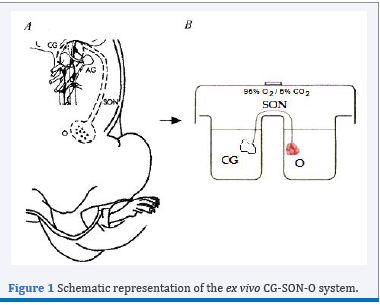
Figure 1 Schematic representation of the ex vivo CG-SON-O system.

Figure 1 Schematic representation of the ex vivo CG-SON-O system.

The neural signals represent additional control of ovarian function in addition to classical endocrine regulation [1]. One of the neural pathways that arrives at the ovary is the Superior Ovarian Nerve (SON), mainly constituted by adrenergic fibres, most of which originate in the coeliac ganglion GC [2]. To study the peripheral neural influence on the ovarian physiology, our laboratory has standardized an integrated ex vivo system known as CG-SON-O [3]. This system is a functional entity which viability has been supported by histological studies at the end of its incubation and it has an own autonomic tone, which allows the use of antagonist without the simultaneous addition of the agonists. This scheme has a singular value for the study of a dynamic gland with complex regulatory mechanisms such as the CL, since it allows preservation of innervations and paracrine and autocrine regulations without the humoral effect. To clarify, a schematic representation of the ex vivo CG-SON-O system may be observed in Figure 1.

The present review summarizes of available evidence of the impact of neural and hormonal agents on the CL of pregnancy through the CG-SON-O system in the rat, focusing on luteal regression.

Action of neural agents on the CL of pregnancy through the CG-SON-O system in the rat

The CG is part of the sympathetic prevertebral chain and it possesses mechanisms that regulate the flow of signals between the gonads and the central nervous system. This ganglion receives fibres of adrenergic nature which come from the medulla and others preaortic ganglia [4,5]. The symphatetic ganglionic neurons have α and β adrenergic receptors [6]. The presence of such receptors was demonstrated physiologically in adult rat during the oestrus cycle and pregnancy [3,7]. Precisely, it was found that in the second half of pregnancy, the addition of noradrenaline (NA) to the ganglionic incubation medium, using the CG-NOS-O system, stimulates the ovarian progesterone release on day 15 and 19. Such effect begins to fall on day 20 and is null at day 21. These data indicate that the steroidogenic response to neural action gradually decreases with the establishment of physiological luteal regression at the end of gestation. The simultaneous addition of LH to the incubation medium ovarian decreases the effect of NA, which reveals an antagonism between the neurotransmitter and the hormone. On the other hand, the presence of isoproterenol, β-adrenergic agonist in culture of isolated luteal cells, obtained from rats at day 15 of gestation, does not modify the release of progesterone [7]. Conversely, a recent study shows that both NA and isoproterenol increased progesterone production through activating β adrenergic receptors in murine cells from luteinized ovaries [8]. It is possible that the different origin of luteal cells may be responsible of disagreement in these results. Taking together the results in our study indicate that the integrity of the system, in which the ovarian and neural structures are preserved, is essential for the ovarian response to neural stimulation. Casais et al. [9], also analyzed the influence of the ganglionic noradrenergic stimulation on the ovarian release of androstenedione (A2 ), hormone with known luteotrophic effect in rat [10-12], and if consequently causes a modification in the antisteroidogenic effect of LH at the end of pregnancy. The study concludes that such stimulus in ganglion reinforces the inhibitory effect induced by LH on ovarian A2 release and that this mechanism of action, in turn, is associated with changes in the release of catecholamines in the ovary. It is possible that in an attempt to favor physiological luteal regression at the end of pregnancy in the rat, the signal coming from the CG may be a fine modulator together LH to limit the production of A2 .

In the sympathetic ganglia, acetylcholine is considered to be the classic preganglion neurotransmitter [13,14] and it possesses nicotinic and muscarinic cholinergic receptors in the ganglionic neurons called principal neurons as well as muscarinic receptors in other neuronal populations [15,16]. The stimulation of such receptors causes modifications in the steroids release in cyclic rats [17-19] and prepubertal rats [20].

Regarding the action of cholinergic agents during the pregnancy, stimulus of the CG with acetylcholine on day 15 of gestation decreases the release of ovarian progesterone and A2 . While on days 19, 20 and 21 of pregnancy, although the same effect remains on the release of androgen, no changes on the progesterone release were observed, indicating that the steroidogenic response to neural action gradually decreases with the establishment of physiological luteal regression. Thus, the ganglionic cholinergic action, mainly in relation to the release of A2 , would act favoring the luteal regression necessary for the delivery to occur [21].

Together, these data suggest that the peripheral neural system may act as a modulator of a dynamic structure as is the CL of pregnancy and in conditions that require a mechanism of fine-tuning such as the luteal regression.

Action of hormonal agents on the CL of pregnancy through the CG-SON-O system in the rat

Several studies support the effect of hormonal agents on the peripheral nervous system [22-25]. The sympathetic ganglia have fenestrated capillaries [26]. In addition, different hormone receptors have been demonstrated in ganglionic neurons [23,27- 30]. Thus, hormones or others bioactives substances may arrive to the ganglionic neurons and regulate the flow of signals through microvasculature.

A2 is the principal circulating androgen in the rat and possesses, in the ovary, a well-known luteotrophic action, which occurs by either its intraluteal conversion to estradiol or also by a direct effect [10,30,31].

Casais et al. [9], observed that in the CG-SON-O system extracted from animals on days 19 and 21 of pregnancy, with previous s.c. administration (48 h) of 10 mg A2 /0.2 ml oil vehicle, the levels of progesterone increased significantly in the ovarian compartment. These results support the idea that, in vivo, the humoral environment is highly influential on the functioning of the CG, thus affecting the ovary by a neural pathway. When the direct ganglionic effect of A2 was analysed in the CG-SON-O system, the results were highly surprising. The levels of progesterone in the ovarian compartment obtained by the application of A2 to the CG in relation to the controls were significantly higher. Thus, A2 can reverse the functional regression of the late pregnancy CL when administered systemically or when added directly to the ganglion compartment via SON.

Then, the mechanisms by which A2 exerts luteotrophic effect from CG were further studied by Vallcaneras et al. [28]. The A2 addition in CG increased 3β-HSD and decreased 20α-HSD activities in ovary, enzymes involved in progesterone synthesis and metabolism, respectively [32,33] and it increased ovarian nitric oxide (NO) release, neurotransmitter found in sympathetic ganglia with influence on ovary physiology [34-36]. We further demonstrated by immunohistochemistry cytoplasmatic androgen receptor immunoreactivity in neural somas in the CG and it was provided evidence for the luteotrophic action of A2 via a neural pathway that may be mediated by these receptors with the use of flutamide, an androgen receptor antagonist [37,31].

On day 4 postpartum, the CL of pregnancy is at advanced regression with reduced capacity to synthesize progesterone and high rate of apoptotic cell death [38]. In addition, expression of regulatory factors involved in cell survival and death such as Bcl-2, Bax and the Fas-FasL system have been reported in the CL regression [39-41]. A2 added in CG from rats on day 4 postpartum without lactation increased A2 and E2 release, while at the same time induced a decline in the expression of proapoptotic FasL. These luteotrophic effects caused by the presence of A2 in CG are associated with increased levels of NA and decreased levels of NO in the ovarian compartment. This suggests that the regressing CL of pregnancy present in postpartum ovary maintain certain responsiveness to stimuli of A2 from CG [42].

Other key hormone in regulating reproductive processes is E2 . During mid-pregnancy, E2 is required for luteal survival, while at the end of pregnancy, E2 levels increase in the ovarian vein and in the general circulation [43,44]. It has been proposed that such increase is responsible for increased expression of prostaglandin F2α receptor, luteolityc factor that induce 20α-HSD in association with CL functional regression [45]. Thus, E2 added directly to the ovarian incubation medium decreased progesterone release as well as 3β-HSD expression and the ratio of Bcl-2/Bax. While, E2 added to CG of CG-SON-O system also decreased progesterone ovarian release but without modification in luteal expression of progesterone metabolizing enzymes, Bcl-2 and Bax [46]. Anesetti et al. [23], demonstrated the presence of estrogen receptors in CG neurons of prepubertal rats, suggesting that the CG is likely a target of E2 . These results would demonstrate that the indirect luteotrophic action of A2 via the SON is an effect of the androgen per se, not mediated through its previous conversion into E2 . E2 either directly in ovary or indirectly from CG has an antiesteroidogenic effect at the end of pregnancy in the rat and this effect is more pronounced when E2 is directly added in ovary.

There are numerous studies that demonstrate the local survival effect of progesterone on the CL, stimulating its own secretion and protecting the CL from cellular death [33,47]. In addition, progesterone is able to perform luteotrophic effect through neural pathway. Ghersa et al. [29], demonstrated the presence of progesterone receptors in CG neurons and they observed that the progesterone addition in CG of CG-SON-O system increased the ovarian progesterone release and the 3β-HSD activity, whereas decreased the expression and activity of 20α-HSD. In addition, a decrease in the number of apoptotic nuclei and a decrease of the expression of FasL were observed. Progesterone may access the ganglionic neurons, bind to the specific receptors and induce release of neurotransmitters via the SON, which would modulate ovarian function.

It is known that prolactin, besides the known trophic effect on the CL, also has a luteolytic effect depending upon the nature of the CL and of the hormonal environment to which they are exposed. Just before parturition, there is a large secretion of prolactin from the pituitary needed to stimulate lactogenesis and to develop maternal behaviour required immediately after parturition [48- 50]. It has been postulated that circulating prolactin at the end of gestation has a role in the regression of the CL of pregnancy from CG through SON [51]. At the moment, according to the literature available, the presence of receptors for prolactin at the level of the peripheral sympathetic nervous system has not been confirmed. However, there is evidence suggesting that prolactin might act on sympathetic ganglions to affect different aspects of their metabolism [52]. The prolactin addition in CG of CG-SON-O system triggered an inhibitory effect on progesterone ovarian release with an increase in the progesterone catabolising capacity marked by the increase in 20α-HSD expression. We also found that prolactin from CG increased ovarian PGF2α release. These results are in agreement with the fact that in rodents a decline in progesterone levels is essential for luteal regression to occur and this is mediated by 20α-HSD, which is increased by PGF2α at the end of pregnancy [53]. Ganglionic prolactin also increased the levels of NO in the ovarian medium and it diminished the luteal expression of Bcl-2 (anti-apoptotic factor) and Bcl-2:Bax ratio, which tilts the balance towards a pro-apoptotic environment [54]. These findings indicate that prolactin may gain access to the CG impacting indirectly on ovarian physiology and inducing the release of PGF2α and NO, which may favour luteal regression at the end of rat pregnancy.

Then, the prolactin effect from CG on the functionality of the ovaries was analyzed on day 4 postpartum in two different endocrine environments: lactating vs. non-lactating animals [55]. Prolactin added in the CG of non-lactating rats increased ovarian progesterone release, which however did not reach the basal levels observed in lactating rats. In addition, prolactin from CG decreased the ratio of Bcl-2:Bax and increased release of ovarian PGF2α, NO and E2 . On the other hand, the ovarian gland in lactating rats is developed in an environment characterized by high levels of prolactin and may be unable to respond to further input of prolactin from the CG mediated via the SON. Goyeneche and Telleria [56] propose that prolactin has different effects on pregnancy CL depending upon the prevailing steroid concentrations. These authors observed in non-lactating postpartum rats receiving subcutaneous E2 that prolactin stimulated luteal apoptosis in the presence of high levels of progesterone. By contrast, during lactation prolactin in presence of high serum levels of progesterone prevents apoptosis in CL and in this physiological situation the E2 levels are low. Thus, our results indicate that prolactin added to the CG via the SON may favor the regression of luteal tissue in non-lactating animals. On the other hand, it is highly possible that in lactating rats the direct prolactin effect prevails over the prolactin effect from CG on the ovaries. Thus, the indirect prolactin effect through peripheral neural pathway may be operational in times of transition when the CL needs to be either abruptly interrupted or rescued from apoptosis

FINAL CONSIDERATIONS

In conclusion, the evidence from this review indicates that neural or endocrine signals may impact the CL of pregnancy through peripheral nervous system modulating its function. Therefore, these mechanisms may become important in disorders related to inadequate progesterone secretion by the CL that have been implicated as etiologic factors for recurrent pregnancy loss such as luteal phase deficiency, hyperprolactinemia, polycystic ovarian syndrome and psychoemotional stress.

ACKNOWLEDGEMENTS

We thank all those who have contributed in the works summarized in the present review.

REFERENCES

1. Gerendai I, Banczerowski P, Halász B. Functional significance of the innervation of the gonads. Endocrine. 2005; 28: 309-318.

2. Baljet B, Drukker J. The extrinsic innervation of the abdominal organs in the female rat. Acta Anat. 1979; 104: 243-267.

3. Sosa ZY, Casais M, Rastrilla AM, Aguado L. Adrenergic influences on coeliac ganglion affect the release of progesterone from cycling ovaries: characterisation of an in vitro system. J Endocrinol. 2000; 166: 307-318.

4. Messenger JP, Furness JB. Distribution of enteric nerve cells that project to the coeliac ganglion of the guinea-pig. Cell Tissue Res. 1992; 269: 119-132.

5. Quinson N, Robbins HL, Clark MJ, Furness JB. Locations and innervation of cell bodies of sympathetic neurons projecting to the gastrointestinal tract in the rat. Arch Histol Cytol. 2001; 64: 281-294.

6. Shivachar AC, Eikenburg DC. Differential effects of epinephrine and norepinephrine on cAMP response and g(i3)alpha protein expression in cultured sympathetic neurons. J Pharmacol Exp Ther. 1999; 291: 258-264.

7. Casais M, Sosa ZY, Rastrilla AM, Aguado LI. Coeliac ganglion adrenergic activity modifies ovarian progesterone during pregnancy: its inter-relationship with LH. J Endocrinol. 2001; 170: 575-584.

8. Wang J, Tang M, Jiang H, Wu B, Cai W, Hu Ch, et al. The role of adrenergic activation on murine luteal cell viability and progesterone production. Theriogenol. 2016; 86: 1182-1188.

9. Casais M, Delgado SM, Sosa Z, Rastrilla AM. Involvement of the coeliac ganglion in the luteotrophic effect of androstenedione in late pregnant rats. Reproduction. 2006; 131: 361-368.

10. Carrizo DG, Rastrilla AM, Telleria CM, Aguado LI. Androstenedione stimulates progesterone production in corpora lutea of pregnant rats: an effect not mediated by oestrogen. J of Steroid Biochem Mol Biol. 1994; 51: 191-197.

11. Goyeneche AA, Calvo V, Gibori G, Telleria CM. Androstenedione interferes in luteal regression by inhibiting apoptosis and stimulating progesterone production. Biol Reprod. 2002; 66: 1540-1547.

12. Telleria CM. Can luteal regression be reversed? Reprod Biol Endocrinol. 2006; 4: 53.

13. Klein CM, Burden HW. Anatomical localization of afferent and postganglionic sympathetic neurons innervating the rat ovary. Neurosci Lett. 1988; 85: 217-222.

14. Berthoud HR, Powley TL. Interaction between parasympathetic and sympathetic nerves in prevertebral ganglia: morphological evidence for vagal efferent innervation of ganglion cells in the rat. Microsc Res Tech. 1996; 35: 80-86.

15. Reid SG, Perry SF. Cholinoceptor-mediated control of catecholamine release from chromaffin cells in the American eel, Anguilla rostrata. J Comp Physiol B. 1995; 165: 464-470.

16. Prud´Homme MJ, Houdeau E, Serghini R, Tillet Y, Schemann M, Rousseau JP. Small intensely fluorescent cells of the rat paracervical ganglion synthesize adrenaline, receive afferent innervation from postganglionic cholinergic neurones, and contain muscarinic receptors. Brain Research. 1999; 821:141-149.

17. Sosa Z, Delgado M, Casais M, Aguado L, Rastrilla AM. Release of ovarian progesterone during the rat oestrous cycle by ganglionic cholinergic influence: the role of norepinephrine. J Steroid Biochem Mol Biol. 2004; 91: 179-184.

18. Daneri C, Orozco AV, Bronzi D, Mohn C, Rastrilla AM, Sosa ZY. Involvement of the ganglion cholinergic receptors in gonadotropinreleasing hormone, catecholamines, and progesterone release in the rat ovary. Fertil Steril. 2013; 99: 2062-2070.

19. Delgado SM, Escudero CG, Casais M, Gordillo M, Anzulovich AC, Sosa Z, et al. Ovaric physiology in the first oestral cycle: influence of noradrenergic and cholinergic neural stimuli from coeliac ganglion. Steroids. 2010; 75: 685-694.

20. Delgado SM, Sosa Z, Dominguez NS, Casais M, Aguado L, Rastrilla AM. Effect of the relation between neural cholinergic action and nitric oxide on ovarian steroidogenesis in prepubertal rats. J Steroid Biochem Mol Biol. 2004; 91: 139-145.

21. Casais M, Delgado SM, Sosa Z, Rastrilla AM. Pregnancy in rats is modulated by ganglionic cholinergic action. Reproduction. 2006; 131: 1151-1158.

22. Shinohara Y, Matsumoto A, Hayashi S, Mori T. Prenatal exposure to diethylstilbestrol decreases the number of estrogen receptor alpha-containing neurons innervating the ovary in rat celiac ganglion. Neuroscience. 2000; 101: 779-783.

23. Anesetti G, Lombide P, Chávez-Genaro R. Prepubertal estrogen exposure modifies neurotrophin receptor expression in celiac neurons and alters ovarian innervation. Auton Neurosci. 2009; 145: 35-43.

24. Jana B, Rytel L, Czarzasta J, Ca?ka J. Reduction of the number of neurons in the caudal mesenteric ganglion innervating the ovary in sexually mature gilts following testosterone administration. J Neuroendocrinol. 2013; 25: 826-838.

25. Jana B, Palus K, Meller K, Ca?ka J. Porcine dorsal root ganglia ovarian neurons are affected by long lasting testosterone treatment. Physiol Res. 2016; 65: 1019-1030.

26. Tanaka K, Chiba T. Microvascular organization of sympathetic ganglia, with special reference to small intensely-fluorescent cells. Microsc Res Tech. 1996; 35: 137-145.

27. Vega Orozco A, Daneri C, Anesetti G, Cabrera R, Sosa Z, Rastrilla AM. Involvement of the oestrogenic receptors in superior mesenteric ganglion on the ovarian steroidogenesis in rat. Reproduction. 2012; 143: 183-193.

28. Vallcaneras SS, Casais M, Delgado SM, Filippa V, Mohamed F, Sosa Z, et al. Androgen receptors in coeliac ganglion in late pregnant rat. Steroids. 2009; 74: 526-534.

29. Ghersa F, Burdisso J, Vallcaneras SS, Fuentes F, de la Vega M, Delgado SM, et al. Neuromodulation of the luteal regression: presence of progesterone receptors in coeliac ganglion. Exp Physiol. 2015; 100: 935-946.

30. Gibori G, Khan I, Warshaw ML, McLean MP, Puryear TK, Nelson S, et al. Placental-derived regulators and the complex control of luteal cell function. Recent Prog Horm Res. 1988; 44: 377-429.

31. Grzesiak M, Knapczyk-Stwora K, Ciereszko RE, Wieciech I, Slomczynska M. Alterations in luteal production of androstenedione, testosterone, and estrone, but not estradiol, during mid- and late pregnancy in pigs: effects of androgen deficiency. Theriogenol. 2014; 82: 720-733.

32. Bowen-Shauver JM, Gibori G. The corpus luteum of pregnancy. In: Leung PCK, dashi EY, editors. The ovary. San Diego: Elsevier Academic Press; 2003; 201-230.

33. Stocco C, Telleria C, Gibori G. The molecular control of corpus luteum formation, function and regression. Endocrine Reviews. 2007; 28: 117-149.

34. Morales MA, Holmberg K, Xu ZQ, Cozzari C, Hartman BK, Emson P, et al. Localization of choline acetyltransferase in rat peripheral pympathetic neurons and coexistence with nitric oxide syntase and neuropeptides. PNAS. 1995; 92: 819-825.

35. Casais M, Delgado SM, Vallcaneras S, Sosa Z, Rastrilla AM. Nitric oxide in prepubertal rat ovary: contribution of the ganglionic nitric oxide synthase system via superior ovarian nerve. Neuroendocrinol Letters. 2007; 3: 9-44.

36. Delsouc MB, Morales LD, Vallcaneras SS, Bronzi D, Anzulovich AC, Delgado SM, et al. Participation of the extrinsic cholinergic innervation in the action of nitric oxide on the ovarian steroidogenesis in the first proestrous in rats. Gen Comp Endocrinol. 2016; 236: 54-62.

37. Kemppainen JA, Lane MV, Sar M, Wilson EM. Androgen receptor phosphorylation, turnover, nuclear transport, and transcriptional activation. Specificity for steroids and antihormones. J Biol Chem. 1992; 267: 968-974.
38. Peluffo MC, Stouffer RL, Tesone M. Activity and expression of different members of the caspase family in the rat corpus luteum during pregnancy and postpartum. Am J Physiol Endocrinol Metab. 2007; 293: E1215-1223.

39. Sugino N, Okuda K. Species-related differences in the mechanism of apoptosis during structural luteolysis. J Reprod Dev. 2007; 53: 977- 986.

40. Yang Y, Sun M, Shan Y, Zheng X, Ma H, Ma W, et al. Endoplasmic reticulum stress-mediated apoptotic pathway is involved in corpus luteum regression in rats. Reprod Sci. 2015; 22: 572-884.

41. Satoh H, Yoshio H, Kawaminami M, Kurusu S. Type-dependent differences in Fas expression and phagocytes distribution in rat corpora lutea during natural regression: an immunohistochemical evidence. J Vet Med Sci. 2016; 78: 1771-1777.

42. Vallcaneras SS, Casais M, Anzulovich AC, Delgado SM, Sosa Z, Telleria CM, et al. Androstenedione acts on the coeliac ganglion and modulates luteal function via the superior ovarian nerve in the postpartum rat. J Steroid Biochem Mol Biol. 2011; 125: 243-250.

43. Shaikh AA. Estrone and estradiol levels in the ovarian venous blood from rats during the estrous cycle and pregnancy. Biol Reprod. 1971; 5: 297-307.

44. Tripathy S, Asaithambi K, Jayaram P, Medhamurthy R. Analysis of 17β-estradiol (E2) role in the regulation of corpus luteum function in pregnant rats: Involvement of IGFBP5 in the E2-mediated actions. Reprod Biol Endocrinol. 2016; 14: 19.

45. Bussmann LE. Prostaglandin F-2 alpha receptors in corpora lutea of pregnant rats and relationship with induction of 20 alpha-hydroxysteroid dehydrogenase. J Reprod Fertil. 1989; 85: 331-341.

46. Casais M, Vallcaneras SS, Campo Verde Arbocco F, Delgado SM, Hapon MB, Sosa Z, et al. Estradiol promotes luteal regression through a direct effect on the ovary and an indirect effect from the celiac ganglion via the superior ovarian nerve. Reprod Sci. 2012; 19: 416-422.

47. Accialini P, Hernández SF, Bas D, Pazos MC, Irusta G, Abramovich D, et al. A link between Notch and progesterone maintains the functionality of the rat corpus luteum. Reproduction. 2015; 149: 1-10.

48. Amenomori Y, Chen CL, Meites J. Serum prolactin levels in rats during different reproductive states. Endocrinol. 1970; 86: 506-510.

49. Grattan DR, Averill RL. Effect of ovarian steroids on a nocturnal surge of prolactin secretion that precedes parturition in the rat. Endocrinology. 1990; 126: 1199-1205.

50. Stocco C. The long and short of the prolactin receptor: the corpus luteum needs them both! Biol Reprod. 2012; 86: 85.

51. Vallcaneras SS, de la Vega M, Delgado SM, Motta A, Telleria C, Rastrilla AM, et al. Prolactin modulates luteal regression from the coeliac ganglion via the superior ovarian nerve in the late-pregnant rat. Reprod Fertil Dev. 2016; 28: 565-573.

52. Gejman PV, Cardinali DP. Hormone effects on muscarinic cholinergic binding in bovine and rat sympathetic superior cervical ganglia. Life Sci. 1983; 32: 965-972.

53. Stocco CO, Zhong L, Sugimoto Y, Ichikawa A, Lau LF, Gibori G. Prostaglandin F2alpha-induced expression of 20alpha-hydroxysteroid dehydrogenase involves the transcription factor NUR77. J Biol Chem. 2000; 275: 37202-37211.

54. Antonsson B. Bax and other pro-apoptotic Bcl-2 family “killerproteins” and their victim the mitochondrion. Cell Tissue Res. 2001; 306: 347-361.

55. Vallcaneras SS, Delgado SM, Motta A, Telleria CM, Rastrilla AM, Casais M. Effect of prolactin acting on the coeliac ganglion via the superior ovarian nerve on ovarian function in the postpartum lactating and non-lactating rat. Gen Comp Endocrinol. 2013; 184: 1-8.

56. Goyeneche AA, Telleria CM. Exogenous estradiol enhances apoptosis in regressing post-partum rat corpora lutea possibly mediated by prolactin. Reprod Biol Endocrinol. 2005; 3: 40.

Vallcaneras S, Casais M (2017) Peripheral Neural Regulation of Pregnancy Corpus Luteum. JSM Invitro Fertil 2(3): 1021.

Received : 11 Sep 2017
Accepted : 05 Oct 2017
Published : 07 Oct 2017
Journals
Annals of Otolaryngology and Rhinology
ISSN : 2379-948X
Launched : 2014
JSM Schizophrenia
Launched : 2016
Journal of Nausea
Launched : 2020
JSM Internal Medicine
Launched : 2016
JSM Hepatitis
Launched : 2016
JSM Oro Facial Surgeries
ISSN : 2578-3211
Launched : 2016
Journal of Human Nutrition and Food Science
ISSN : 2333-6706
Launched : 2013
JSM Regenerative Medicine and Bioengineering
ISSN : 2379-0490
Launched : 2013
JSM Spine
ISSN : 2578-3181
Launched : 2016
Archives of Palliative Care
ISSN : 2573-1165
Launched : 2016
JSM Nutritional Disorders
ISSN : 2578-3203
Launched : 2017
Annals of Neurodegenerative Disorders
ISSN : 2476-2032
Launched : 2016
Journal of Fever
ISSN : 2641-7782
Launched : 2017
JSM Bone Marrow Research
ISSN : 2578-3351
Launched : 2016
JSM Mathematics and Statistics
ISSN : 2578-3173
Launched : 2014
Journal of Autoimmunity and Research
ISSN : 2573-1173
Launched : 2014
JSM Arthritis
ISSN : 2475-9155
Launched : 2016
JSM Head and Neck Cancer-Cases and Reviews
ISSN : 2573-1610
Launched : 2016
JSM General Surgery Cases and Images
ISSN : 2573-1564
Launched : 2016
JSM Anatomy and Physiology
ISSN : 2573-1262
Launched : 2016
JSM Dental Surgery
ISSN : 2573-1548
Launched : 2016
Annals of Emergency Surgery
ISSN : 2573-1017
Launched : 2016
Annals of Mens Health and Wellness
ISSN : 2641-7707
Launched : 2017
Journal of Preventive Medicine and Health Care
ISSN : 2576-0084
Launched : 2018
Journal of Chronic Diseases and Management
ISSN : 2573-1300
Launched : 2016
Annals of Vaccines and Immunization
ISSN : 2378-9379
Launched : 2014
JSM Heart Surgery Cases and Images
ISSN : 2578-3157
Launched : 2016
Annals of Reproductive Medicine and Treatment
ISSN : 2573-1092
Launched : 2016
JSM Brain Science
ISSN : 2573-1289
Launched : 2016
JSM Biomarkers
ISSN : 2578-3815
Launched : 2014
JSM Biology
ISSN : 2475-9392
Launched : 2016
Archives of Stem Cell and Research
ISSN : 2578-3580
Launched : 2014
Annals of Clinical and Medical Microbiology
ISSN : 2578-3629
Launched : 2014
JSM Pediatric Surgery
ISSN : 2578-3149
Launched : 2017
Journal of Memory Disorder and Rehabilitation
ISSN : 2578-319X
Launched : 2016
JSM Tropical Medicine and Research
ISSN : 2578-3165
Launched : 2016
JSM Head and Face Medicine
ISSN : 2578-3793
Launched : 2016
JSM Cardiothoracic Surgery
ISSN : 2573-1297
Launched : 2016
JSM Bone and Joint Diseases
ISSN : 2578-3351
Launched : 2017
JSM Bioavailability and Bioequivalence
ISSN : 2641-7812
Launched : 2017
JSM Atherosclerosis
ISSN : 2573-1270
Launched : 2016
Journal of Genitourinary Disorders
ISSN : 2641-7790
Launched : 2017
Journal of Fractures and Sprains
ISSN : 2578-3831
Launched : 2016
Journal of Autism and Epilepsy
ISSN : 2641-7774
Launched : 2016
Annals of Marine Biology and Research
ISSN : 2573-105X
Launched : 2014
JSM Health Education & Primary Health Care
ISSN : 2578-3777
Launched : 2016
JSM Communication Disorders
ISSN : 2578-3807
Launched : 2016
Annals of Musculoskeletal Disorders
ISSN : 2578-3599
Launched : 2016
Annals of Virology and Research
ISSN : 2573-1122
Launched : 2014
JSM Renal Medicine
ISSN : 2573-1637
Launched : 2016
Journal of Muscle Health
ISSN : 2578-3823
Launched : 2016
JSM Genetics and Genomics
ISSN : 2334-1823
Launched : 2013
JSM Anxiety and Depression
ISSN : 2475-9139
Launched : 2016
Clinical Journal of Heart Diseases
ISSN : 2641-7766
Launched : 2016
Annals of Medicinal Chemistry and Research
ISSN : 2378-9336
Launched : 2014
JSM Pain and Management
ISSN : 2578-3378
Launched : 2016
JSM Women's Health
ISSN : 2578-3696
Launched : 2016
Clinical Research in HIV or AIDS
ISSN : 2374-0094
Launched : 2013
Journal of Endocrinology, Diabetes and Obesity
ISSN : 2333-6692
Launched : 2013
Journal of Substance Abuse and Alcoholism
ISSN : 2373-9363
Launched : 2013
JSM Neurosurgery and Spine
ISSN : 2373-9479
Launched : 2013
Journal of Liver and Clinical Research
ISSN : 2379-0830
Launched : 2014
Journal of Drug Design and Research
ISSN : 2379-089X
Launched : 2014
JSM Clinical Oncology and Research
ISSN : 2373-938X
Launched : 2013
JSM Bioinformatics, Genomics and Proteomics
ISSN : 2576-1102
Launched : 2014
JSM Chemistry
ISSN : 2334-1831
Launched : 2013
Journal of Trauma and Care
ISSN : 2573-1246
Launched : 2014
JSM Surgical Oncology and Research
ISSN : 2578-3688
Launched : 2016
Annals of Food Processing and Preservation
ISSN : 2573-1033
Launched : 2016
Journal of Radiology and Radiation Therapy
ISSN : 2333-7095
Launched : 2013
JSM Physical Medicine and Rehabilitation
ISSN : 2578-3572
Launched : 2016
Annals of Clinical Pathology
ISSN : 2373-9282
Launched : 2013
Annals of Cardiovascular Diseases
ISSN : 2641-7731
Launched : 2016
Journal of Behavior
ISSN : 2576-0076
Launched : 2016
Annals of Clinical and Experimental Metabolism
ISSN : 2572-2492
Launched : 2016
Clinical Research in Infectious Diseases
ISSN : 2379-0636
Launched : 2013
JSM Microbiology
ISSN : 2333-6455
Launched : 2013
Journal of Urology and Research
ISSN : 2379-951X
Launched : 2014
Journal of Family Medicine and Community Health
ISSN : 2379-0547
Launched : 2013
Annals of Pregnancy and Care
ISSN : 2578-336X
Launched : 2017
JSM Cell and Developmental Biology
ISSN : 2379-061X
Launched : 2013
Annals of Aquaculture and Research
ISSN : 2379-0881
Launched : 2014
Clinical Research in Pulmonology
ISSN : 2333-6625
Launched : 2013
Journal of Immunology and Clinical Research
ISSN : 2333-6714
Launched : 2013
Annals of Forensic Research and Analysis
ISSN : 2378-9476
Launched : 2014
JSM Biochemistry and Molecular Biology
ISSN : 2333-7109
Launched : 2013
Annals of Breast Cancer Research
ISSN : 2641-7685
Launched : 2016
Annals of Gerontology and Geriatric Research
ISSN : 2378-9409
Launched : 2014
Journal of Sleep Medicine and Disorders
ISSN : 2379-0822
Launched : 2014
JSM Burns and Trauma
ISSN : 2475-9406
Launched : 2016
Chemical Engineering and Process Techniques
ISSN : 2333-6633
Launched : 2013
Annals of Clinical Cytology and Pathology
ISSN : 2475-9430
Launched : 2014
JSM Allergy and Asthma
ISSN : 2573-1254
Launched : 2016
Journal of Neurological Disorders and Stroke
ISSN : 2334-2307
Launched : 2013
Annals of Sports Medicine and Research
ISSN : 2379-0571
Launched : 2014
JSM Sexual Medicine
ISSN : 2578-3718
Launched : 2016
Annals of Vascular Medicine and Research
ISSN : 2378-9344
Launched : 2014
JSM Biotechnology and Biomedical Engineering
ISSN : 2333-7117
Launched : 2013
Journal of Hematology and Transfusion
ISSN : 2333-6684
Launched : 2013
JSM Environmental Science and Ecology
ISSN : 2333-7141
Launched : 2013
Journal of Cardiology and Clinical Research
ISSN : 2333-6676
Launched : 2013
JSM Nanotechnology and Nanomedicine
ISSN : 2334-1815
Launched : 2013
Journal of Ear, Nose and Throat Disorders
ISSN : 2475-9473
Launched : 2016
JSM Ophthalmology
ISSN : 2333-6447
Launched : 2013
Journal of Pharmacology and Clinical Toxicology
ISSN : 2333-7079
Launched : 2013
Annals of Psychiatry and Mental Health
ISSN : 2374-0124
Launched : 2013
Medical Journal of Obstetrics and Gynecology
ISSN : 2333-6439
Launched : 2013
Annals of Pediatrics and Child Health
ISSN : 2373-9312
Launched : 2013
JSM Clinical Pharmaceutics
ISSN : 2379-9498
Launched : 2014
JSM Foot and Ankle
ISSN : 2475-9112
Launched : 2016
JSM Alzheimer's Disease and Related Dementia
ISSN : 2378-9565
Launched : 2014
Journal of Addiction Medicine and Therapy
ISSN : 2333-665X
Launched : 2013
Journal of Veterinary Medicine and Research
ISSN : 2378-931X
Launched : 2013
Annals of Public Health and Research
ISSN : 2378-9328
Launched : 2014
Annals of Orthopedics and Rheumatology
ISSN : 2373-9290
Launched : 2013
Journal of Clinical Nephrology and Research
ISSN : 2379-0652
Launched : 2014
Annals of Community Medicine and Practice
ISSN : 2475-9465
Launched : 2014
Annals of Biometrics and Biostatistics
ISSN : 2374-0116
Launched : 2013
JSM Clinical Case Reports
ISSN : 2373-9819
Launched : 2013
Journal of Cancer Biology and Research
ISSN : 2373-9436
Launched : 2013
Journal of Surgery and Transplantation Science
ISSN : 2379-0911
Launched : 2013
Journal of Dermatology and Clinical Research
ISSN : 2373-9371
Launched : 2013
JSM Gastroenterology and Hepatology
ISSN : 2373-9487
Launched : 2013
Annals of Nursing and Practice
ISSN : 2379-9501
Launched : 2014
JSM Dentistry
ISSN : 2333-7133
Launched : 2013
Author Information X