Histological Basis in the Study of Cyprinus Carpio Sp. Operculum Bone Scaffold as a Novel Scaffold Material for Cranio-Maxillofacial Defects
- 1. Department of Ophthalmology, Universitas Padjadjaran/Cicendo National Eye Hospital, Indonesia
- 2. Department of Ophthalmology, Royal Hobart Hospital, Australia
- 3. National Nuclear Energy Agency of Indonesia, Indonesia
- 4. Department of Anatomy, Physiology and Cell Biology, Universitas Padjadjaran, Indonesia
- 5. Department of Biochemistry, Universitas Padjadjaran, Indonesia
- 6. Department of Oral and Maxillofacial Surgery, Universitas Padjadjaran, Indonesia
- 7. Department of Chemistry, Universitas Padjadjaran, Indonesia
Abstract
Introduction: Carp operculum (Cyprinus carpio sp.) is being proposed as a candidate for xenograft. In this animal study using rats with bony defects, the histological response of osteoconduction with carp operculum grafts are compared with standard bovine graft.
Methodology: Carp operculum bone and bovine ribs were implanted on calvarial bone of white male rats (Rattus novergicus). After 15 days, the calvarian bone was harvested and histologic preparations were made. Foreign-body giant cells, osteoblasts and polymorphonuclear cells were observed for under a light microscope.
Results: Foreign-body giant cells, osteoblasts and polymorphonuclear cells were qualitatively noted to be of similar response in both carp operculum and bovine graft groups.
Conclusion: Histological study of carp operculum bone xenograft implant showed osteoconductive ability and similar healing process as the standard bovine xenograft implant. Further quantitative studies and evaluation of other aspects of the carp operculum as a viable xenograft material need to be carried out.
Keywords
• Xenograft
• Operculum
• Bovine
• Fractures
• Facial fractures
Citation
Kartiwa A, Yang V, Abbas B, Pandansari P, Atik N (2020) Histological Basis in the Study of Cyprinus Carpio Sp. Operculum Bone Scaffold as a Novel Scaffold Material for Cranio-Maxillofacial Defects. JSM Oro Facial Surg 4(1): 1012
INTRODUCTION
Cranio-maxillofacial bone defects result commonly from congenital defects, periodontal disease, trauma and surgical excision or cranioplasty. Anatomical considerations in this part of the body include the complex three-dimensional structure, and the neurovascular bundle that is closely related to it, as well as the aesthetic outcome [1]. Physiological functions in the craniofacial region would require the grafts to protect internal organs and be of suitable mechanical strength for mastication and other movements [2].
A bone scaffold is essential for the tissue engineering process. The bone scaffold provides a surface to stimulate bone healing through the process of osteogenesis, osteoinduction, osteoconduction, osteovascularization, osteoincorporation and osteomineralization [3].
The graft options broadly include autografts, allografts and xenografts. Xenografts have been widely studied and utilised in Orthopaedic surgery and are commonly bovine or porcine [4].
In this study, we investigate and compare Cyprinus carpio sp. Operculum with bovine ribs as an implant material in an animal study. Specifically, white male rats (Rattus novergicus) with calvarial fractures were used and the number of new osteoblasts and foreign-body giant cells were studied histologically in each group.
MATERIALS AND METHODS
The study had been approved by the Research Ethics Committee of the Faculty of Medicine, Universitas Padjadjaran.
This was an animal experimental study of bone fracture model of white male rats (Rattus novergicus), of the Wistar strain, which were treated with onlay graft implantation on the calvaria bone either by bovine graft or operculum bone.
The bone grafts of 500 grams were harvested from the operculum bone of the carpio sp and processed. Bovine ribs of 500 grams were obtained and underwent the same processing methods. The process included deproteinization, demineralization, lyophilization and sterilization at a radiation at a dose of 25 kGy. The process has been previously validated [5].
Histological preparations of the only graft of carp operculum bone and bovine ribs on the calvaria bone of rats were each stained with Haematoxylin Eosin (H&E) looking to identify osteoblast cells, polymorphonuclear cells and foreign-body giant cells of in the area of implantation.
RESULTS AND DISCUSSION
Results
The analysis of histologic study from both implants by H&E staining using a light microscope with 4x and 40x magnification were obtained.
In the first group, the rat model with the fracture was implanted with carp operculum bone graft for 15 days (Figure 1).
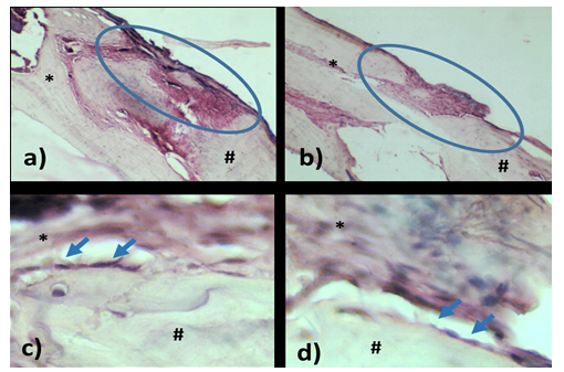
Figure 1: Inflammatory reaction and osteoblast formation in rat fracture model implanted with carp operculum bone graft (*graft, # host).
The rats’ bones were stained with H&E. Microscopic images of 4x magnification from two independent animal subjects (a and b) showed inflammatory reaction at insertion area of bone plate after injury (blue ellipse). Microscopic images of 40x magnification from two independent animal subjects (c and d) showed osteoblast formation in donor area (blue arrows).
In the second group, the rat model with the fracture was implanted with standard bovine ribs for 15 days (Figure 2).
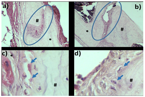
Figure 2: Inflammatory reaction and osteoblast formation in rat fracture model implanted with bovine ribs graft (*graft, # host).
Similarly, the tissues were prepared with H&E staining. Microscopic image of 4x magnification from two independent animal subjects (a and b) showed inflammatory reaction at insertion area of bone plate after injury (blue ellipse). Microscopic image of 40x magnification from two independent animal subjects (c and d) showed osteoblast formation in donor area (blue arrows).
Macroscopic and microscopically, the fractures in the animal models healed equally well with the use of carp operculum or bovine.
Microscopically, we see qualitative evidence of osteoblasts, polymorphonuclear cells and foreign-body giant cells in both groups of carp operculum and bovine. However, a statistical analysis was not carried out in this preliminary study.
DISCUSSION
In fracture healing, a bony scaffold is the matrix that stimulates the attachment and proliferation of cells responsible for osteoblastic properties. Factors considered in choosing and designing bone scaffolds include: 1) bio-compatibility of the material to induce inflammatory reactions; 2) bio-compatibility of the material with no toxicity; 3) mechanical properties especially in weight-bearing bones; 4) suitable architecture for viability e.g. porosity for nutrient exchange [6].
From previous studies conducted by the group studying the use of Cyprinus carpio sp. operculum bone as a graft option, Cyprinus carpio sp. operculum is biochemically supportive of the fracture healing process [7] and has sufficient porosity that would enable diffusion and angiogenesis which are both important for tissue healing and viability.
In this study, we specifically are looking for the preliminary histological basis to further the prospects of carp operculum as a xenograft candidate. From this study, histologically, operculum bone has shown similar osteoblasts, polymorphonuclear and foreign-body giant cells in a similar fashion to the standard of bovine grafts, at least in rat models. However, for carp operculum to be further considered as a xenograft implant, specific studies to quantify the histological osteoconductive response. The potential toxicities especially in the long-term need to be carried out.
CONCLUSION
Histological evidence of carp operculum bone xenograft implant showed osteoconductive ability and similar ability to improve bone healing process as the bovine xenograft implant. Further studies need to be conducted to statistically compared the eligibility of carp operculum bone as a promising xenograft implant in orbital floor fracture.
ACKNOWLEDGEMENTS
Acknowledgements to the anatomical laboratory technicians of Universitas Padjadjaran for their guidance and technical expertise.


