A Case of Cutaneous Diffuse Large B-Cell Lymphoma and Concurrent Follicular Lymphoma In-Situ
- 1. Department of Dermatology, MD Anderson Cancer Center, Houston, Texas, USA
- 2. Texas College of Osteopathic Medicine, University of North Texas Health Science Center, Fort Worth, Texas, USA
Abstract
Diffuse large B-cell lymphoma and follicular lymphoma are the most common non-Hodgkin lymphomas seen in adults. Follicular lymphomas are derived from germinal centers of lymphatic tissue and are composed of centrocytes and centroblasts arranged into secondary lymphoid follicles with a germinal center. Follicular lymphomas can transform into diffuse large B-cell lymphoma. Follicular lymphoma in situ has been identified as a risk factor for several B-cell neoplasms, including diffuse large B-cell lymphoma. We report a case of a 62 year old man with diffuse large B-cell lymphoma on his scalp, germinal center phenotype, who was incidentally found to have a nodal follicular lymphoma in situ on his chest wall. This case highlights the clinical implications of in situ follicular lymphoma.
Keywords
Non-Hodgkin, B-cell neoplasm, Germinal center, FLIS
Citation
Wang C, Tran C, Duvic M (2015) A Case of Cutaneous Diffuse Large B-Cell Lymphoma and Concurrent Follicular Lymphoma In-situ J Dermatolog Clin Res 3(1): 1038.
ABBREVIATIONS
DLBCL: Diffuse Large B-cell Lymphoma; NHL: Non-Hodgkin’s Lymphoma; FL: Follicular Lymphoma; R-CHOP: Rituximab, Cyclophosphamide, Hydroxydaunorubicin, Vincristine, Prednisone; FLIS: Follicular Lymphoma In Situ; LDH: Lactate Dehydrogenase; PET: Positron Emission Tomography; CT: Computed Tomography; FDG: Flu Deoxyglucose; PCLBCL LT: Primary Cutaneous Diffuse Large B-cell Lymphoma Leg Type
INTRODUCTION
Diffuse large B-cell lymphoma (DLBCL) makes up 30%-40% of non-Hodgkin lymphoma (NHL) diagnoses making it the most common NHL in adults. Incidence of DLBCL peaks in the sixth and seventh decades [1]. DLBCL can arise de novo or transform from indolent lymphomas like chronic lymphocytic leukemia/small lymphocytic lymphoma, marginal zone lymphoma, and follicular lymphoma [1]. Histologically, DLBCL is composed of large cells with nuclei that are two times the size of a normal lymphocyte, and in the most common form, the tumor cells grow diffusely, affecting the normal organ structure [1]. The standard treatment for DLBCL patients is rituximab (anti-CD20 monoclonal antibody) in addition to cyclophosphamide, hydroxydaunorubicin, vincristine, and prednisone (R-CHOP) [1,2].
Follicular lymphoma (FL) is an indolent lymphoma which arises from normal germinal center B-cells and is the second most common NHL in the United States and Western Europe [3,4]. The median age at diagnosis is 60 years [3,4]. Eighty five percent of patients have at (14:18) translocation resulting in the over expression of BCL-2 protein which normally functions as an inhibitor of apoptosis [3]. The hallmark on pathology is the presence of centrocytes and their immature precursors, centroblasts, which mimic arrangement of secondary lymphoid follicles with a germinal center [3]. An increased number of centroblasts is related to greater clinical aggressiveness [3]. Follicular lymphoma can be managed conservatively with observation provided the patient is asymptomatic with no cytopenias. For patients who need treatment, R-CHOP can be used [3]. Follicular lymphoma in situ (FLIS), on the other hand, has uncertain clinical significance because a significant proportion of affected patients do not develop overt FL [5].
We report a unique case of cutaneous DLBCL with an incidentally discovered follicular lymphoma in situ arising concurrently in a 62 year old man, raising the possibility that the DLBCL transformed from the FLIS.
CASE PRESENTATION
A 62 year old male initially noticed an enlarging nodule on his posterior scalp over the course of one year. An initial biopsy of the scalp nodule showed DLBCL prompting referral to our institution. Review of systems was negative for B-symptoms. There was no palpable supraclavicular, cervical, axillary, or inguinal lymphadenopathy on physical exam. The skin exam was significant for a 2.5cm x 2.5cm x 0.5cm violaceous nodule on the left posterior scalp (Figure 1).
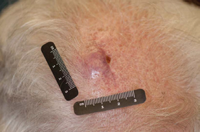
Figure 1 Cutaneous diffuse large B-cell lymphoma presenting as a2.5cm x 2.5cm x 0.5cm violaceous nodule on the left posterior scalp.
Laboratory testing revealed normal lactate dehydrogenase (LDH), beta-2 micro globulin levels, and complete blood count. A detailed immune phenotypic examination of the patient’s scalp biopsy revealed numerous large atypical cells that were positive for CD20, CD19, CD10, BCL2, and BCL-6 and negative for CD5 and CD30 and MUM-1. The ki-67 proliferation rate was 50-60%. These findings support the diagnosis of diffuse large B-cell lymphoma with germinal center B-cell immune phenotype and immunoblastic morphology. CD21 stain showed rare, intact, minute follicular dendritic mesh works. Analysis for a clonal IgH rearrangement was not performed.
Subsequent staging positron emission tomography-computed tomography (PET/CT) scan showed additional flu deoxyglucose (FDG) uptake on the deep right chest wall. Computed tomography (CT) of the chest with contrast showed an enlarged right sub pectoral node (36mm x 16mm) and a right axillary node (19mm x 16mm). A core needle biopsy of the right anterior chest wall lymph node revealed follicular B-cell lymphoma in situ, characterized by atypical B-cells colonizing pre-existing follicles. The tumor cells were positive for CD10, CD19, CD20, CD200, BCL2, and BCL-6. The right axillary lymph node was not biopsied nor was it seen on the initial PET-CT scan. The patient received 6 cycles of R-CHOP, and a restaging PET/CT of the chest 5 months later showed no FDG-avid lesions in the chest or axilla. He is in complete remission at follow-up one year later.
DISCUSSION
Our patient presented with a clinically apparent primary cutaneous DLBCL and was incidentally found to have a nodal FLIS on staging. Primary cutaneous DLBCL is uncommon though not rare. Castillo et al found that in 68.4% of 25,992 patients with DLBCL, the primary site of diagnosis of DLBCL was the lymph nodes [6]. Of the 8,204 cases which occurred in extra nodal sites, 11% of cases were in the skin and soft tissue. PET-CT is a wellestablished method of staging and response-assessment for both Hodgkin and non-Hodgkin lymphomas. Large studies have seen FDG avidity in >95% of patients presenting with FL [7]. The frequency of FDG avidity in FLIS is unknown.
Primary cutaneous B-cell lymphomas are divided into three main groups: marginal zone lymphoma, follicle center cell lymphoma and diffuse large B-cell lymphoma [8]. Leg type primary cutaneous diffuse large B-cell lymphoma (DLBCL LT) is a subset of primary cutaneous DLBCLs that is characterized by high BCL-2 expression and MUM-1 positivity. Clinically, it exhibits a propensity to appear on the lower extremities and is associated with an aggressive course [9]. Our patient’s cutaneous DLBCL is not consistent with DLBCL LT as it does not express MUM-1, classifying it as DLBCL, other.
nterestingly, the patient’s DLBCL has histologic features that raise the possibility that it transformed from the FLIS on his chest wall. Follicular lymphoma transformingto DLBCL is a frequently reported phenomenon. A study of 281 cases of follicular lymphoma showed that 37 (13%) eventually transformed into diffuse large B-cell lymphoma [11]. In an analysis done by Maeshima et al, it was shown that Bcl-2 and Bcl-6 expression was retained in most situations of DLBCLs that transformed from FLs, which is the case with our patient [11]. Also, the DLBCL was of germinal center B-cell immune phenotype, and in a study done by Davies et al, 89% of follicular lymphomas that transformed had this phenotype [12].
The germinal center B-cell immune phenotype and the presence of intact follicular mesh works in the DLBCL together raise the possibility that the DLBCL arose from follicular lymphoma. However the patient did not have overt FL, but FLIS. FLIS is distinguishable from overt FL by the relatively intact lymph node architecture surrounding the atypical germinal centers in which the tumor cells proliferate [5]. Many cases of FLIS are identified incidentally in reactive nodes. In a study of 34 patients with FLIS, six had prior or concurrent FL, and five had another histologically distinct (or composite) lymphoma [13]. In another study of 13 cases, FLIS was found to precede or coexist with FL or DLBCL [5]. These studies do not suggest that FLIS is a premalignant condition, but rather an indicator of risk for other B-cell neoplasms, possibly due to genetic instability. Our patient’s case is unique because the lack of evidence of overt FL and the histology of his DLBCL suggests that his case of FLIS may have transformed to DLBCL, implicating it as a premalignant condition.


