Acquired Trichorrhexis Nodosa in a Girl: The Use of Trichoscopy for Diagnosis
- 1. Department of Internal Medicine, University of Brasília, Brazil
Abstract
Trichorrhexis nodosa (TN) is a common hair shaft abnormality. It is classified as congenital or acquired. Clinically, brittle and broken hair can be noted. We present here one case of acquired TN, which was diagnosed using trichoscopy, a non-invasive method. Trichoscopy shows nodular thickenings along the hair shafts at low magnification, and at higher magnification we can see hair shafts which split longitudinally into numerous small fibers, producing a picture resembling two brooms or brushes aligned in opposition.
Keywords
Trichorrhexis nodosa, Trichoscopy, Acquired trichorrhexis nodosa
Citation
Pinheiro AM (2016) Acquired Trichorrhexis Nodosa in a Girl: The Use of Trichoscopy for Diagnosis. J Dermatolog Clin Res 4(1): 1064.
ABBREVIATIONS
TN: Trichorrhexis Nodosa
INTRODUCTION
Trichorrhexis nodosa (TN) is a common hair shaft abnormality, although it is not often reported and may be congenital or acquired. The acquired form results most commonly from repeated trauma, combing habits and use of heat or weathering in the hair [1]. But it can be associated with some disorders like hypothyroidism, argininosuccinicaciduria and iron deficiency [2,3]. The congenital form usually appears in the first year of life and can be associated with metabolic disorders or ectodermal defects [4].
Clinical findings and light microscopy have been used in the diagnosis of TN, but nowadays trichoscopy using low and high magnification can allow a diagnosis to be made, showing the characteristic aspects: “two brushes with their ends pushed together [5].
We report one case of acquired TN, and its trichoscopy features.
CASE PRESENTATION
A 16-year-old black girl presented with a history of very small white nodes in the distal end of the hair, distributed all over the scalp, for 6 months. The patient had no systemic medical illnesses and no family history of a similar condition, but she used a hair dryer daily at high temperatures.
Clinical examination showed small whitish points or nodes all over the scalp (Figure 1).
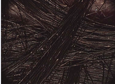
Figure 1 Whitish nodes distributed irregularly along the shaft (20x).
The damaged hair sample were examined using videodermoscope FotofinderR, at 20- and 70-fold magnification, which showed breaks in the hair shaft at irregular intervals with fraying of the cut edges (Figure 2 & Figure 3).
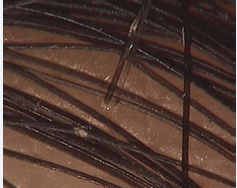
Figure 2 Area of nodular formations causes transverse fissures, where the hair can break off completely (70x).
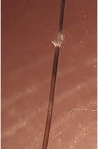
Figure 3 Hair shafts which split longitudinally into numerous small fibers, producing a picture that resembles two brushes aligned in opposition (70x).
DISCUSSION
Trichorrhexis nodosa (TN) is a common hair shaft disorder [1]. It is also known as trichonodosis and was first described by Wilks in 1852 [2]. It is classified as congenital or acquired, and the latter type is by far the most common one [2,6].
Clinically, acquired TN is characterized by nodular swelling of the hair shaft accompanied by frayed fibers and loss of the cuticle, which leads to dry, dull and brittle hair with varying numbers of small grayish-white or yellowish nodules distributed irregularly along the shaft. This area of nodular formations causes transverse fissures, where the hair can break off completely [2,3,5].
Acquired TN can be seen most as a result of excessive or repeated trauma caused by frequent application of hair permanent liquid, hair dryers and sprays and repeated ultraviolet exposure [1,6]. Excessive brushing, stressed hairstyles, heat application and scratching can contribute to the damage, as can manipulation involving shampooing and straightening [7,8]. Presumably, the decreased tensile strength of chemically treated hair in African Americans, heat exposure and other drying agents, play a role in the development of breakage. The hair breaks in areas stressed by heating or combing [7,8].
Acquired TN can be classified as proximal, distal and localized. In the first type, the whitish nodes can be seen a few centimeters from the skin surface in areas with physical damage, like friction from combing or use of caustic chemicals [9]. Distal TN can be observed several centimeters from the scalp surface, as a result of excessive brushing, hair styling and heat application. The localized type is seen as a patch and can occur as a result of chemical or physical trauma.
Although the scalp is the most common localization, TN can be seen on the face and genital areas [1,6], associated with pruritus and physical trauma.
The diagnosis of TN is clinical, and the standard technique is examination of a cut or plucked sample of hair under light microscopy associated with the use of cross-polarized filters [10]. The use of electron microscopy provides details of the surface characteristics, but is an expensive method and mainly used for research purposes [4,11].
Rakowska et al., described the use of trichoscopy in genetic hair shaft abnormalities without cutting or pulling the hair for diagnoses. Trichoscopy, the use of a videodermoscope, in the examination of the hair and scalp with low and high magnifications at 20 to 160-fold, allows us to distinguish some of the different types of hair shaft abnormalities [12,13].
Hair diseases as pili annulati, pediculosis or white piedra can be clinically similar to TN, observing whitish nodules on the hair shaft, but dermoscopy shows alternating white and dark bands, nits fixed to the hair shaft and small hair casts, respectively [12,13].
In TN, trichoscopy at higher magnification shows hair shafts which split longitudinally into numerous small fibers, producing a picture that resembles two brushes aligned in opposition [5,13].
In trichorrhexis nodosa, the use of trichoscopy allows medical practitioners to reach a diagnosis easily, without the need to pluck or cut hairs
REFERENCES
2. Lagrán ZM, González-Hermosa MR, Diaz-Pérez JL. Localized Trichorrhexis nodosa. Actas Dermosifiliogr. 2009; 100: 511-525.
4. Bartels NG, Blume-Peytavi U. Hair Loss in children. In: Hair Growth and Disorders. 1st edition. Berlin-Heidelberg, Germany: SpringerVerlag. 2008; 274-290
6. Camacho-Martínez F. Localized trichorrhexis nodosa. J Am Acad Dermatol. 1989; 20: 696-697.
11.Hillmann K, Blume-Peytavi U. Diagnosis of hair disorders. Semin Cutan Med Surg. 2009; 28: 33-38.


