Juvenile Xanthogranuloma with Central Hyperkeratosis
- 1. Department of Dermatology, Affiliated Hospital of Guizhou Medical University, China
Citation
Zhang W, Shen X, Liu Q, Lu H (2019) Juvenile Xanthogranuloma with Central Hyperkeratosis. J Dermatolog Clin Res 7(1): 1125.
CLINICAL IMAGE
A 11-month-old Chinese female infant presented with an erythematous plaque on the left forearm, without trauma. Her parents reported noticing it 5 months after birth, and that it progressively enlarged. Previously, a dermatologist suspected it as insect bites and treated with topical steroids but with little effect. Her birth history was unremarkable, and her growth and development were normal. Physical examination revealed a red flat-topped plaque, 15 mm in diameter, with a yellowish, keratotic center and thin scales, located in the left forearm (Figure 1).
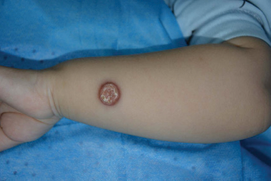
Figure 1 Rash of the left forearm, with a red flat-topped plaque and a yellowish, keratotic center and thin scales.
Following excision of the mass, histopathological examination revealed a proliferation of histiocytic cells admixed with Touton giant cells (Figure 2,3) characterized by polynuclei or wreath nuclei and eosinophils.
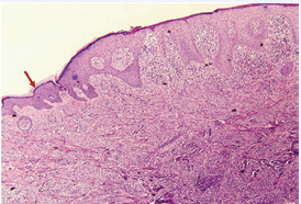
Figure 2 Histopathologic findings of under low-power magnification (hematoxylin-eosin, original magnification ×40), low-powered histological view showing epidermal hyperkeratosis (red arrow).
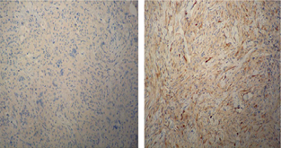
Figure 3 The histiocytic cells were negative for CD1a (Left) and S100 (Right) by immunohistochemical staining.
The histiocytic cells were negative for S100 and CD1a via immunohistochemical staining. These features were consistent with juvenile xanthogranuloma [1]. At 6 months post-excision, the lesion had not recurred. Similar lesions were not present elsewhere. Juvenile xanthogranuloma is the variant of non-Langerhans cell histiocytoses commonly occurring in infants and small children, which typically presents as yellowish, smooth, firm, dome-shaped papules or nodules on the head and trunk [2]. According to previous study, Juvenile xanthogranuloma occurs more frequently in Caucasians than in African-Americans [3], and this disease was rare among Chinese (non-Caucasian) in our clinical practice. In this case, hyperkeratosis of the lesion surface with yellowish-white scale, especially on the forearm was unusual, which may cause diagnostic confusion with some skin diseases such as pyogenic granuloma, eccrine poroma and viral verruca.


