Phytophotodermatitis in Children: A Difficult Diagnosis Mimicking other Dermatitis
- 1. Department of Dermatology, CHU Caen, France
- 2. Department of Dermatology, CHU Caen, France
Abstract
Phytophotodermatitis are phototoxic cutaneous reactions that are linked to a photosensitising plant in conjunction with exposure to the sun. They most often take the form of a rash consisting of vesicles or bullae, sometimes very marked, and residual hyperpigmentation.
We report four cases of phytophotodermatitis in children with varied clinical presentations. The plant thought responsible was identified in one of the cases – Heracleumgiganteum (Giant Hogweed), which is rich in photosensitisingfurocoumarins.
Diagnosis of these phytophotosensitisations can be difficult because it is easy to confuse with herpes infection, bullous impetigo, cutaneous allergies and even child abuse. It is when the patient or the parents are questioned on contact with certain plants in sunny conditions, often not spontaneously reported, that the diagnosis can be made. Evolution is generally positive and spontaneous after brief local corticotherapy.
Keywords
Phytophotodermatitis, Phototoxicity, Phytodermatitis, Photodermatitis, Heracleumgiganteum
Citation
Picard C, Morice C, Moreau A, Dompmartin A, Stefan A, et al. (2017) Phytophotodermatitis in Children: A Difficult Diagnosis Mimicking other Dermatitis. J Dermatolog Clin Res 5(3): 1101.
ABBREVIATIONS
UV: Ultraviolet
INTRODUCTION
Cutaneous reactions to plants are frequent in dermatological practice. The mechanisms involved may be immuno-allergic, irritant or toxic. Some of these reactions may be triggered by ultraviolet light: these are known as phytophotodermatitis, which can be divided into phototoxic reactions, the most common, and photoallergic reactions which are extremely rare. Clinical presentations may be spectacular, and puzzle a non-specialist, so that it is useful to know how to recognise them.
Here we report four cases of phytophotodermatitis occurring in children.
CASE PRESENTATION
Case 1
The first child, aged one, without any previous history of cutaneous reactions, was addressed after the discovery of skin lesions on the face when he woke in the morning. The lesions were erythematous and pustular, in linear formation on both cheeks, accompanied by oedema of the left cheek and vesicles on the nose and forehead (Figure 1A).
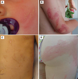
Figure 1 Phytophotodermatitis in our four patients. A: Linear vesicular lesions (Case 1) B: Erythematous bullous plaques, linear in places, linked to contact with plant of the Herculaneum genus (Case 2) C: Sequellar linear post-inflammatory hyperpigmentation after a phototoxic cutaneous reaction (Case 3) D: Urticaria plaque on the left buttock (Case 4).
There were no lesions on the rest of the body. When questioned, the parents reported that the evening before the child had handled plants and flowers in sunshine. These different elements yielded a diagnosis of phototoxic reaction to one of the components of these plants. Treatment with topical steroids yielded a cure within a few days.
Case 2
The second child, aged four, presented a weeping erythematous rash with burning sensations on the arms and forearm, rapidly producing pustules. Some of the lesions were in linear formation (Figure 1B). The rash persisted despite administration of antibiotics on the basis of suspected impetigo. After questioning, it emerged that the child had bathed in a swimming pool the day before the rash appeared, and had pulled up weeds in his swimming tarunks in the sun with no sunscreen. The clinical presentation, with bullous eczema-like lesions in linear distribution, localised on zones of possible contact with the plants, suggested a phototoxic cutaneous reaction. The rash disappeared after a few days on topic steroids. The plant incriminated belongs to the Apiaceae family and the Heracleum genus, commonly known as hogweeds (Figure 1B). There are two species in France, Common Hogweed (Heracleumsphondyllium) and Giant Hogweed (Heracleumgigantium), which is know for its phototoxicity, linked to the high levels of furocouramins in its sap.
Case 3
The third child aged 6 presented partially linear hyperpigmented lesions on the trunk and left arm (Figure 1C). These lesions had evolved from an erythematous vesicular rash that appeared the day following exposure to sunlight during a bicycle ride, bare-chested, in the course of which non-identified plants had brushed his back. The lesions were regressing, and no treatment was decided.
Case 4
The fourth child was a Canadian aged 11 with no history of skin reactions, consulting in the course of a holiday in France. She presented a weeping rash which had appeared on the day she arrived in France, starting at the top of the thigh a secondarily spreading over the buttocks, legs and abdomen. The rash was evocative of urticaria, with numerous pustules sometimes in linear formation on the lower limbs, with a large area on the right buttock (Figure 1D). In Canada the day before travelling she had done some gardening in the sun in shorts on the edge of the swimming pool. The plant responsible was not identified. The evolution was positive under topic steroid treatment.
DISCUSSION
Photosensitisation reactions to plants known as phytophotodermatitis are not uncommon, and can involve both children and adults [1]. Assessment requires careful clinical examination, and attention to risk factors such as leisure activities, travel, and in adults the profession, in particular the handling of certain fruit, vegetables or cosmetics (essential oils) [2]. The photosensitisation reaction implies the concomitant action of a chemical substance and artificial or solar rays. The wavelength is generally 320 nm or more [3].
Phytophotodermatitis are mainly linked to phototoxic reactions. These are not governed by an immuno-allergic mechanism, but by a photo-chemical mechanism. In theory they can appear in any individual without any particular predisposition, provided that the photosensitising substance is in sufficient concentrations and the light rays sufficiently strong. A humid atmospheric environment, as for three of our four patients, favours the cutaneous spread of the photosensitising molecule [4]. The cutaneous reaction appears at the time of the first exposure, without refractory period, and is always the same in later exposures. The cause is a decrease in the sensitivity threshold to UV light (in particular UVA) linked to the action of phototoxic agents present in certain plants, among which are furocouramins [3]. Furocouramins are a group of substances, including psoralens, which when stimulated by UVA radiation form covalent bonds with pyrimidines and interact with oxygen, resulting in the release of oxydantradicals , which cause lesions of the epidermis, the dermis and the endothelial cells [3]. In fact numerous plants are involved in these phototoxic reactions (Figure 2),
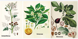
Figure 2 The three main families of plants responsible for phytophotodermatitis.
in particular those belonging to the Apiaceae family (umbellifers) which comprises over 3000 species, among which there are wild plants, food plants like aniseed, carrot, coriander celery, or parsnip, and numerous garden plants [5]. Hogweed (Heracleum sphondyllium), which is found across most European countries and responsible for the rash in our second case, is the main cause of phototoxic reactions in Europe and North America [6]. Cases of phototoxicity have also been reported with the Rutaceae (rue) family to which the citrus species belong. For instance, numerous cases of phytophotodermatitis have been reported on the upper limbs of Mexican barmen serving cocktails made from beer and green lemons [7], and among consumers of mojitos after receiving squirts of lemon juice [8]. Officinal rue, which is a fairly common wild plant used as an insect repellant also causes classic phototoxic reactions, as do certain plants in the Miraceae family, including Ficus [9].
Clinically, cutaneous phototoxic reactions to plants are restricted to areas exposed to sunlight and having been in contact with the photosensitising substance. Classically, phytophotodermatitis, known in France as “dermite des près”, develop rapidly in subjects sitting or lying in grass in sunny weather, and 24-48 hours later translate into the appearance of an erythematous-vesicular or bullous weeping rash, reproducing the outlines of a plant or leaf. The lesions often have a linear distribution. In the majority of cases they are localised, but highly inflammatory, bullous and even widespread lesions can occur. Evolution is generally spontaneous cure in a few days. Aesthetic consequences are possible (hyperpigmentation) and photosensitivity may last several months [3,10]. The diagnosis is clinical, and cutaneous histology and photobiogical explorations are not usually required. In the acute stage, the lesions may evoke several possible diagnoses such as herpes lesions when vesicles are clustered, or bullous impetigo, as these infections are frequent in children, or again intentional burns in a setting of child abuse [1] (Figure 3).
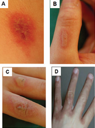
Figure 3 Differential diagnoses for phytophotodermatitis A. Herpes on the neck. B and C: bullous impetigo on the hands. D: Abuse by burns.
These phytophotodermatitis are also difficult to differentiate from photoallergic reactions from contact with plants, which are far rarer, and clinically resemble eczema. Unlike phototoxic reactions, these photoallergic reactions involve an immuno-allergic hypersensitivity reaction mediated at cellular level, and they affect subjects who are already sensitised. The allergic reaction will only occur on the occasion of a second interaction between the photosensitising plant and a particular UV wavelength on the skin. The triggering of the interaction is independent from the concentration of the photosensitising agent and the dose of radiation received. Aggravation is observed for successive each exposure, as the triggering threshold progressively lowers. Clinically, the lesions consist in eczema initially localised in the contact zone, and then exhibiting the particularity of spreading beyond the zones exposed to UV lightand to the plant [3,10]. The most frequently implicated plants are the Frullaniaceae, the Apiaceae (Heracleumgiganteum) and the Asteraceae (chrysanthemums) [3].
The therapeutic care for phytophotodermatitis consists in immediate decontamination of the exposed zones by thorough washing with soapy water. Topic steroids and emollients are recommended in case of moderate lesions, in association with H1 antihistamines for sedative or antipruritic purposes. Oral antibiotics are only indicated in case of bacterial superinfection, and oral corticotherapy may be required in rare severe forms [1,2].
CONCLUSION
A bullous, linear, photo-distributed rash in children should primarily suggest the diagnosis of phytophotodermatosis.
ACKNOWLEDGEMENTS
Mrs Angela Swaine reviewed the English language
REFERENCES
4. Booy O, Cock M, Eckstein L. Manuel pratique de la Berce Géante. Hoersholm: Forest & Landskape Denmark. 2005.
7. Schmutz J-L, Trechot P. Citron vert, bière et phytophotodermatose. Ann Dermatol Vénéréologie. 2012; 139: 81.


