Unusual Clinical Presentation of Cutaneous Tuberculosis Caused by Mycobacterium bovis
- 1. Department of Dermatology, Military Hospital, Tunis, Tunisia
Abstract
Cutaneous tuberculosis (CT) is rare despite the frequency of tuberculosis worldwide. CT is caused by the invasion of the skin by Mycobacterium tuberculosis, and less commonly Mycobacterium bovis or bacillus Calmette-Guérin (BCG). CT manifests a wide spectrum of clinical presentations. Lupus vulgaris (LV) is the most prevalent form of CT in Tunisia and has the most widely variable presentation of all forms of CT. Therefore, LV is considered to be one difficult diagnosis to make, especially in developed countries. We report a case of a 44 year old woman with multiple, annular LV caused by Mycobacterium bovis (M bovis) with emphasis on the diagnostic challenges of the disease.
Keywords
Cutaneous tuberculosis , Lupus vulgaris , Mycobacterium bovis , Annular plaque , Tunisia
Citation
Litaiem N, Youssef S, Jaber K, Dhaoui MR, Doss N (2014) Unusual Clinical Presentation of Cutaneous Tuberculosis Caused by Mycobacterium bovis. J Dermatolog Clin Res 2(1): 1010.
ABBREVIATIONS
CT: Cutaneous Tuberculosis; LV: Lupus Vulgaris;BCG: Bacillus Calmette-Guérin; M. bovis: Mycobacterium bovis
INTRODUCTION
Cutaneous tuberculosis remains a serious health problem throughout the world. According to the World Health Organization, one third of the world’s population is estimated to carry tuberculosis microorganisms, and thus the risk of developing the disease later in life [1]. CT represents around 2% of all forms of tuberculosis. Lupus vulgaris (LV) is the most prevalent form of CT in Tunisia and has the most widely variable presentation of all forms of CT. Recognizing rare clinical presentations of LV is important to prevent missed or delayed diagnosis. Moreover, this case is unusual as it is caused by M. bovis.
CASE PRESENTATION
We present the case of a 44 year old woman, living in a rural area, correctly vaccinated with BCG, with no particular personal history, presenting with progressive skin lesions, at neck and groin, for the past two years. There was no history of deterioration of her general condition, or night sweats. Physical examination revealed two plaques arranged in an annular pattern in the back of the neck and groin, measuring 10 cm and 8 cm respectively. The borders were erythematous and brownish with markedly elevated soft skin surface. Hyperpigmentation was seen in the center of the lesions. Diascopy revealed apple-jelly nodules.
The standard Mantoux test was highly positive. Skin biopsy revealed epithelioid granuloma, with multinucleate giant cells and minimal caseation necrosis. The culture of the skin biopsy was positive to Mycobacterium bovis.
The chest x-ray, the computed tomography of the chest and the ultrasonography of the lymph nodes did not show signs of active tuberculosis.
On the basis of these findings, the diagnosis of lupus vulgaris caused by M. bovis was established. Quadruple treatment (isoniazid, pyrazinamide, ethambutol and rifampicin) was initiated. After two months, treatment was continued with isoniazid and rifampicin during eight months. The response to treatment was good.(Figure 1,2)
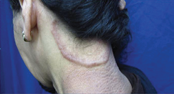
Figure 1 Large plaque of the back of the neck with elevated borders measuring 10 cm.
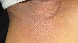
Figure 2 Annular plaque of the groin measuring 8 cm.
DISCUSSION
Four forms of LV disease are frequently described and consist of plaque or keratotic (classic), ulcerative, nodular, and vegetative or hypertrophic forms [2]. Typically, plaque LV presents as a slowly enlarging plaque with slightly elevated borders [3]. In this case, active borders are markedly elevated, and center is healing and hyperpigmented.
LV usually occurs in previously sensitized patients (vaccination, infection) who have a delayed, strongly positive hypersensitivity reaction to the tuberculin skin test [4]. Skin lesions are usually solitary, involving the head, neck, and extremities [2]. Groin localization is uncommon. Multiple lesions may be caused by inmunosuppression [3]. In this case, no clinical or personal history of inmunosuppression was found.
LV usually occur trough lymphatic, or hematogenous dissemination. Hematogenous dissemination is implicated in cases of facial LV or disease involving multiple sites. Therefore, we believe that LV in this particular case is caused by hematogenous dissemination of M. bovis.
CT caused by M. bovis is a clinically indistinguishable of CT caused by M. tuberculosis. M. bovis infection is most frequently contracted through infected milk products [1]. Active measures to eradicate M. bovis are taken in Tunisia. These include milk pasteurization and screening for tuberculosis in cattle. Therefore, cases of CT caused by M. bovis are becoming rare.
There are only 5 cases of LV caused by M. bovis reported in the literature in the past 20 years.(4) For most of these cases, the disease is characterized by the long evolution of the lesions (4 to 60 years), affecting at risk persons living in rural areas, or in contact with cattle.
The diagnosis of LV is challenging, especially in cases of atypical forms or locations of the disease. Histological examination in LV varies depending on the stage and form of disease with demonstration of abscess, ulceration, atrophy, or scarring [2]. The Ziehl-Neelsen stain of the skin biopsy specimen is usually negative, because the lesion is granulomatous and the number of bacilli is often small. Culture is considered to be the gold standard for the diagnosis of tuberculosis, but skin culture is only positive in 6% of cases of LV [5]. Polymerase chain reaction technique (PCR) permits rapid diagnosis. Nonetheless, PCR sensitivity in LV diagnosis is not better than culture [3]. [5] PCR is still not available in many developing countries where the prevalence of tuberculosis is higher. Other tests including The ELISA test for diagnosis of tuberculosis are not a routine diagnosis tests. Despite all these advances, LV diagnosis is still challenging. Many physicians still rely on the standard Mantoux test and therapeutic trials, as diagnostic tools [3].
Missed or delayed diagnosis of LV can cause important scars or disfigurement. Some locations are at risk especially cartilage of the nose or ears. Furthermore, squamous cell carcinoma can develop in long lasting LV [3].
In conclusion, we believe that physicians should be highly suspicious of CT caused by M. bovis especially in at risk population (farmers, rural living...).
REFERENCES
2. Ramarao S, Greene J, Casanas B, Carrington M, Rice J, Kass J. Cutaneous Manifestation of Tuberculosis. Infect Dis Clin Pract. 2012; 20: 376-383
3. Bravo F, Gotuzzo E. Cutaneous tuberculosis. Clinics in Dermatology. 2007; 25: 173-180.


