The Role of Transposable Elements Activity in Gene Instability and Their Relationship to Aging Process
- 1. Undergraduate student at Shanghai Jiao Tong University School of Medicine, China
- 2. Cornell University, College of Agriculture and Life Science, Ithaca 14853 NY, USA
- 3. King Abdullah University of Science and Technology, KAUST, Biological and Environmental Sciences and Engineering Division, Thuwal 23955-6900, Kingdom of Saudi Arabia
- 4. School of Public Health, Shanghai Jiao Tong University School of Medicine, Shanghai 200025, China
- 5. Department of Urology, Huashan Hospital, Fudan University, 12 Urumqi Road (M), Shanghai, 200040, China
- 6. Xinjiang Key Laboratory of Biological Resources and Genetic Engineering, College of Life Science & Technology, Xinjiang University, Xinjiang, China
Abstract
Transposable elements are mobile DNA sequences capable of self-replication within the genome, which may lead to various forms of DNA damage. The introduction encompasses the diverse classes and subclasses of TEs, particularly emphasizing the most active TEs present in the human genome. An analysis of the retrotransposition process of TEs is presented, illustrating how this mechanism can result in DNA damage and gene rearrangements. Furthermore, the review meticulously examines the implications of TE insertions on gene expression and genomic organization, which may contribute to the development of various diseases, including cancer. The relationship between TE activation and the aging process is also explored, emphasizing that epigenetic modifications associated with aging can lead to the derepression of TEs, thereby promoting genomic instability and inflammation. These factors may play a significant role in the pathogenesis of age-related diseases, such as cancer, cardiovascular disorders, and neurodegenerative conditions. Finally, the review considers potential therapeutic approaches aimed at targeting TE activity to alleviate the impacts of aging and associated diseases.
Keywords: DNA Damage; Transposon; Aging; Arrangement
Highlights
1. One of the principal mechanisms through which transposons induce DNA damage is by causing double-strand breaks (DSBs) in the DNA molecule.
2. The level of recombination has been established as being positively correlated with the abundance of TEs.
3. Additionally, transposable element insertions can also alter mRNA splicing and cause aberrant alternative splicing, leading to the skipping of exons or retention of introns. Consequently, aberrant gene expression ensues.
4. An imbalance in TE repression often occurs during aging or other processes, leading to TE activation and subsequent DNA damage.
5. TE activation-induced DNA damage contributes to aging and age-related diseases
Introduction
Transposons, also referred to as transposable elements (TEs) (colloquially known as “jumping genes”), are mobile DNA sequences that have the ability to replicate independently of the host cell's DNA [1]. A most suprising result discovered by the Human Genome Project is that nearly half of the human genome originates from transposons. However, only very small amounts of transposons are still jumping and thus able to propagate. The retrotransposition process will induce different kinds of DNA damage in the host cell genome, such as insertions and double-strand breaks. Furthermore, according to the twelve hallmarks of aging [2], genome instability induced by TEs is likely to play a significant role in the aging process and the development of age-related diseases. So it is important to figure out TE-induced DNA damage and how it could be prevented to slow down the ageing process. To summarize the different kinds of TE-induced DNA damages so as to help the readers with an overview of this field, this article includes a summary of different sorts of TE-induced DNA damages for researchers. The first part of the article introduces the classification and retrotransposition processes of TEs followed by the rate determining factors of TE activity, and then details how TE-activated elements lead to these types of DNA damages. Later parts cover the different types of DNA damages caused by TEs and a summary of TE-induced aging and age-related diseases
The overview of transposon classifications and structures
There are many different classification systems, but no worldwide standard committee was formed based on those put forward by Finnegan proposal [3], to Repbase proposal [4,5] and Wicker proposal [6], and finally to Curcio and Derbyshire proposal [7]. Of all the classification systems proposed thus far for transposable elements, the ones most commonly used are those presented by Repbase and Wicker. The systems proposed by these two research groups will be explained in detail below. Based on their mechanisms of transposition, TEs have been classified as class I elements (retrotransposons) and class II elements (DNA transposons) [8]. These two classes can be further subdivided into multiple subclasses, (or orders [6]), primarily distinguished by their replication and chromosomal integration mechanisms, and then into superfamilies and families, which are more accurately characterized on the basis of phylogenetic relationships [9] (Figure 1A). Briefly, most class II transposable elements, although not all, mobilize through a “cut-and-paste” mechanism, whereby the transposon itself is excised and moved to a new genomic location [10]. Thus, in contrast to retrotransposons, these elements typically do not accumulate in copy number. Instead, they adopt strategies of either transposing from replicated to unreplicated sites during host DNA synthesis or utilizing the error-prone homologous recombination repair to elevate their copy number [1]. This usually causes internal deletions in transposons [11] (nonautonomous elements lacking the coding capability but carrying transposase binding site for autonomous element proliferation at cost), and the progeny are thus highly possibly to form large families of MITEs (miniature inverted-repeat TE) [12]. DNA Transposons can be divided into TIRs (Terminal Inverted Repeats), Cryptons, Helitrons and Mavericks (also known as Polintons). TIRs are distinguished by the presence of terminal inverted repeats and are responsible for encoding a transposase (DDE transposases, named after the catalytic residues of aspartic acid and glutamic acid [13]) that mediates excision and integration by binding to TIRs. The precise mechanism by which DDE transposase transposes varies from superfamily to superfamily, but in the case of all members of the eukaryotic organisms studied thus far, the process commences with a nucleophilic attack of a water molecule close to the ends of each TIR, eventually resulting in direct excision and subsequent relocation of the transposon DNA [14]. DDE transposons represent the most diverse and extensively distributed category of TEs, comprising at least 17 large superfamilies that are characterized by phylogenetically distinct transposases [15]. Cryptons share similar characteristics with DDE transposons, consisting of a single open reading frame (ORF) that produces tyrosine recombinase (YR) rather than DDE transposases enclosed within short terminal inverted repeats (TIRs). Helitrons are mainly non-autonomous elements lack TIRs but encode large Rep/Hel proteins, which feature DNA helicase domains fused with HUH nuclease domains [16]. They replicate by means of a“peel-and-paste”mechanism that leads to the formation of a covalently closed circular double-stranded DNA intermediate during rolling of the circle towards the 3’end of the Helitron while separating its sense strand and, possibly, synthesizing the other strand [17]. Mavericks are substantial DNA transposons that encode up to twenty protein-coding genes, which are flanked by long TIRs [18]. Maverick/Polintons exhibit similarities with various groups of double-stranded DNA (dsDNA) viruses. These include a protein-primed family-B DNA polymerase (pPolB) that is most similar to that of adenovirus, suggesting that they replicate by directly synthesizing copies of DNA (hence the term “self-synthesizing transposons”[19] ). They also encode a DDE nuclease that is most closely related to retroviral INs, which aligns with the fact that they generate 5- or 6-base pair target site replication during the process of chromosomal integration [19]. It is possible that numerous Maverick and Polinton elements could be encoded for double as well as single jelly-roll cap-like protein domains, leading people to think they may represent potential endogenous viruses or virophages [20]. Since DNA transposons have been inactive for so long that they became inactive by truncation and mutation [21] and retrotransposons outnumber DNA transposons 13 to 1 in the human genome [22], we will limit our attention below to retrotransposons only. Unlike class II elements, which go through a process of replication using RNA as an intermediary?class I elements go through the following steps after they are replicated: the RNA is then reversed transcribed into DNA; the newly formed DNA copy replaces the original template element, thus named“copy-and-paste”elements. Because the original template element remains intact, these retrotransposons (RTPs) are also called “copy-and-paste” elements [10]. Retrotransposons were later classified into four subclasses based on the structure and the mechanism they used for their own replication: long terminal repeat (LTR), non-LTR, tyrosine recombinase (YR)-mobilized and Penelope-like elements (PLEs) [1]. LTR elements are distinguished by the presence of 5’ and 3’ non-coding long terminal repeats that regulate the expression of “retroviral genes”, reflecting their evolutionary connection to retroviruses. Autonomous LTR elements typically encompass a minimal collection of two distinct genes, namely gag and pol, which are generally expressed as a single polycistronic RNA transcribed from a Pol II promoter situated within the LTR itself. Both gag and pol genes code for polyproteins, which are further divided into different proteins via protease (PR) produced from the pol gene. Besides, the Pol gene codes important enzymes like reverse transcriptase (RT), RNase H and integrase (IN) [1]. Both the replication and integration processes of LTR elements share similarities to those of retroviruses, with the only difference being that retroviruses possess the fusogenic env gene. This env gene is frequently lost, leading to the endogenization of retroviruses that are active in the germline [23] (a classic example of this is IAP in mice [24]). LTR elements are further categorized into five superfamilies: Copia, Gypsy, Bel-Pao, Retrovirus, and ERV, among ERVs being the most active and extensively studied [25]. Incidentally, HERVs (human ERVs) can be further divided into various families according to two distinct classification systems. The International Committee on the Taxonomy of Viruses (ICTV) classifies ERVs based on the similarity and phylogenetic relationship to exogenous retroviruses into class I, II, and III [26]. And traditionally, HERV family designations have been assigned letters corresponding to the specific type of the amino acid of human tRNA that binds to the primer binding site (PBS) during reverse transcription, such as HERV-W, HERV-K, and HERV-H [27]. Two types of non-LTR elements exist: long interspersed nuclear elements (LINEs) and short interspersed nuclear elements (SINEs). LINEs are characterized by the presence of a 5' untranslated region (5'UTR) and a polyadenylation signal, and they encode the necessary proteins for retro-transposition(specifically open reading frames ORF1 and ORF2) [8], on the other hand, the role of ORF1 remains a mystery in certain classifications of non-LTR elements and is dispensable for non-LTR elements such as the R2 element, which lacks an ORF1 [28]. In cases where ORF1 is necessary, such as in L1 elements, the proteins encoded by ORF1 assemble into an oligomeric structure that plays a role in the recognition and transport of the template RNA to the nucleus [29]. ORF2 is responsible for encoding both an endonuclease (EN) and a reverse transcriptase (RT), with the latter of which is essential for target-primed reverse transcription (TPRT) [30].Within the LINE category, several superfamilies exist, including R2, RTE, Jockey, l, and L1, with L1 elements being the most active and extensively researched [25]. SINEs are nonautonomous transposable elements whose retrotransposition relies on LINE-encoded machinery, specifically its ORF2 (but not ORF1 enzyme) [31]. Of all the types of transposable elements, SINEs alone use RNA polymerase III to make RNAs and all known SINE superfamilies appear to have descended from one of the three RNAs transcribed by pol III (tRNA, 7SL RNA or 5S rRNA) [32]. Therefore, SINEs are subdivided into three superfamilies: tRNA, 7SL and 5S, based on their source RNA. YR retrotransposons are the third main subclass of class I elements, yet they have received considerably less study than other subclasses [7]. They possess very similar genomic organization to LTR elemnets; however, they encode YR instead of IN, which causes YR elements to function and process terminal repeat sequences differently based on the type of superfamily of YR retroelements to which it belongs: for instance, DIRS elements contain inverted repeats that do not match their partner repeats (unlike true LTRs), whereas Ngaro, VIPER, and TATE elements appear to use direct repeats in a “split-repeat” pattern [33,34]. The functions of the terminal repeats as well as how YR elements replicate remains to be elucidated. However, it has been proposed that replication proceeds as follows: reverse transcription of an mRNA template occurs, then circularization of the single-stranded cDNA copy that is primed by the pairing of the terminal repeat, synthesis of the complementary cDNA strand, followed by YR-mediated integration into the genome [35,36]. Penelope elements were noted as the first group of elements proven to be mutagens with action similar to activation-induced cytidine deaminase (AID) in a large eukaryotic insect, Drosophila virilis. However, these remained the only members of this class of element for a long time [37]. Penelope-like elements (PLEs) have open reading frames encoding PLE enzyme with RT and EN domains fused as one polypeptide(8). The characteristic features of PLE elements are: first, the pseudo-LTR sequence, and second, GIY-YIG (amino acid motif) endonuclease domain, distinct from that of other subclasses of retroelement classes [38]. At last, the four most active TE groups that we bring up again and again in the manuscript itself, these make up approximately 8% of the human genome (Figure 1B). Despite the limited evidence for ongoing retrotransposition of ERVs in human—primarily associated with HERV-K elements [39], these elements may still play a significant role in oncogenesis and other various diseases [40]. Secondly, L1s are the only autonomous retrotransposons that have been discovered to be able to jump into human genomes—about 17% of the human genomes according to the sequence in Figure 1B [22]. According to the current estimate, there are between 80-100 human endogenous retroposons capable of movement and about 10% of the complete L1s in the human genome exhibit very high mobility [41]. Moreover, the human genome includes two other active, non-autonomous, non-LTR retrotransposons, Alu elements (Figure 1B) [42], and SVA elements, which are the primate-specific members of the SINE family comprising ~11% and ~0.2%, respectively of the human genome (see Figure 1B)[43].
The retrotransposition process
The retrotransposition of L1
Lifecycle of L1 transposons starting from their transcription is shown in Figure 2. It starts when host RNA polymerase II mediate transcription, while its repetitiveness could result in blocked transcription bubbles, which would then cause DNA damage. Then, the two proteins encoded by the capped and polyadenylated bicistronic L1 mRNA are translated into the cytoplasm and interacts in cis with L1 mRNAs to form ribonucleoprotein particles (RNP) [55]. These L1 RNPs are composed of multiple ORF1 trimers [56] that coat the mRNA, with perhaps only one or two ORF2 molecules bound to the A-rich tail of the 3' untranslated region(UTR) [46]. Upon entering the nucleus, L1 RNPs initiate the reverse transcription of RNA, predominantly during mitosis when the nuclear envelope is broken [57]. Mechanistically, typical LINE-1 retrotransposition occurs through target-primed reverse transcription (TPRT) [30,58]. In this model, the endonuclease activity of ORF2p generates a DNA nick at a flexible target sequence (3' -AA/TTTT-5') to release a 3' OH [59]. This process reveals a short segment of polyT single-stranded DNA that can bind complementarily to the polyA tail of the LINE-1 transcript, thereby forming a primer-template structure. However, the generation of primers may induce the single-strand breaks(SSB) DNA or even double-strand breaks(DSB) due to the action of ORF2p endonuclease. Subsequently, ORF2p reverse transcriptase utilizes the LINE-1 RNA as a template to synthesize the first strand of LINE-1 cDNA [58]. This process is characterized by the fact that the reverse transcription is generally truncated in 5'-direction before it can be completed, and non-LTR retrotransposons encode their own internal Pol II promoters at their 5' ends that are required for their expression, thereby enabling these truncated retrotransposons to fail to further insert new copies [29]. After reverse transcription, a new copy of the LINE-1 is inserted at the genomic loci as a chimeric DNA flanked by TSDs with less than 20 bp [60,61], at this time, the life cycle of a LINE-1 unit has been completed, and its end product is therefore an inserted LINE-1 that has both polyA tails at the ends and is sandwiched between short TSDs. The result of L1 retrotransposition is an inserted LINE-1 sequence that has polyA tails on the ends, and short TSDs around it. Slippage replication during the LINE-1 ORF2p RT producing cDNA from pre-existing LINE-1 RNA might result in point mutations, small insertions/deletions and even large genomic rearrangements, leading to genomic instability. Still, how the line-1 single-strand cDNA intermediates become double-strand insertions remains unclear and might require the assistance of host DNA repair factors. The apparent lack of RNase H activity on ORF2p may indicate that this protein helps the line-1 RNA template move across the proposed L1 RNA: cDNA intermediate or degrade it [62,63]. Furthermore, the presence of TSDs indicates that the top strand DNA is cut, near and downstream from the first nick, either by ORF2p EN itself or host factors, so that there is a pair of nicked DNA ends, called staggered DNA nicks. This scenario may be responsible for generating short TSDs. It is unclear what leads to the synthesis of the second-strand LINE-1 cDNA; however, it’s feasible that ORF2p RT is responsible. The remaining steps are unclear as well but may involve the ligation of the 5' end of a double-stranded LINE-1 cDNA to genomic DNA to complete the insertion. The 5' junctions of complete LINE-1 insertions sometimes contain non-templated nucleotides [60,64], yet the origin and function of these nucleotides in resolving the insertion remain ambiguous. Lastly, the specific role of ORF1p in the TPRT process is still unclear, although its involvement in the LINE-1 life cycle may be limited to the cytosolic environment. According to C. Mendez-Dorantes et al. [45], the sequence structures resulting from LINE-1 retro transposition encompass full-length insertions, 5' truncations, 5' inversions, 3' transductions, and EN-independent insertions.
The retrotransposition of Alu and SVA
Both Alu and SVA elements use LINE-1 ORF2p to amplify these elements within the human genome (see Figure 2), so like the LINE-1 element, formation of DSBs/SSBs during the retrotransposition of Alu and SVA elements by virtue of ORF2p activity is also expected to be very high. Additionally, transcriptional activity of Alu elements is dependent on RNA polymerase III-dependent, and has a poly-A sequence at its 3’end, which helps bind the RNA of another cell to ORF2p in trans [46]. Moreover, transposons Alu and SVA have excessively repetitive elements of AT sequences and CG sequences, respectively, both of which are believed to interfere with the transcription, causing the stall during the transcription, and leading to DNA damage. Perhaps the formation of RNPs is facilitated by the interaction between Alu transcripts and SRP9-14 proteins within the ribosome, which together inhibit the translation of ORF2p in close proximity to Alu RNAs [65]. However, although the mechanisms underlying SVA retrotransposition remain poorly understood, it appears to be reliant on RNA polymerase II. Importantly, unlike Alu elements, the retrotransposition of SVAs necessitates both ORF1p and ORF2p [66]. Incidentally, mRNAs can also be reverse transcribed via ORF2p, which has produced about 8000 processed pseudogenes (retrocopies) in our genome [67]. It is crucial to note that these retrocopies lack promoters, and as a result, most exhibit no transcriptional activity.
The retrotransposition of ERV
ERV is transcribed from proviral mRNAs, primarily from the 3′ end of the 5′LTR [68], giving rise to a Gag-Pro-Pol fusion protein, which is cleaved through the action of a protease to produce virus-like particles, accompanied by a Gag-Pro-Pol fusion protein and the original ERV mRNA. One specific type of transfer RNA (tRNA) binds to the primer binding site (PBS), aiding in reverse transcription and the subsequent generation of complementary DNA (cDNA) within the cytoplasm. Then, the cDNA forms a complex with integrase (IN), which induces a double-strand DNA break, followed by integrating a new copy of ERV in the genome [69] (Figure 3). As a result, IN-induced DSB can lead to the occurrence of chromothripsis on the chromosomal level, resulting in structural rearrangements like chromosomal inversions and resuffling of chromosomes. It is worth pointing out that, as mentioned previously, HERVs appear unable to undergo retrotransposition as they do not show evidence of de novo mobilization in humans, nor have polymorphic HERV insertion sites and/or HERV-K family members containing intact ORFs been observed [70]. However, immobile ERVs, especially their LTRs, can contain promoters [71] enhancers [72], and numerous binding sites for transcription factors [73], recruit complexes responsible for DNA and histone modifications [74], and endow ERVs with considerable potential to alter the expression of gene networks necessary for normal cellular function. To know the molecular basis of retrotransposition is indispensable to unravel the control of TE expression as well as the two major concerns (DNA damage and inflammation) that lurk after TEs successfully invade the genome.
Figure 1
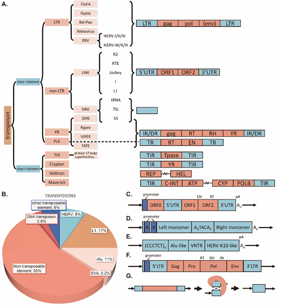
Figure 1. The classification and structure of TEs
- The classification of TEs
- The proportion of TEs in the human genome
- A small ORF of unknown function, ORF0, has recently been identified in the antisense direction of the 5’UTR(44). 5’UTR also contains a promoter. ORF1 protein s form an oligomeric product involved in the recognition and transport of the template RNA to the nucleus(29). ORF2 encodes both endonuclease (EN) and reverse transcriptase (RT). And finally, 3’ UTR, polyA signal (pA), and an oligo dA-rich tail.
- Alu elements are heterodimers of two non-identical monomers joined by an adenosine-rich linker. In the left monomer, Alu elements contain an internal promoter (A box and B box) that is mediated by RNA polymerase III to initiate transcription but lacks a terminator sequence, instead using downstream T-rich genomic DNA to terminate transcription(45). The 3’ end of the Alu sequence also contains an oligo dA-rich tail, which is required for the trans-binding of Alu RNA to ORF2p(46).
- SVA is a composite element consisting of five repeats: a hexameric repeat [(CCCTCT)n], two antisense Alu-like fragments, a variable number tandem repeat (VNTR), a SINE derived from the LTR of an ERV (HERV-K10), and a polyA signal, which is followed by an oligo-dA-rich tail(47). Due to the differences in their VNTRs, the full-length sizes of SVAs vary considerably, ranging from 50 bp to 2 kb, though the majority of SVAs are 2 kb in length(48, 49). It is now generally accepted that SVA elements are transcribed by Polymerase II. However, SVA elements apparently do not contain internal promoters, and they may be at least partially dependent on promoter activity in flanking regions(50).
- An intact HERV provirus consists of at least a 5′ LTR (containing a promoter) and a 3′ LTR flanking the internal Gag-Pro-Pol multiprotein-encoding sequence(51). Gag is cleaved by the protease (Pro) to generate the viral particle structural proteins, whereas Pol encodes the enzyme activities of reverse transcriptase, ribonuclease H, and integrase(52). Although HERVs may contain residual envelope (env) genes and other accessory genes, they are generally not infectious(53) and also lack the potential for intracellular mobility (probably with the exception of HERV-K) due to germline ORF mutations. Importantly, many mammalian ERVs have lost the ORF for env, which is essential for infecting new target cells. When ERVs lose the env gene (e.g. IAP in mice), they typically exhibit enhanced retrotransposition activity, resulting in a dramatic increase in their proliferation within the genome — about a 30-fold increase(23).
- Incidentally, at least 85% of the reference genome ERV instances are solitary (or “single”) LTRs(54). Single LTRs originate from homologous recombination between ancestral 5’ and 3’ proviral LTRs, in which the protein-coding sequence in the middle is deleted(54).
Figure 2
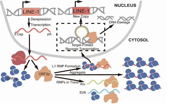
Figure 2: The L1 retrotransposition process
TEs cause DNA damage in diverse ways
Transposons move within the genome through various mechanisms, which may directly or indirectly lead to DNA damage and genomic instability, with profound impacts on biological functions that cannot be overlooked. Mostly, unsuccessful transposition events are responsible for inducing DNA damage. This review aims to present a comprehensive examination of the diverse mechanisms through which transposons cause DNA damage, alongside relevant insights into the association between transposons and the integrity of DNA.
TE can cause DSBs and SSBs directly or indirectly
One of the main ways bring about DNA damage is by cutting or breaking the double strands in the DNA molecule. A non-LTR retrotransposons has two open reading frames, one of them being ORF2p that encodes an endonuclease (EN), and a reverse transcriptase (RT). In the particular process, ORF2p cause many cuts in the chromosome DNA before every successful integration event [75]. Indeed, previous studies demonstrated that point mutations at the EN domain of the LINE-1 ORF2 sequence resulted in complete loss of γ-H2AX, a DSB damage signal in HeLa cells[75], indicating that the ORF2 encoded EN is the source for DSBs. Another line of evidence indicates that following transposition initiation, there is an exponential rise in the progeny number of active elements, resulting in the accumulation of insertions at rates that quickly surpass those associated with classical spontaneous mutations. It can therefore be surmised that the activation of LINE-1 transposition may result in the formation of a considerable number of DSBs within the genome of human cells [75,76], with the potential to account for one in every 250 pathogenic mutations observed in human diseases [77,78]. It is important to note that a larger number of L1-induced DSBs were found relative to the estimated number of successful integrations, indicating that this process is relatively inefficient. Furthermore, evidence exists to suggest that the damage associated with the activation of LINE-1 may contribute to the increased accumulation of neurons expressing features similar to senescence throughout the brain during aging or neurodegeneration. Moreover, it might be hypothesized that senescent-like neurons produce senescence-associated secretory phenotype (SASP), which then causes other nearby neurons to become senescent-like as well, causing chronic inflammation to further develop into neurodegenerative diseases (NDs), and there are many studies aimed to show that removing senescent cells helps prevent NDs [79]. Another potential source of DSBs associated with TEs is HERVs with HERV-K loci. HERV-K loci contain open reading frames for integrase [80]. Assuming that the integrase enzyme is active during HERV retrotransposition, whereby it can create what is referred to as a pre-integration complex in conjunction with the reverse-transcribed double-stranded DNA and host proteins. The integrase facilitates the preparation of the linear dsDNA ends for the integration process and covalently links these ends to the host DNA via a strand transfer reaction, resulting in DNA lesions that require subsequent repair by the host's DNA repair mechanisms [81]. During this process, aberrant IN activity or a failure in DNA repair renders the cell prone to the formation of DSBs [82]. Taken together, this theoretical framework of possible HERV retrotransposition activity implies that IN-induced genomic instability might be possible, yet it is uncertain whether this genomic instability stems from the expression of IN proteins or TE mobilization [83]. It is noteworthy that there is a chain reaction induced by transposition-nmediated DSBs and it is reported that transposon-mediated DSB may feed chromothripsis pathway leading to the formation of large-scale structural variant. This may lead to chromosome inversions and chromosome reshuffling [84]. Furthermore, TEs can elevate the number of R-loops to cause gene instability, which can subsequently result in DSBs. Both the synthesis of DNA and transcription of RNA necessitate the accessibility of chromatin-associated DNA and the unwinding of the double helix, which ultimately results in the formation of DNA-RNA hybrids. These three-stranded structures, designated as R-loops, are frequently observed in regions exhibiting elevated transcriptional activity and repetitive sequences [85,86]. The increased transcriptional activity and the activation of transposable elements are tightly coupled, and R-loop upregulation might be an indicator of genome integrity [85]. The production and accumulation of inappropriate R-loops may threaten cell integrity. Recently, the R-loop was recognized as the origin of genome instability as well as a main source of DNA damage. A substantial body of evidence demonstrates that genomic instability can be attributed to genome aberration by R-loop, but replication forks will become slowly due to R-loop. Recently, a few specific types of R-loop including the DNA damaged sites was associated with genomic instability [87-90]. It was observed that exposing ssDNA within R-loop served as a target site for the nuclease ORF2p, XPG, and XPF to cleave ssDNA and producing SSBs and DSBs [85,87,89]. Additionally, the process of TE-inducing DSBs can be mediated by acute oxidative stress. As expected, acute oxidative stress damages DNA within mouse dopaminergic neurons, and research has shown that it also elevates expression levels of LINE-1 [91]. Furthermore, experiments using chromatin immunoprecipitation reveals that Engrailed can bind specifically to the promoter region of LINE-1 elements and functions as a transcriptional repressor of LINE-1 in vivo in murine models [83, 92]. By repressing the expression of LINE-1, Engrailed ameliorated oxidative stress induced DNA damage and the apoptosis in dopaminergic neuronal cells from stressed mice [83,92]. Besides, prior to DSB appearance the transcripts from LINE-1 increased [83]. These phenomena seem to hint at a possible role of LINE-1 as sensor of oxidative stress damage [91]. Secondly, oxidative stress increases the incidence of DNA damages not only because it generates DNA damaging products such as ROS inside the cell but also because it augments levels of ROS already existing within the cell [93-95]. The oxidative products generated inside the cell may damage DNA in different ways such as double-strand breakage or nucleotide oxidation generating a great variety of substrates including T-endorphin or 8-oxo-dGuo among others [93,96-98]. In addition to double-strand breaks (DSBs), single-strand breaks (SSBs) may also occur. The protein subunits encoded by L1 elements have been observed to prefer cis association with L1 mRNA to form a ribonucleoprotein (RNP) complex [85]. This complex can then be translocated to the nucleus where the SSB formed within the genomic DNA can act as a primer for initiation of reverse transcription of RNA [85]. The presence of TE sequences may also result in the formation of DSBs, even in the absence of transposition. TE-enriched sequences may cluster within the vicinity of DSBs in cancer cells.Likewise, it has been suggested that short inverted repeats (e. g., Alu elements) form secondarily structured hairpins during DNA replication, which may contribute to replication stalling or double-strand break formation [99,100].
TE insertion can cause gene structural rearrangement
Firstly, cleavage that occurs during the transposition process facilitates gene rearrangement to a certain degree. To illustrate, a potential pathway of DNA damage involving piggyBac transposase would entail cleavage by transposase at chromosomal sites bearing resemblance inverted terminal repeats (ITRs; triangles) [101-103]. Such events resemble reactions at one side of the integrated transposon during excision. This could introduce two break points into two double strand-breaks, thereby potentially giving rise to a swapped chromosomal arrangement - a chromosomal rearrangement [101-103]. Another mechanism by which the transposase might induce chromosomal damage is by interacting with regions in chromosomes corresponding to the regular ITR binding sites on either side of a transposon, and cleaving the chromosomal DNA directly without going through a transcription reaction [101]. Besides, as soon as a transposition event by a transposon has occurred, numerous DSBs are introduced into the DNA in large quantities. Following the emergence of these breaks in the cells’DNA, cellular DNA repair pathways can then be activated to fix them up [104-106]. Usually, non-homologous end joining(NHEJ) is utilized to repair the DSBs that arise from such an activity [107]. However, in light of the fact that the error-prone process of NHEJ is more likely to result in frameshift mutations,which may lead to the occurrence of insertions or deletions, as well as substantial genomic rearrangements [104]. Given that NHEJ operates independently of sequence homology, it has the potential to introduce errors during the DNA repair process [108]. Such errors may result in genetic mutations or alterations in the structural integrity of the genome. In certain instances, NHEJ may erroneously join non-adjacent DNA fragments [106,108,109], leading to substantial genomic rearrangements [107]. Besides, DSBs in L1s are repaired more slowly, likely as a result of histone-driven euchromatinization [110]. Another factor leading to genome recombination is slippage replication driven by ORF2p. As mentioned earlier, the ORF2 protein has both endonuclease and reverse transcriptase activity. Therefore, LINE-1 elements make use of ORF2p for the reverse transcriptase reaction that occurs in the LINE-1 retrotransposition event, generating longer telomeric DNA sequence via slippage during DNA replication [111]. Two-dimensional sequence alignment shows that replication slippage may generate a significant proportion of all transversion substitutions [112-114]. During the process of DNA replication, DNA polymerases may undergo a phenomenon known as "slippage" when encountering repetitive sequences. This slippage can result in the replication fork either advancing or retracting by one or more repeat units along the template strand. Re-alignment of the molecule with the template strand causes the polymerase to resume replication with possible incorporation of one or more extra repeat units and/or deletion(s) of already present repeat units into the newly replicated daughter DNA. This phenomenon known as replication slippage leads to genomic instability by introducing single-base substitutions, small indels and large genomic rearrangements [112-114]. The level of recombination has been established as being positively correlated with the abundance of TEs [115]. Highly active TEs are capable of facilitating the relocation of highly homologous regions to distant loci within the genome. This process ultimately contributes to extensive genomic alterations, including large-scale deletions, duplications, and inversions that occur during recombination events [116,117]. An illustrative instance of such recombination involving two non-allelic insertions is presented in Figure 5A. In this example, Alu-Alu recombination transpires between adjacent AluYa5 insertions located at the ubiquitin E2 T conjugating enzyme (UBE2T) gene, which is associated with Fanconi anemia. This rearrangement results in an interstitial deletion that spans exons 2 to 6 of the paternal allele, accompanied by a corresponding duplication of the maternal allele in the affected individual [118].
A. Insertion of Alu elements and recombination. Example of recombination between two AluYa5 insertions in the parental alleles of the UBE2T gene (a) leading to one recombination allele with deletion (b) leading to one recombination allele with duplication (c) The exons are represented by grey boxes and the AluYa5 insertions by blue boxes.
B. Example of SVA element insertion causing X-linked dystonia with parkinsonism in the TAF1 gene. TAF1 consists of 38 exons (vertical bars; red bars are exons flanking the insertion) with an SVA insertion in reverse orientation in intron 32 (orange triangle). This insertion leads to the retention of intron 32 in the mRNA and the variable number of repeats of the hexanucleotide (CCCTCT)n affects the disease.
C. Inserting a LINE-1 element into the APC gene. Insertion of a LINE-1 element causes a new polyadenylation site to be created in the last exon of the APC gene if it is truncated and partially inverted.
The numerous structural genomic variants may be ascribed to the marked similarity across TEs within families, which occurs particularly when neighboring sequences such as inverted terminal repeats are identical. These areas of sequence identity increase the chances for NAHR events, where non-allelic homologous recombination can occur. This increases the chances for large chromosomal changes (e.g., inversions, duplications, translocations and deletions) to happen [119]. For example, the high abundance of LINE-1 and Alu repeats favors ectopic recombination, i.e., recombination between non-homologous loci. Retrotransposons, such as Alu, can impart nontemplated homology to the invader [120]. Furthermore, TE contains microhomologies: short stretches (typically 2–10 bp long) with high identity or strong similarities in position within the genome. These break down the correct-pairing, template-switching pathways, allowing non-base pair effects during the error correction step of DNA synthesis, generating copy number variation, insertion deletion polymorphism (IDP), and DNA damage [121].
TE insertions can cause abnormal gene expression
Transposable elements (TEs) can cause structural or functional harm to the genome of the host, leading to gene silencing and gene expression. As well as their ability to induce genomic damage through insertion, TEs might affect the genome through alternative mechanisms. As previously described, there is ample evidence that TEs play an active part in the regulation of the host genome, acting as modulators and switching the cells between alternative epigenetic states [91,122-124]. In developmental process, gene expression can be controlled in part by these repeat sequences, influencing not only the host genes but also neighboring sequences [125-127]. In this sense, these non-canonical transcriptional regulators are usually functioned as alternative promoters, enhancers, or silencers [128-130]. Increased evidence has shown that the promoter and enhancer/silencer regions of human endogenous retrovirus (HERV) long terminal repeats (LTRs) has particularly strong regulatory potential. For instance, in X-linked Opitz syndrome, a HERV-E LTR functions as a tissue-specific promoter and transcriptional activator of the MID1 gene, contributing to disease pathogenesis [131]. Another example of TE-mediated gene regulation involves intronic LINE-1 (L1) elements, which can alter host gene splicing via exonization—a process wherein the L1 sequence is incorporated as an additional exon into the mature transcript [43,132]. Such aberrant splicing events frequently disrupt normal gene expression, either by introducing frameshifts, generating premature termination codons, or reducing transcript stability, occurring in approximately 79% of documented cases [132, 133]. Moreover, the impact that insertion has on causing aberrant gene expression is likely the area most comprehensively researched in the field. To illustrate, several points will be made. A recent observation concerning epigenetics includes nuclear repositioning of TEs. In D. melanogaster, epigenetically silenced TEs inserted in euchromatic chromosome arms have been seen to relocate to interact physically with pericentromeric heterochromatin. While this spatial reorganization could theoretically shift neighboring genes from transcriptionally active to repressive nuclear compartments, its functional consequences remain unexplored. Intriguingly, TEs exhibiting such epigenetic effects—whether through heterochromatin spreading or nuclear repositioning—appear to undergo stronger negativction than other TEs, suggesting their modifications are generally deleterious, though the precise mechanisms remain unclear. A key mechanism involves the silencing of euchromatic TE insertions via the accumulation of trimethylated histone H3 at lysine 9 [134]. These H3K9me3 domains can spread beyond TE boundaries, potentially silencing adjacent genes [134]. Additionally, SINE transcripts can localize to target genes and inhibit transcriptional elongation by restricting RNA polymerase II (RNAPII) mobility, actively slowing down or stopping gene expression. but when SINE RNA binds to EZH2, it can promote ribozyme self-cleavage, releasing RNAPII and restoring transcriptional elongation—particularly for stress-responsive genes [134]. In addition to transcriptional interference, cytoplasmic intrinsic immune sensors RIG-I and MDA5 detect LINE-1 transcripts. After RIG-I and MDA5 bind to LINE-1 RNA, they recruit mitochondrial MAVS, triggering a signaling cascade that phosphorylates IRF3 and activates interferon-stimulated genes [134]. This represents an abbreviated expression of IRF3 in this context. The polyadenylation signal found in the 3'untranslated region (3'UTR) of LINE-1 elements can affect the transcriptional elongation of the host gene and lead to a reduction in full-length transcript production, which can result in truncation of the protein product or total abrogation of protein expression [91]. Additionally,stably integrated but nonfunctional TEs can be involved in silencing host genes and dysregulated TEs could promote transposition, resulting in further increases in genomic instability that may lead to cancer progression [135,136].
(a) Insertion transposable elements (TEs) into euchromatic regions often results in the transcriptional silencing; the localized enrichment of H3K9me3 contributes to transcriptional repression mediated by TEs, and sometimes even extend to nearby euchromatic regions, resulting in downregulation of nearby proximal genes.
(b) Short interspersed nuclear elements (SINEs) produce transcripts that localize to target genes, where they inhibit the elongation phase of transcription by restricting the mobility of RNA polymerase II (RNAPII). The interaction of SINE RNA with the protein EZH2 enhances the inherent self-cleaving activity of the ribozyme, facilitating the release of RNAPII and promoting effective transcriptional elongation at genes involved in stress responses.
(c) Transcripts of human endogenous retrotransposon Long interspersed nuclear element 1 (LINE-1) are recognized and bound to by human cytoplasmic receptors of innate immunity such as RIG-I and MDA5 upon contact with RNA transcripts from LINE-1. This causes RIG-I and MDA5 to then bind to the mitochondrial antiviral signaling protein (MAVS) which is anchored on the mitochondrial membrane, which then initiates a signaling cascade to cause the phosphorylation and activation of IRF3. Upon activation, IRF3 activates IRF3 target genes.
Also transposable element insertions often cause aberrant alternative splicing events (Figure 5B) like exon skipping or intron retention. This disrupts the patterns of the mRNA splicing leading to dysregulated gene expression which has a pathological consequence. One striking example of this phenomenon is illustrated in Figure 5B, where an SVA retrotransposon insertion within intron 32 of the TAF1 (TATA box-binding protein-associated factor 1) gene disrupts the correct splicing of the mRNA leading to intron retention in mature RNA and thereby promoting a deleterious mutation by producing a long aberrant RNA molecule. This molecular defect was associated with X-linked dystonia-parkinsonism (XDP), a disorder which causes movement disorders and is highly prevalent among Filipino population [137]. Figure 5C tells another critical mechanism that ectopic polyadenylation signals such as a canonical polyadenylation signal (AATAAA) appear naturally in LINE-1, and are quite common on the A-rich tails of SINE elements, including HERV LTRs that bear endogenous polyadenylation signals. They can be interposed into an internal region of a gene, thereby creating a novel intragenic polyadenylation site. This site may prompt premature termination during transcription that initiates downstream gene truncation to form a shortened mRNA isoform. Such shortening will, without doubt, cause the loss of crucial exon regions and would thus be expected to result in a nonfunctional protein product. Importantly, this phenomenon where altered polyadenylation sites arise from these RNA elements appears to be a hallmark of various human genetic diseases.
The insertion of retrotransposons in proximity to genes can also significantly interfere with gene expression, which potentially result in to the activation of oncogenes such as Ostf1 and Ret [138]. When such insertions occur within a gene's coding region, they may result in frameshift mutations and premature termination codons, which ultimately lead to a loss of gene functionality. Furthermore, retrotransposon insertions located within introns can facilitate alternative splicing processes of the associated genes [139,140]. For example, Alu and LINE-1 can induce alternative splicing to alter transcript integrity [140,141]. When TEs get inserted into DNA repair genes such as breast cancer type 2 susceptibility protein (BRCA2) [142], tumor suppressor genes like adenomatous polyposis coli protein (APC) [143], or retinoblastoma protein 1 (RB1) [144], the genome stability will be disrupted or tumorigenesis will occur respectively. Retrotransposons also have the capability to induce structural variation in the form of inversions, deletions, insertions, or amplifications [145]. By recruiting epigenetic modifiers and subsequently altering chromatin accessibility, retrotransposons also could influence neighbouring gene expression [146]. Chromosome end shortening triggers chromatin opening leading to the activation of LINE1 retrotransposons and other active elements. For example, LINE1 elements can act as alternative promoters (intronic insertions), donators of splice sites (intronic insertions), or alternative enhancers/elongation factors (enhancer insertions) in genes. As a result of these insertions, point mutations/SNPs, deletions, insertions/translocations, recombination events, inversions, and duplications could happen.
Other Supplementary facts related to TE and DNA damage
As illustrated previously, HERVs can trigger IN-induced DSBs in cell lysis, which leads to gene recombination. Moreover, there is also another route of HERVs causing DNA damage response throught Np9. Np9 is a protein encoded by the 9-kD locus on HERV-K elements, and it is described as oncogenic. In particular, it has been reported that Np9 is expressed ubiquitously in various kinds of tumors and transformed cells. Meanwhile?the ectopic expression of Np9 protein could also stimulate the occurrence of a DNA damage response in the host cells especially through upregulation of γH2AX [147]. In addition, HERVK may affect genome stability indirectly by assembling into virus-like particles and spreading to other cells while inducing cellular senescence. The HERVK-encoded env protein can promote cellular proliferation and oncogenesis by interacting with various oncoproteins. HERVW-derived proteins can mediate cell fusion, thereby eventually causing polyploidy or aneuploidy. The previously mentioned findings underscore the significant contribution of endogenous retroviruses (ERVs) to the induction of genomic instability.
Besides, an interesting fact is that DNA damage involving TE can be mediated by SIRT6. SIRT6 (sirtuin 6) is widely recognized as a crucial repressor of LINE-1 elements, while also playing a significant role in promoting longevity through various mechanisms, including the enhancement of DNA repair processes, the attenuation of inflammation, and the inhibition of tumorigenesis. Nonetheless, the availability of SIRT6 is limited. As the incidence of DNA damage rises due to transposition events or the aging process, SIRT6 accumulates at sites of DNA strand breaks to facilitate the coordination of DNA repair. This accumulation consequently leads to a depletion of SIRT6 at LINE-1 promoters, resulting in their downregulation [148]. Consequently, the inhibition of LINE-1 may lead to the emergence of novel DNA damage, creating a cyclical phenomenon.
A recent study has presented that transposon and CRISPR technology can be used in conjunction to induce DNA damage. The CRISPR-induced suicide switch (CRISISS) serves as an innovative tool for the targeted elimination of genetically modified cells in a controlled and efficient manner. This mechanism operates by directing Cas9 to highly repetitive Alu retrotransposons within the human genome, leading to severe genomic fragmentation caused by the Cas9 nuclease, ultimately resulting in cell death [149-151]. The key components of the suicide switch were expressed cassettes encoding Cas9 under transcriptional and post-translational induction, along with an Alu-specific sgRNA integrated via SB-mediated transposition into the genome of target cells. Notably, the transgenic cells exhibited no adverse effects showed no negative fitness effects in the absence of induction, as there were no signs of unintended background expression, DNA damage response, or cell death. Conversely, after induction, there was enhanced Cas9 expression, DNA damage responses, cessation of cell proliferation rapidly and near-complete cell death within 4 d following induction. As proof-of-concept, we propose this novel switch, which opens new possibilities in developing robust suicide switches with high probability to eliminate potential danger from knocking out certain genetic determinants in gene and cell therapy [152-154].
Regulation imbalance in aging cause TE activation induced DNA damage
TEs bring great benefits to cellular systems, including mediating the regulation of gene expression and accelerating evolution under environmental stress; however, during this process, transposition may be detrimental and induce such situations as DNA damage and activation of cGAS-Sting signaling pathways, which may cause the occurrence of inflammatory responses. Cells thus need to establish their own regulatory mechanisms to maintain a delicate equilibrium between the expression and repression of TEs [155]. The intracellular regulatory network for the TE's transcription is highly complicated, which involves not only at the epigenetic level but also comprises post-transcriptional, translational, and nuclear importing of the corresponding RNA molecules. However, an imbalance in TE repression often occurs during aging or other processes, resulting in TE activation and subsequent DNA damage. Understanding regulation algorithms will help in controlling DNA damage and solving aspects of the aging mechanism. Hence, it is necessary to describe all the essential regulatory modes in detail so that imbalance mechanisms due to the decrease or increase of some genes can be figured out.
Molecular recognition
The first important “identifier” introduced, is KRAB/KAP1 (also known as TRIM28). Krüppel-associated box with proteins containing zinc-finger-like motifs (KRAB-ZFPs) has specific DNA binding capacity via C-terminal zinc fingers [84,85], so that they can be utilized to identify retroTEs. More than 400 KZFPs exist in the human genome and co-evolve with retrotransposons [86]. When identified by any mechanism, KRAB-ZFPs interact with KRAB-associated protein 1 (KAP1), this complex then starts interacting with epigenetic elements such as the auto-SUMOylation bromodomain of SETDB1 [87], or other components such as DNA methyltransferase 3, nucleosome remodeler, and deacetylation complex [88,89]. Another potential partner of KAP1 is the human silencing hub (HUSH) complex, formed by TASOR, Mpp8, and periphilin 1, which later on recruits SETDB1 together with ATP-dependent chromatin remodeller MORC2 [90,91].
Secondly, another “identifier” of TEs is piRNA. The mature piRNAs inhibit TE expression by two ways. In one way, it induces TE transcription and gene silencing called transcriptional gene silencing (TGS); in the other, it leads to TE RNA degradation and gene silencing known as post-transcriptional gene silencing (PTGS) [92,93]. For the aforementioned TGS process in mouse male germline, two MIIW2-associated proteins: SPOCD1 and TEX15 are indispensable for inducing de novo DNA methylation of a subset of TEs [94,95]. Over than this, TGS can be also mediated by histone modification of H3K9me2/3 [96]. In germline cells, not only the primary PiRNA pathway is involved in the biogenesis of the initial PiRNA pool in germ cells, but the second stage PiRNA amplification pathway known as the‘ Ping-Pong ’model can be detected which is controlled by Aub-TDRD(Tudor domain containing proteins)interaction loop amplifying the piRNA pool by creating PTGS against TEs [92,93, 97,98] Incidentally, the phosphorylation of TOFU-4 (twenty-one-U fouled-up 4) by CK2 (casein kinase 2) will promote the assembling of USTC (upstream sequence transcription complex), which is used for piRNA transcription [99]. However, some studies indicate that small RNA pathways are age-related and may play an important role in regulating retrotransposon expression [156]. Researchers have also observed that mature piRNAs and piRNA precursor abundance gradually declined over the time period of aging. Research findings showed that mature piRNA reporter was significantly derepressed in aging and levels of line2h and turmoil1 transposons were substantially elevated. CK2 enzymatic activity decreases during aging, leading to USTC disassembly, reduced piRNA generation, and defective piRNA-mediated gene silencing, which includes defects in transposon silencing [157]. DNA integrity deteriorates accordingly.
Chromatin modifications in transposable element regulation
1. DNA Methylation as a Repressive Mark
Thus far, DNA methylation is accepted as a typical negative epigenetic mark: the site-specific methylation of the first seven CpG dinucleotides in the retrotransposon promoter region represses the somatic retrotransposition and insertion of L1 [100]. However, it has been shown that not all transposable elements require DNA methylation for suppression. Compared with DNMT1 (DNA methyltransferase 1)-deficient or DNMT inhibitors treated cells, the state of DNMT1-dependent SINEs retrotransposons barely changes, which is mainly controlled by histone methylation [101]. The most distinctive DNA methylation mark is 5-methylcytosine (5mC), while other marks such as N6-methyladenosine (6mA) and N4-methylcytosine may also contribute to TEs' repression but their functions have not yet been clarified sufficiently [102-104].
2. Age-Associated DNA Methylation Changes and TE Activation
Global DNA hypomethylation is a hallmark of aging [158]. For instance, certain L1 subfamilies display highly decreased methylation degrees in replicative senescent human epithelial cells with increased L1 and Alu RNAs content and accumulated TE-derived DNA present in the cytosol [159]. In addition, age-related DNA methylation changes show different patterns: in gene-poor, heterochromatin-rich regions dominated by repetitive sequences, cytosine methylation gradually declines, whereas CpG islands near genes show more dynamic, tissue-specific shifts [160,161]. Remarkably, a "regression to the mean" trend in methylation change is found in aged mouse skeletal muscle, where the weakly methylated retroelement copies become hypermethylated and the highly methylated ones are hypomethylated [162]. Some of these modifications exist regardless of age, which has allowed for the creation of epigenetic "methylation clocks," correlated with biological aging [163]. The age-associated disruption of DNA methylation patterns is thought to contribute to stochastic retrotransposon activation [164]. However, L1 hypomethylation alone is often insufficient to induce L1 expression due to redundant silencing mechanisms [165]. And interestingly, CpG islands (CGIs) exhibit context-dependent regulatory effects, either enhancing or suppressing gene expression depending on genomic location and cellular stimuli, and they can also influence alternative splicing [166].
3. RNA epigenetic modifications in TE regulation
Beyond DNA, RNA molecules undergo dynamic epigenetic modifications, including N6-methyladenosine (m6A) and 5-hydroxymethylcytosine (5hmC). The m6A modification is deposited during transcription by the methyltransferase complex, usually composed of METTL3 and METTL14 or METTL16 [152,153]. A newly discovered enzyme ZCCHC4 also facilitates m6A deposition [154]. On the contrary, m6A could be removed either through the m6A demethylase, fat mass and obesity-associated protein (FCO) [155] or AlkB homolog 5 (ALKBH5) [156].
Additionally, many proteins detect m6A modifications, for example, the YTH family proteins and insulin-like growth factor-2 mRNA-binding protein (IGF2BP) family proteins [157]. The genome-encoded Ythdc1 is abundant in retrotransposons like IAP and LINE1, co-enriching wih m6A marks, H3K9me3 and SETDB1 at these loci [158]. Interestingly, m6A marks decrease IAP transcript stability but seems to protect against the degradation of L1 RNA [159]. Further, m6A likely controls the formation or maintenance of retrotransposon-associated heterochromatin at some level possibly by interaction with chromatin-associated RNA, at certain retrotransposon loci [158]. SAFB/SAFB2 acts as an RNA binding protein that negatively controls m6A enriched L1 expression and retrotransposition and weakens the L1 mediated transcriptional interference [160]. Another RNA modification, 5hmC, is catalyzed by TET2, a member of the Ten-eleven translocation (TET) family. The RNA-binding protein PSPC1 recruits TET2 to actively transcribed MuERVL retrotransposon RNA, where it mediates 5hmC modification and subsequent transcript destabilization [161].
Histone modification
1. Histone Modifications: Methylation and Acetylation
Unlike DNA modification, histones undergo a diverse array of post-translational modifications, including methylation, acetylation, phosphorylation, ubiquitination, and, more recently, SUMOylation [8] .Additional modifications such as crotonylation, butyrylation, propionylation, tyrosine hydroxylation, biotinylation, neddylation, O-GlcNAcylation, ADP-ribosylation, N-formylation, proline isomerization, and citrullination have also been reported [105]. This review will focus primarily on the two most well-studied modifications: histone methylation and acetylation.
2. Histone Methylation: Functional Diversity and Genomic Distribution
Histone methylation occurs on many lysine residues, each of which gives a particular chromatin state or regulation capability. Generally speaking, H3K9 and H3K27 methylation usually contribute to the gene transcriptional silencing, but the two types of histone methylation distributions are distinct. H3K9me3 tends to exist in peri-centromeric heterochromatin as well as transposable elements; however, H3K27me3 is found mainly in facultative heterochromatin, where it silences genes in a cell-type-specific manner [106]. Other repressive marks, including H4K20me3, H3K64me3, and H3K56me3, are also associated with heterochromatic regions. In contrast, euchromatin is characterized by activating methylation marks such as H3K4me, H3K79me, and H3K36me. Genome-wide studies reveal that H3K4me3 localizes to the transcriptional start site (TSS) of active genes, while H3K4me2 and H3K4me1 form a methylation gradient downstream [107]. Moreover, H3K4me1 is also enriched in enhancers [108]. And methylated H3K79 and H3K36 are normally enriched in gene bodies [109].
3. Methyltransferases and Their Functional Specificity
Multiple methyltransferases contribute to H3K9 methylation, including SUV39H, SETDB1, SETDB2, G9a, and GLP, each exhibiting distinct substrate specificities [8,110,111]. These enzymes not only establish H3K9 methylation but also facilitate the recruitment of heterochromatin protein 1 (HP1) to constitutive heterochromatin [112]. Intriguingly, DNA methyltransferases have been found to form complexes with H3K9 methyltransferases, suggesting a cooperative mechanism that reinforces chromatin compaction and DNA inaccessibility [113]. For H3K27 methylation, the primary methyltransferase is EZH2 (enhancer of zeste homolog 2), a core component of Polycomb repressive complex 2 (PRC2) [114]. This complex mediates H3K27 trimethylation and maintains facultative heterochromatin, with additional contributions from the human silencing hub (HUSH) complex [115,116] and retinoblastoma protein (RB1) [114]. Notably, H3K27me3 may serve as a compensatory silencing mechanism for TEs when H3K9 methylation or DNA methylation is impaired [117], suggesting that H3K27 methylation represents an evolutionarily ancient TE suppression system [8]. H3K36 methylation exhibits context-dependent functions: H3K36me3 is enriched in gene bodies, where it suppresses cryptic transcription, whereas H3K36me2 localizes to TSS and intergenic regions, where it may act as an activating mark [118]. However, recent evidence suggests that H3K36me2 can also contribute to TE repression, possibly through crosstalk with heterochromatin machinery [119].
4. Demethylases and Age-Related Epigenetic Alterations
Histone demethylation is mainly mediated by enzymes such as the Jumonji -domain-containing family, including JMJD2/KDM4 (targeting H3K9me2/me3) and JMJD1/KDM3 (acting on H3K9me2/me1 [120-122]. Lysine-specific demethylase 1 (LSD1/KDM1A), initially identified as an H3K4 demethylase, can also remove methyl groups from H3K9me2/me1 under specific conditions [123]. Epigenetic landscape modulates significantly during aging process leading to loss of repressive histone marks like H3K9me3 and H3K27me3. Senescent cells have lower level of RB1 binding with L1 elements and deficient of H3K9me3 and H3K27me3 binding at L1 elements locus similarly during Drosophila’s aging there is reduction of H3K9 me3 and HP1 marking on pericentromeric region and heterochromatin islands [167,168].
5. Repressive Modifications on Histone H4
Histone H3 is one of the well-characterized histone modification sites, whereas histone H4 holds important repressive marks as well. Most commonly, the H4K20me3 mark is introduced at a locus via SUV420H1/2 methyltransferase activity and follows the patterning of H3K9 methylation, but given that H4K20 trimethylation is completely abolished in cells lacking H3K9 methyltransferase activity [125], certain genomic loci still have H4K20me3 when H3K9me3 is missing [126]. Mechanistically, DNMT1 recognizes H4K20me3 through its BAH1 domain, enhancing DNMT1 activity and reinforcing LINE1 repression [127]. Another repressive mark, H4R3me2, deposited by PRMT5, mediates silencing of retrotransposons including LINEs, SINEs, and LTR elements [128,129].
6 Bivalent chromatin and activating histone marks in TE regulation
Emerging evidence suggests that some transposable elements (TEs) in embryonic stem cells exhibit bivalent chromatin domains, simultaneously harboring both repressive (e.g., H3K27me3) and activating (e.g., H3K4me3) marks [128]. This epigenetic configuration may maintain TEs in a transcriptionally poised state, enabling rapid response to environmental cues. Intriguingly, TE silencing may sometimes result from loss of activating marks rather than acquisition of repressive modifications [130]. As the definitive form of a transcription activation tag, H3K4me is placed by 6 methyltransferases (SETD1A/B, MLL1–4) and is subsequently erased by KDM5 and LSD1 demethylases [118,113,132]. Nevertheless, LSD1 can also physically interact with the silencing factor KAP1 [133] suggesting there may be communication between the activation and repression pathways. Similarly, although histone acetylation is usually a mark for active transcription, some TEs also bear it [128,134]. In the opposite direction, deacetylases like HDA6 (Arabidopsis) and Rpd3 (Drosophila) have been linked to TE silencing [135-137].
7 Non-Canonical Regulatory Mechanisms
In addition to d covalent modification, histone variant H3.3 also has the role in TE regulation. Although H3.1/H3.2 deposited during replication is co-coupled with replication at cells, while H3.3 incorporated into chromatin all along cell cycle through ATRX/DAXX chaperones [138,139]. Although generally associated with active chromatin, recent work reveals H3.3 deposition precedes H3K9me3 at certain TE loci, implicating it in silencing initiation [140]. Furthermore, the "SUMO spray" hypothesis proposed that collective SUMOylation of multiple proteins (including KAP1 in humans and Su(var)2-10 in Drosophila) facilitates SetDB1 recruitment and TE repression [141,142]. This mechanism may involve widespread SUMOylation of proteins containing consensus motifs, creating a repressive platform for effector complex assembly.
Regulatory function of the nucleus
The nuclear architecture of the nucleus itself is vital. Iwasaki Y.W. et al. proposed a theory that piwi triggers the spatial regulation of TE loci, which is based on piwi–piRNA-mediated TE silencing occurring in a stepwise manner, whereby the relocalization in the nucleus is followed by a change in chromatin conformation [143]. Furthermore, in humans and mice, TE-derived CTCF (the CCCTC-binding factor) sites contribute to loop formation, and TE retrotransposition would provide redundant CTCF motifs to ensure the stability and robustness of the three-dimensional fold [144]. Incidentally, during the formation and function of heterochromatin, topological stress may arise in both DNA and RNA, which require multiple topoisomerases to resolve [145]. Depletion of lamin A/C was also associated with L1 expression/activity [146]. And according to the study that using human embryonic stem-cell-derived neurons recapitulating cellular aging in vitro, the loss of B-type lamins inducing resurrection of ERVs as an initiating event of the aging-bound cascade in post-mitotic neurons was verified [147]. During aging neuronal-specific lamina degeneration, coupled with the induction of active ERVs and the accumulation of ERV particles in both the frontal lobe and cerebrospinal fluid of aged non-human primate models, were confirmed [169]. PIN1 (the only phosphorylation-specific cis/trans prolyl isomerase) maintains the B-type lamins structure and also the anchoring function for HP1α [148]. Lamins can directly physically interact with transposons and epigenetic regulators such as SIRT7, BAF, and microRNA can also serve as mediaries for their interactions. Moreover, lamins are part of the NURD (nucleosome remodeling deacetylase complex) and interact with histone deacetylases [149]. There is also a reverse regulation between lamins and TEs as transposons are the source of miRNAs that affect lamins [149]. Interestingly, the SWI/SNF family of chromatin-remodeling complexes (e. g., SMARCAD1), which play an important role in controlling nucleosome positioning by using the energy derived from the ATP hydrolysis to slide, eject, or move nucleosomes, and thus modify chromatin accessibility by altering the nucleosomal environment either by activating or repressing transcription [150, 151].
DNA repair factors
DNA repair factors also affect TE mobilization. Firstly, NER protein. It has been reported that core proteins of the nucleotide excision repair (NER) pathway, XPD and XPA, the lesion-binding protein, XPC, and the endonuclease complex ERCC1-XPF, can restrict LINE-1 retrotransposition. However, it’s likely that the NER proteins play a certain degree role during normal occasions when inserting the LINE-1 element [162]. Secondly, ATM. Deficiency or substantial reduction in ATM kinase results in increased LINE-1 activity in neural stem cells [163]. Thirdly, BRCA1. BRCA1 prevents LINE-1 insertion into replication forks by initiating double-strand cleavage, excision, and protective coating of the DNA ends by replication protein A, leading to the targeted site replication formation [164]. In addition, BRCA1 in the cytosol can inhibit the translation of L1 ORF2 and thus the formation of functional L1 RNPs [164]. Lastly, Fanconi anemia factors. LINE-1 expression and activity have been reported to increase in case of defects in Fanconi anemia factors SLX4/FANCP and FANCD2 [165]. In addition, FANCD2 activates the ubiquitin ligase RAD18, which mono-ubiquitinates proliferating cell nuclear antigen proteins. These proteins interact with ORF2 during retro transposition and may inhibit LINE-1 insertion [166].
Telomere length
Likewise, when telomeres are critically short, the heterochromatin marks such as H3K9me3, present around the subtelomeric areas, are likely to be reduced and instead, the chromatin accessibilities around the subtelomeric retrotransposons become high enough to activate them [167]. Additionally, the link between the telomeric retrotransposon HeT-A ribonucleoprotein complex and the cell cycle regulators (like Polo and Cdk1 mitotic kinases) helps the cells respond to any problems that might arise with telomere functionality [168]. Other specific mechanisms include direct control. For example, the recruitment of Daxx and Atrx complex via Suv39h silences telomeres and retrotransposons [169] and Hdac5 promotes H3K9me3 deposition and telomere maintenance [170]. In addition, some indirect control exists. For instance, CLOCK regulates telomeres by controlling the expression of Tert mRNA while simultaneously inhibiting L1 through H3K9me3 [171,172]; SIRT7 can promote the expression of TR4, which is involved in the process of ALT (an alternative way to extend telomere) [173,174], inhibiting LINE1 and counteracts cellular senescence [175,176]; DGCR8 controls the stability of human telomerase RNA and is essential for maintaining H3K9me3-marked heterochromatin, including LINE1 loci [177,178]. Telomere length and transposons have an obvious negative feedback loop: when the telomeres get critically short, the subtelomeric regions will be activated to enhance the expression of Klf4 and cMyc, and subsequently trigger the upregulation of Tert, leading to telomere elongation [179]. However, in the absence of maintenance mechanisms with aging, telomeres naturally shorten with each cell division due to internal damage and replication issues that prevent the synthesis of the lagging-strand sequence at the ends of chromosomes during DNA replication [170]. Besides, due to the loss of factors that facilitate the replication of telomeric DNA and the shelterin complex components such as Trf1, Trf2, and Tin [171], telomeres tend to shorten. For example, Consequently, telomere shortening with age leads to chromosomal instability and rearrangements, which in turn activate L1 [172].
Other regulatory modes
Lastly, there exist many more related regulatory modes that we have not listed due to space constraints. However, they are very important: nuclear transportation [180,181], APOBEC transcript editing [182,183], ADAR (RNA-dependent Adenosine DeAminase) [184], TREX1 (three-prime repair exonuclease 1) [185,186], RNA helicase MOV10 [82], nucleotide triphosphate hydrolase SAMHD1 [187], the human silencing hub (HUSH) complex [188,189], Morc1 [190], interferon-induced genes [82,191], siRNA mechanism [192], Argonaute protein to form stress granules (environmental stimuli) [193,194], the lifespan-regulating protein sirtuin 6 (SIRT6) [195,196], SIRT7 [176], TNF-α pathway [197], Tau protein [21,198], TDP-43 [199, 200], P53 (a tumor protein) [201], lncRNA [202], histone chaperones (CHAF1A and ASF1) [203-206], lack of polyadenylation signal in some TEs [207], negative dosage effect [208], and progerin [209]. Furthermore, there are several other closely linked regulatory components such as TIP60 [173], FACT [173], ZSCAN4C [173], DUX4 [173], female sex hormones [173], TERT, the catalytic subunit of telomerase [173], heat-shock protein-90 (HSP90) [174], 4.5SH RNA [175], U2AF65 [176], KLF5 [177], as well as developmental transcription factors [178], host proteins (AID, ZAP ...) [179], hormones [180] etc. With age, TREX1 is downregulated, and the transcription factor FOXA1 is upregulated and binds to the L1 5’UTR [167] in senescent cells, which activates the TE expression. It is worth noting, however, that senescent cells make up only a small fraction of cells in aged tissues [181] and are unlikely to be responsible for all of the retrotransposon activation that has been observed [182]. One possible mechanism of aging independent of senescent cells is SIRT6. In aged mouse tissues, SIRT6 is lost from L1 elements, possibly due to increased recruitment to DNA oxidative damage sites [183]. A similar mechanism is found in mouse SIRT1 [184]
Figure 3
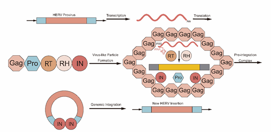
Figure 3: The HERV transposition process
Figure 4
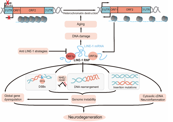
Figure 4: The potential detrimental impact of LINE-1 on the genetic material of cells
Figure 5
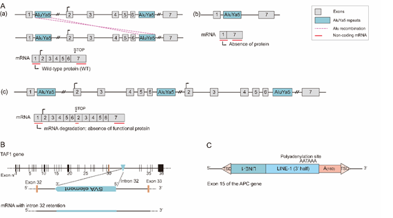
Figure 5: Some examples of TE insertion genes [207]
Figure 6
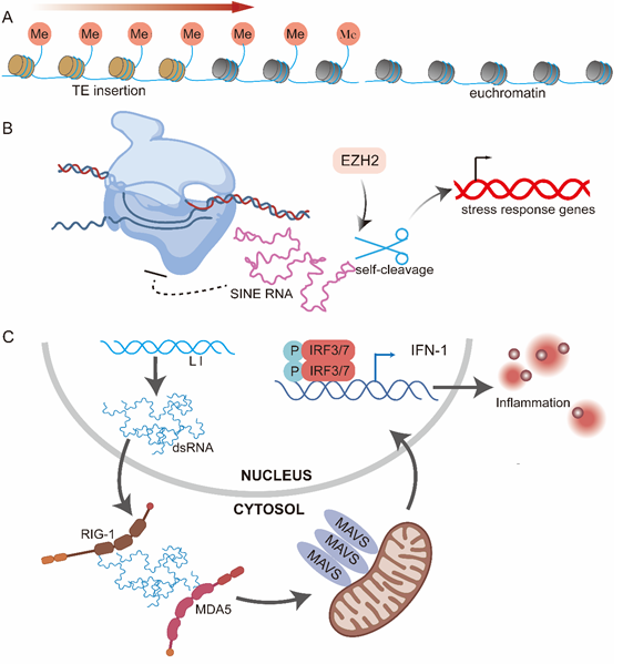
Figure 6: Aberrant Gene Expression Following Transposable Element Insertions
Figure 7
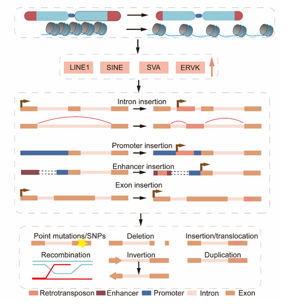
Figure 7: Activation of retrotransposons upon telomere shortening destabilizes the host genome by insertion into various genomic loci [170].
Figure 8
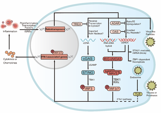
Figure 8: TE activation-induced inflammation in aging and age-related diseases
TE activation-induced DNA damage contributes to aging and age-related diseases
When we look at those long-lived animals, the genome of the longest-lived rodent, the naked mole rat, contains fewer retrotransposons than other rodent [185]. Interestingly, although long-lived bats have genomes rich in TEs [186], they have evolved dampened responses to cytoplasmic DNA [187]. Moreover, in higher termites, relatively long-lived reproductive queens have lower TE activity than short-lived sterile workers [188]. It's worth mentioning that there were an increasing number of TEs but decreasing TE expression levels in longer-lived Drosophila genomes which could be due to increased opportunities for newly inserted TEs to be passed on to future generations [189]. In the middle-aged ovaries of the naturally shorter-lived African turquoise killifish, PIWI-pathway components were temporarily reduced, piRNA levels fell and TEs were expressed at higher levels, indicating that the egg quality has lowered [190]. Cellular senescence is an irreversible proliferative arrest that can be caused by a variety of stresses, including DNA damage [181]. Although senescence has beneficial functions (such as tumor suppression), senescent cells accumulate in most tissues with age, and they are an important part of the overall aging process [79]. A good example of this is Drosophila. DNA damage-mediated cell death is probably the main reason for the deleterious effects of retrotransposons on Drosophila, which lacks an interferon system [191]. Furthermore, in Drosophila, “toxic Y chromosomes”, with more repeats than X, are believed to reduce the health of males [192]. However, let me say again?for mammals, retrotransposons, DNA damage, interferon, and inflammation all interconnect [193]. For instance, retrotransposons help create DNA damage, but on the other hand, DNA damage leads to retrotransposon activation; this forms a vicious positive cycle [194]. Inflammation causes different types of DNA damage through various mechanisms, but in turn, DNA damage can induce an inflammatory response. Retropsectons also lead to the triggering of innate immune sensors, but interferons also have control on retrotransposons (discussed below) [195]. And given that we are unaware of how frequently retrotransposition-induced damage occurs, it might well be of little consequence to our cells overall [193] and similarly, a recent study found that the number of transposon insertions did not increase significantly with age and suggested that transposon expression rather than insertion plays a key role in regulating lifespan [196]. Nonetheless, transposons have been shown to cause mutations in cells. At least 124 diseases are known to result from transposable element insertions [197], 118 mutations in human disease are due to retrotransposons [198] and more than 120 L1-mediated germline insertions have been associated with human genetic diseases [197]. Subsequent efforts have identified multiple de novo somatic insertions and associated rearrangements in human cancers, which in some cases can number in the hundreds in a single tumor [199-202]. Upregulated TEs could induce genome instability, activate oncogenes, or inhibit tumor suppressors, leading to cancer initiation and progression [203]. For example, TEs could insert in important suppressor genes, leading to poor prognosis and cancer development [203]. Notably, whole genome sequencing (WGS) of 43 cancer samples from either colorectal, prostate, ovarian, multiple myeloma, and glioblastoma origin, and their matched normal blood samples, found that LINE-1 and Alu somatic insertions were more common in cancers of epithelial origin (e.g., colorectal, prostate, and ovarian), as compared to blood or brain cancers [199]. However, most de novo insertions in cancers are located in non-coding sequences and do not show hotspots near tumor suppressor or oncogene loci, so there is little current evidence that de novo L1 insertions play an important role in tumorigenesis [204]. Similarly, retrotransposition events in brain malignancies are either absent or extremely rare compared to normal brain tissues [205].
Some examples:
Examples of cancers and leukemias related to Alu and LINE-1 insertion -- see [207]
Examples of non-cancerous diseases (hemoglobinopathies, metabolic and neurological diseases as well as common diseases) related to TE insertion -- see [207]
Some examples of studies associating brain-related diseases with Transposable elements (TEs) activity – see [205]
To determine the relationship between aging and DNA damage, it is indispensable to measure the rate of DNA damage. Retrotransposition during development or adulthood would lead to somatic mosaicism, and has been estimated that adult neurons undergo retrotransposition with varying (but generally low) frequency. Only a small fraction of L1 transcripts in the cytoplasm end up being retrotransposed in somatic cells [208]. The discrepancies in measurements may arise due to different methods and different samples used to quantify retrotransposition events [205]:
|
Methods |
Samples |
Findings |
References |
|
qPCR |
postmortem human tissue from brain regions, liver, and heart
|
the ORF2 copy number increases by about 80 copies per cell |
Coufal et al., 2009[209] |
|
retrotransposon capture sequencing (RC-seq) |
hippocampus and caudate nucleus of three individuals
|
thousands of putative somatic L1, Alu and SVA insertions |
Baillie et al., 2011[210] |
|
single neuron sequencing |
300 single neurons from the cortex and caudate regions of the human brain
|
less than 0.6 unique somatic insertions per neuron in these regions |
Evrony et al., 2012[211] |
|
RC-seq |
neurons and glia of the human hippocampus as well as cortical neurons
|
13.7 somatic L1 insertions per hippocampal neuron |
Upton et al., 2015 [212] |
|
-- |
-- |
the rate of L1 insertion is about 0.2 per hippocampal neurons |
Evrony et al., 2016[213] |
|
high-throughput sequencing method and machine learning-based analysis |
in bulk tissue and single nuclei from the frontal cortex and hippocampus of three healthy individuals |
approximately 0.58–1 somatic event per cell occurred in both glia and neurons |
Erwin et al., 2016[214] |
|
human active transposon sequencing (HAT-seq) |
in the prefrontal cortex neurons |
estimated 0.63–1.66 L1 somatic insertions per prefrontal cortex neuron in healthy people |
Zhao et al., 2019[215] |
|
Methods |
Samples |
Findings |
References |
|
qPCR |
postmortem human tissue from brain regions, liver, and heart
|
the ORF2 copy number increases by about 80 copies per cell |
Coufal et al., 2009[209] |
|
retrotransposon capture sequencing (RC-seq) |
hippocampus and caudate nucleus of three individuals
|
thousands of putative somatic L1, Alu and SVA insertions |
Baillie et al., 2011[210] |
|
single neuron sequencing |
300 single neurons from the cortex and caudate regions of the human brain
|
less than 0.6 unique somatic insertions per neuron in these regions |
Evrony et al., 2012[211] |
|
RC-seq |
neurons and glia of the human hippocampus as well as cortical neurons
|
13.7 somatic L1 insertions per hippocampal neuron |
Upton et al., 2015[212] |
|
-- |
-- |
the rate of L1 insertion is about 0.2 per hippocampal neurons |
Evrony et al., 2016[213] |
|
high-throughput sequencing method and machine learning-based analysis |
in bulk tissue and single nuclei from the frontal cortex and hippocampus of three healthy individuals |
approximately 0.58–1 somatic event per cell occurred in both glia and neurons |
Erwin et al., 2016[214] |
|
human active transposon sequencing (HAT-seq) |
in the prefrontal cortex neurons |
estimated 0.63–1.66 L1 somatic insertions per prefrontal cortex neuron in healthy people |
Zhao et al., 2019[215] |
Notably, these detection methods were presumably re-tuned over time. The earliest qPCR method comes with drawbacks. First, copies of L1 introduced as plasmid should provide incorrect copy number [209]. Second, it may give rise to over-estimate due to the reverse transcription of L1 present in cytoplasm and possibly not integrated into the genome [216]. Likewise, the latter RC-seq method also has some shortcomings, such as sequencing error and insufficient sequencing depth, in contrast to single neuron sequencing, which has a higher resolution of cellular differences and can reveal genetic heterogeneity of cell populations [211]. Finally, the high-throughput sequencing method and machine-learning-based analysis offer several advantages over previous methods, including higher sensitivity and efficiency, use of non-PCR based method of fragmentation and adaptor ligation, more confident detection of novel insertions, and machine-learning-based prediction of variants [214]. Incidentally, Evrony et al. [213], identified different factors affecting single-cell sequencing results during their study that led to thousands of artifacts being interpreted as somatic L1 insertion events. Furthermore, the rate of retrotransposition is also influenced by tissue specificity. Although the frequency of retrotransposition in other tissues has not been investigated in detail, they appear to be significantly lower, with the highest expression of retroelement-derived sequences in the healthy brain compared with other somatic tissues [217]. In both the human and Drosophila brains, some retrotransposons are not only expressed but also actively transposed, and it has been suggested that they contribute to the diversification of neuronal cell populations [210, 218]. Additional analyses also demonstrated that age-dependent derepression of TEs observed in somatic cells of aged flies has not been detected in their germline (possibly due to a higher expression level of piRNAs), which may explain the preservation of older germline genomes in general. Somewhat interestingly, TEs appear to be enriched with more enhancers compared with Somatic Lineages in hemopoietic lineage [219]. Importantly, it isn’t only TE-induced DNA damage, but also many mechanisms that result from TEs that affect aging and age-related diseases, like inflammation (Figure 7). It is well-known that free DNA or dsRNA in the cytosol is often regarded as an invading pathogen and, as we mentioned, is produced by TEs during the retrotransposition process. For cDNAs that are present in the cytosol due to the reverse transcription occurring in the absence of a standard DNA template [220] or the export from the nucleus [221], they can be degraded by the exonuclease TREX1[167]. If the level of cDNA is too high, it will be sensed by cGAS(183), which then triggers the synthesis of cyclic GMP-AMP (cGAMP) [222] and subsequently the sequential activation of STING (stimulator of interferon genes), TBK1 (TRAF family member-associated NF-kappa-B activator (TANK)-binding kinase 1), and IRF3 (interferon regulatory factor 3) [193]. For dsRNAs that are present in the cytosol due to the upregulation of retroTEs [223,224], they are mainly inverted Alu repeats [225] and can be inhibited by the deaminase ADAR [225] or the oligo adenylate synthetase (OAS) family [226, 227]. dsRNAs or RNA:DNA hybrids [228] can be sensed by either MDA5 or RIG-I [229, 230], which then trigger the aggregation of MAVS, and subsequently the sequential activation of TBK1 and IKK?, and IRF3/IRF7 and NF-κB [231]. Hence, L1 dysregulation is a common theme among diseases with chronic induction of type I IFN signalling through cGAS-STING, such as Aicardi–Goutières syndrome, Fanconi anemia, and dermatomyositis. In addition to the IFN-I pathway, dsRNA can also do damage to cells through other pathways such as STAU1-mediated mRNA decay (SMD) [232] and ZBP1-dependent necroptosis [233]. In addition, proteins can also induce inflammation. Certain HERVs are able to encode an intact envelope protein (env) and assemble into virus-like particles, which have been observed in some aging-related diseases [234-237]. Importantly, virus-like particles constitute a transmissible message that can make young cells appear senescent [238]. The env is now considered a “superantigen” that can trigger polyclonal activation of lymphocytes [239] and nucleic acids of the virus-like particle in the endosome can be sensed by TLRs (toll-like receptors) [240]. All in all, these signals from either cDNA, dsRNA, or proteins are propagated to the nucleus, where they initiate the type-I interferon (IFN-I) response that generates a variety of cytokines and chemokines [193], leading to inflammation. Notably, the inflammatory may reciprocally enhance ERVK transcription, as certain ERVK promoters contain interferon-stimulated response elements (ISREs), while others contain conserved binding motifs to proinflammatory transcription factors such as NF-κB [241].Other than DNA damage and inflammation, there exist many other possible mechanisms induced by TEs that may contribute to aging. For example, TE-derived miRNAs and lncRNAs [242-244], mitochondrial genome dysfunction [245], circular RNAs (emerging species of regulatory RNAs relevant to aging) [246], translational repression [247], and so on. In conclusion, there is indeed much evidence for TE-induced DNA damage in aging and age-related diseases. However, the frequency of TE retrotransposition in senescent cells (instead of healthy cells) from different tissues remains unknown. Thus a new detection method may be required to accurately measure TE-induced DNA damage, not just insertions in senescent cells. What’s more, to what extent is DNA damage correlated with aging? Even if DNA damage occurs, does it rarely occur in critical regions of the genome? Are other mechanisms more relevant to aging, such as inflammation? Importantly, whether the de-repression of TEs is a cause or consequence of aging still remains unclear. The ERV de-repression due to intracellular and extracellular effects is reported to be seemingly merely complementary to major stressful events, such as DNA damage [82] or activation of the underlying inflammatory state [248]. And although L1 copy number increases in senescent cells of humans [249], changes in health or longevity of transgenic animals with hyperactive L1 or Alu elements have not yet been adequately studied [250]. Thus, the role of TE-induced DNA damage in aging remains to be further investigated.
Therapies opportunities
The role of retrotransposons in aging and age-related diseases provides an opportunity to develop novel therapies since retrotransposons may causally contribute to the aging process and interventions that inhibit retrotransposon activity may improve healthy longevity [193]. First of all, the Nucleoside Reverse Transcriptase Inhibitors (NRTIs), such as Abacavir, Lamivudine, Azidothymidine (AZT), and Zidovudine Stavudine (STV). Treatment with NRTIs that inhibit L1 reverse transcriptase reduces cytoplasmic L1 cDNAs and ameliorates cGAS and IFN-I activation [167]. In mice, treatment with NRTIs doubles the lifespan of progeroid SIRT6-deficient mice and improves overall health including bone density, muscle mass, intestinal function, and exercise performance [183]. Treatment of middle-aged normal mice with NRTIs retards the progression of aging biomarkers, particularly DNA methylation age and p16INK4A expression [183]. In addition, NRTIs can be beneficial for cancer treatment, by reducing the survival rate to anticancer drug therapy, since L1s expression increases the rate of mutation and thus develops drug resistance [251]. Interestingly, beyond their effects on inhibiting reverse transcription, NRTIs also limit the production of retrotransposon-derived RNA or protein products [167]. However, NRTIs are not without side effects. Chronic NRTI treatment has been shown to induce adverse side effects in human patients, such as hepatotoxicity, as it also inhibits the mitochondrial DNA polymerase γ [252, 253]. Moreover, NRTIs also inhibit telomerase and are thought to contribute to accelerated aging phenotypes associated with HIV patients [254, 255]. Therefore, more targeted interventions need to be developed to safely and effectively treat conditions caused by uncontrolled L1 activity. For example, strategies to promote cytoplasmic L1 cDNA degradation [256]. Incidentally, two non-nucleoside reverse transcriptase inhibitors (NNRTIs), nevirapine and efavirenz, have also been found to inhibit the dedifferentiation in various cancer cells [257]. What’s more, anti-inflammatory drugs can be used to treat autoimmune disorders, such as small molecule inhibitors of STING [258]. and cGAS inhibitors are being developed [259]. The disadvantage of such treatments that over-target immunity mechanisms is that they may increase susceptibility to infections. The most direct approach to inhibiting retrotransposons may be to enhance the epigenetic mechanisms responsible for their silencing, especially those that are impaired with age. One such epigenetic regulator is SIRT6, as male mice overexpressing SIRT6 have an extended lifespan [260]. Small molecule activators of SIRT6 are currently being developed and may have a range of health benefits, including improved retrotransposon silencing [193]. Other anti-aging interventions and medications include caloric restriction, acarbose, rapamycin [261], and metformin [262], which could reduce TE overexpression and be implemented to postpone aging and cancer. In addition, SINE B1 antisense RNA can inhibit the aging process by enhancing antioxidant activity and regulating the expression of aging-associated genes [263]. also, interventions such as CRISPR-mediated inhibition of ERV activation or the use of antibodies to block HERV-K env have shown the ability to counteract ERV-mediated pro-senescence effects [264]. Antibodies against HERV-K env or MSRV env have been employed in preclinical investigations as a means of treating age-related disorders and multiple sclerosis, respectively [238,265], which highlights the potential of HERV env proteins as promising targets for antibody-based therapy. As an increasing number of diseases are associated with HERVs, vaccination against HERV proteins offers an exciting opportunity to prevent a wide range of diseases and it has been proven safe and immunogenic [266-268]. Moreover, engineering chimeric antigen receptors (CARs) that recognize HERVs would enable T cells to limit tumor growth [269,270]. Notably, the potential for using HERV-specific T cells in therapy is not limited to cancer treatment. It may also be used to treat aging and age-related diseases since HERV-K env is highly expressed in senescent cells [238]. Notably, apart from inhibiting TEs, sometimes unleashing them also has benefits. It has been proposed that DNA methyltransferase inhibitors (DNMTis) treatment can sensitize patients with a variety of cancer types to immune checkpoint therapy [224]. Unleashing the expression of TEs by DNA Hypomethylating Agents (DHAs) and histone deacetylase inhibitors (HDACi) may reawaken the immune response to senescent and cancerous cells and enhance immune surveillance against these life-threatening cells [203]. Several environmental factors affect TE activation and aging as well. Five months of voluntary wheel running downregulates the expression of the LINE-1 gene in rat skeletal muscle [271]. Deficiencies of folic acid (vitamin B9), B6, or B12 can lead to loss of methyl donor and hypomethylation of L1, resulting in genomic instability [166]. Alu methylation may be affected by fiber-rich foods and antioxidants, or circulating LDL subfractions and plasma copper [272]. Trans fatty acids may cause hypomethylation, which may be counteracted by extra virgin olive oil [158]. What’s more, retroTEs can be used as biomarkers. Biological changes in retroTE expression that occur with physiological aging may be useful marks for selecting the appropriate timing of therapeutic interventions (e.g. selectively removal of senescent cells) [273]. and the L1-encoded ORF1p may serve as noninvasive and multicancer biomarkers [274]. Incidentally, some genes associated with longevity have been reported. Alu deletions in LAMA2 and CDH4 genes are key components of polygenic predictors of longevity [275]. and recently, new allelic variants of SIRT6 (N308K/A313S) have also been associated with longevity [276].
Conclusion
There is a close relationship between TE and the aging process and age-related diseases. Although transposable elements (TEs) play a positive role in the process of genetic diversity and evolution of organisms, TE activity may lead to the DNA damage and genomic instability, thus causing the features of aging. There are different types of TE-induced DNA damage mechanisms. These include the direct generation of double- and single-strand breaks, structural rearrangements, and alterations in gene expression. All these result in loss of normal cellular function, leading to oncogenic, cardiopathogenic or neuropathogenic effects. Regulation of TE activity occurs via numerous epigenetic checks and balances, including DNA and RNA methylation, histone modifications, and nuclear architecture. When regulation is unbalanced, TEs can get activated, resulting in further DNA damage. In short, TEs activation creates an infinite loop of damage/inflammation, which may also induce more TE activation by constantly affecting epigenetics. Targeting TE activation and its associated molecular processes might be beneficial in preventing age-associated decline, aging, and age-related diseases. Nucleoside RTIs, anti-inflammatories, and epigenetic modifiers represent potential interventions on TE expression and TE-induced damage of DNA. Developing novel TE detection methodologies and biomarkers will greatly enhance our ability to pinpoint TE activity in vivo, decipher TE activity in driving the aging process, and ultimately lead to tailored therapy.
Declaration of Competing Interest
The author declare that there are not conflicts of interests.
Acknowledgements
Xianli Wang conceived and designed the study; Jingran Hu, Tianhao Mao and Kainan Huang wrote the manuscript and drew the figures; Jingran Hu, Tianhao Mao, Wenrui Yu, Jiacheng Huang, Haocheng Sun and Shangzhi Yang handle project and administration and resoures.
References
- Wells JN, Feschotte C. A Field Guide to Eukaryotic Transposable Elements. Annu Rev Genet. 2020; 54: 539-561.
- López-Otín C, Blasco MA, Partridge L, Serrano M, Kroemer G. Hallmarks of aging: An expanding universe. Cell. 2023; 186: 243-278.
- Finnegan DJ. Transposable elements. Curr Opin Genet Dev. 1992; 2: 861-867.
- Jurka J, Kapitonov VV, Pavlicek A, Klonowski P, Kohany O, Walichiewicz J. Repbase Update, a database of eukaryotic repetitive elements. Cytogenet Genome Res. 2005; 110: 462-467.
- Kapitonov VV, Jurka J. A universal classification of eukaryotic transposable elements implemented in Repbase. Nat Rev Genet. 2008; 9: 411-412.
- Wicker T, Sabot F, Hua-Van A, Bennetzen JL, Capy P, Chalhoub B, et al. A unified classification system for eukaryotic transposable elements. Nat Rev Genet. 2007; 8: 973-982.
- Curcio MJ, Derbyshire KM. The outs and ins of transposition: from mu to kangaroo. Nat Rev Mol Cell Biol. 2003; 4: 865-877.
- Di Stefano L. All Quiet on the TE Front? The Role of Chromatin in Transposable Element Silencing. Cells. 2022; 11: 2501.
- Arkhipova IR. Using bioinformatic and phylogenetic approaches to classify transposable elements and understand their complex evolutionary histories. Mob DNA. 2017; 8: 19.
- Finnegan DJ. Eukaryotic transposable elements and genome evolution. Trends Genet. 1989; 5: 103-107.
- Engels WR, Johnson-Schlitz DM, Eggleston WB, Sved J. High-frequency P element loss in Drosophila is homolog dependent. Cell. 1990; 62: 515-525.
- Naito K, Cho E, Yang G, Campbell MA, Yano K, Okumoto Y, et al. Dramatic amplification of a rice transposable element during recent domestication. Proc Natl Acad Sci U S A. 2006; 103: 17620-17625.
- Doak TG, Doerder FP, Jahn CL, Herrick G. A proposed superfamily of transposase genes: transposon-like elements in ciliated protozoa and a common "D35E" motif. Proc Natl Acad Sci U S A. 1994; 91: 942-946.
- Hickman AB, Dyda F. DNA Transposition at Work. Chem Rev. 2016; 116: 12758-12784.
- Feschotte C, Pritham EJ. DNA transposons and the evolution of eukaryotic genomes. Annu Rev Genet. 2007; 41: 331-368.
- Kapitonov VV, Jurka J. Rolling-circle transposons in eukaryotes. Proc Natl Acad Sci U S A. 2001; 98: 8714-8719.
- Grabundzija I, Hickman AB, Dyda F. Helraiser intermediates provide insight into the mechanism of eukaryotic replicative transposition. Nat Commun. 2018; 9: 1278.
- Feschotte C, Pritham EJ. Non-mammalian c-integrases are encoded by giant transposable elements. Trends Genet. 2005; 21: 551-552.
- Kapitonov VV, Jurka J. Self-synthesizing DNA transposons in eukaryotes. Proc Natl Acad Sci U S A. 2006; 103: 4540-4545.
- Krupovic M, Bamford DH, Koonin EV. Conservation of major and minor jelly-roll capsid proteins in Polinton (Maverick) transposons suggests that they are bona fide viruses. Biol Direct. 2014; 9: 6.
- Ochoa Thomas E, Zuniga G, Sun W, Frost B. Awakening the dark side: retrotransposon activation in neurodegenerative disorders. Curr Opin Neurobiol. 2020; 61: 65-72.
- Lander ES, Linton LM, Birren B, Nusbaum C, Zody MC, Baldwin J, et al. Initial sequencing and analysis of the human genome. Nature. 2001; 409: 860-921.
- Magiorkinis G, Gifford RJ, Katzourakis A, De Ranter J, Belshaw R. Env-less endogenous retroviruses are genomic superspreaders. Proc Natl Acad Sci U S A. 2012; 109: 7385-7390.
- Ribet D, Harper F, Dupressoir A, Dewannieux M, Pierron G, Heidmann T. An infectious progenitor for the murine IAP retrotransposon: emergence of an intracellular genetic parasite from an ancient retrovirus. Genome Res. 2008; 18: 597-609.
- Piégu B, Bire S, Arensburger P, Bigot Y. A survey of transposable element classification systems--a call for a fundamental update to meet the challenge of their diversity and complexity. Mol Phylogenet Evol. 2015; 86: 90-109.
- Coffin J, Blomberg J, Fan H, Gifford R, Hatziioannou T, Lindemann D, et al. ICTV Virus Taxonomy Profile: Retroviridae 2021. J Gen Virol. 2021; 102: 001712.
- Jern P, Sperber GO, Blomberg J. Use of endogenous retroviral sequences (ERVs) and structural markers for retroviral phylogenetic inference and taxonomy. Retrovirology. 2005; 2: 50.
- Burke WD, Calalang CC, Eickbush TH. The site-specific ribosomal insertion element type II of Bombyx mori (R2Bm) contains the coding sequence for a reverse transcriptase-like enzyme. Mol Cell Biol. 1987; 7: 2221-2230.
- Richardson SR, Doucet AJ, Kopera HC, Moldovan JB, Garcia-Perez JL, Moran JV. The Influence of LINE-1 and SINE Retrotransposons on Mammalian Genomes. Microbiol Spectr. 2015; 3.
- Luan DD, Korman MH, Jakubczak JL, Eickbush TH. Reverse transcription of R2Bm RNA is primed by a nick at the chromosomal target site: a mechanism for non-LTR retrotransposition. Cell. 1993; 72: 595-605.
- Mills RE, Bennett EA, Iskow RC, Luttig CT, Tsui C, Pittard WS, et al. Recently mobilized transposons in the human and chimpanzee genomes. Am J Hum Genet. 2006; 78: 671-679.
- Kramerov DA, Vassetzky NS. Origin and evolution of SINEs in eukaryotic genomes. Heredity (Edinb). 2011; 107: 487-495.
- Goodwin TJ, Poulter RT. A new group of tyrosine recombinase-encoding retrotransposons. Mol Biol Evol. 2004; 21: 746-759.
- Ribeiro YC, Robe LJ, Veluza DS, Dos Santos CMB, Lopes ALK, Krieger MA , et al. Study of VIPER and TATE in kinetoplastids and the evolution of tyrosine recombinase retrotransposons. Mob DNA. 2019; 10: 34.
- Cappello J, Handelsman K, Lodish HF. Sequence of Dictyostelium DIRS-1: an apparent retrotransposon with inverted terminal repeats and an internal circle junction sequence. Cell. 1985; 43: 105-115.
- Poulter RTM, Butler MI. Tyrosine Recombinase Retrotransposons and Transposons. Microbiol Spectr. 2015; 3.
- Evgen'ev MB, Zelentsova H, Shostak N, Kozitsina M, Barskyi V, Lankenau DH, et al. Penelope, a new family of transposable elements and its possible role in hybrid dysgenesis in Drosophila virilis. Proc Natl Acad Sci U S A. 1997; 94: 196-201.
- Evgen'ev MB, Arkhipova IR. Penelope-like elements--a new class of retroelements: distribution, function and possible evolutionary significance. Cytogenet Genome Res. 2005; 110: 510-521.
- Mills RE, Bennett EA, Iskow RC, Devine SE. Which transposable elements are active in the human genome? Trends Genet. 2007; 23: 183-191.
- Kurth R, Bannert N. Beneficial and detrimental effects of human endogenous retroviruses. Int J Cancer. 2010; 126: 306-314.
- Brouha B, Schustak J, Badge RM, Lutz-Prigge S, Farley AH, Moran JV, et al. Hot L1s account for the bulk of retrotransposition in the human population. Proc Natl Acad Sci U S A. 2003; 100: 5280-5285.
- Batzer MA, Deininger PL. Alu repeats and human genomic diversity. Nat Rev Genet. 2002; 3: 370-379.
- Cordaux R, Batzer MA. The impact of retrotransposons on human genome evolution. Nat Rev Genet. 2009; 10: 691-703.
- Denli AM, Narvaiza I, Kerman BE, Pena M, Benner C, Marchetto MC, et al. Primate-specific ORF0 contributes to retrotransposon-mediated diversity. Cell. 2015; 163: 583-593.
- Mendez-Dorantes C, Burns KH. LINE-1 retrotransposition and its deregulation in cancers: implications for therapeutic opportunities. Genes Dev. 2023; 37: 948-967.
- Doucet AJ, Wilusz JE, Miyoshi T, Liu Y, Moran JV. A 3' Poly(A) Tract Is Required for LINE-1 Retrotransposition. Mol Cell. 2015; 60: 728-741.
- Hancks DC, Kazazian HH Jr. SVA retrotransposons: Evolution and genetic instability. Semin Cancer Biol. 2010; 20: 234-245.
- Wang H, Xing J, Grover D, Hedges DJ, Han K, Walker JA, et al. SVA elements: a hominid-specific retroposon family. J Mol Biol. 2005; 354: 994-1007.
- Chu C, Lin EW, Tran A, Jin H, Ho NI, Veit A, et al. The landscape of human SVA retrotransposons. Nucleic Acids Res. 2023; 51: 11453-11465.
- Ostertag EM, Goodier JL, Zhang Y, Kazazian HH Jr. SVA elements are nonautonomous retrotransposons that cause disease in humans. Am J Hum Genet. 2003; 73: 1444-1451.
- Löwer R, Löwer J, Kurth R. The viruses in all of us: characteristics and biological significance of human endogenous retrovirus sequences. Proc Natl Acad Sci U S A. 1996; 93: 5177-5184.
- Jansz N, Faulkner GJ. Endogenous retroviruses in the origins and treatment of cancer. Genome Biol. 2021; 22: 147.
- Jern P, Sperber GO, Blomberg J. Definition and variation of human endogenous retrovirus H. Virology. 2004; 327: 93-110.
- Thomas J, Perron H, Feschotte C. Variation in proviral content among human genomes mediated by LTR recombination. Mob DNA. 2018; 9: 36.
- Kulpa DA, Moran JV. Cis-preferential LINE-1 reverse transcriptase activity in ribonucleoprotein particles. Nat Struct Mol Biol. 2006; 13: 655-660.
- Khazina E, Truffault V, Büttner R, Schmidt S, Coles M, Weichenrieder O. Trimeric structure and flexibility of the L1ORF1 protein in human L1 retrotransposition. Nat Struct Mol Biol. 2011; 18: 1006-1014.
- Mita P, Wudzinska A, Sun X, Andrade J, Nayak S, Kahler DJ, et al. LINE-1 protein localization and functional dynamics during the cell cycle. Elife. 2018; 7: e30058.
- Cost GJ, Feng Q, Jacquier A, Boeke JD. Human L1 element target-primed reverse transcription in vitro. EMBO J. 2002; 21: 5899-5910.
- Feng Q, Moran JV, Kazazian HH Jr, Boeke JD. Human L1 retrotransposon encodes a conserved endonuclease required for retrotransposition. Cell. 1996; 87: 905-916.
- Symer DE, Connelly C, Szak ST, Caputo EM, Cost GJ, Parmigiani G, et al. Human l1 retrotransposition is associated with genetic instability in vivo. Cell. 2002; 110: 327-338.
- Beck CR, Garcia-Perez JL, Badge RM, Moran JV. LINE-1 elements in structural variation and disease. Annu Rev Genomics Hum Genet. 2011; 12: 187-215.
- Mathias SL, Scott AF, Kazazian HH Jr, Boeke JD, Gabriel A. Reverse transcriptase encoded by a human transposable element. Science. 1991; 254: 1808-1810.
- Malik HS, Burke WD, Eickbush TH. The age and evolution of non-LTR retrotransposable elements. Mol Biol Evol. 1999; 16: 793-805.
- Kojima KK. Different integration site structures between L1 protein-mediated retrotransposition in cis and retrotransposition in trans. Mob DNA. 2010; 1: 17.
- Weichenrieder O, Wild K, Strub K, Cusack S. Structure and assembly of the Alu domain of the mammalian signal recognition particle. Nature. 2000; 408: 167-173.
- Hancks DC, Goodier JL, Mandal PK, Cheung LE, Kazazian HH Jr. Retrotransposition of marked SVA elements by human L1s in cultured cells. Hum Mol Genet. 2011; 20: 3386-3400.
- Esnault C, Maestre J, Heidmann T. Human LINE retrotransposons generate processed pseudogenes. Nat Genet. 2000; 24: 363-367.
- Kovalskaya E, Buzdin A, Gogvadze E, Vinogradova T, Sverdlov E. Functional human endogenous retroviral LTR transcription start sites are located between the R and U5 regions. Virology. 2006; 346: 373-378.
- Gerdes P, Richardson SR, Mager DL, Faulkner GJ. Transposable elements in the mammalian embryo: pioneers surviving through stealth and service. Genome Biol. 2016; 17: 100.
- Mager DL, Henthorn PS. Identification of a retrovirus-like repetitive element in human DNA. Proc Natl Acad Sci U S A. 1984; 81: 7510-7514.
- Cohen CJ, Lock WM, Mager DL. Endogenous retroviral LTRs as promoters for human genes: a critical assessment. Gene. 2009; 448: 105-114.
- Chuong EB, Rumi MA, Soares MJ, Baker JC. Endogenous retroviruses function as species-specific enhancer elements in the placenta. Nat Genet. 2013; 45: 325-329.
- Bourque G, Leong B, Vega VB, Chen X, Lee YL, Srinivasan KG, et al. Evolution of the mammalian transcription factor binding repertoire via transposable elements. Genome Res. 2008; 18: 1752-1762.
- Wolf G, Yang P, Füchtbauer AC, Füchtbauer EM, Silva AM, Park C, et al. The KRAB zinc finger protein ZFP809 is required to initiate epigenetic silencing of endogenous retroviruses. Genes Dev. 2015; 29: 538-554.
- Gasior SL, Wakeman TP, Xu B, Deininger PL. The human LINE-1 retrotransposon creates DNA double-strand breaks. J Mol Biol. 2006; 357: 1383-1393.
- Belgnaoui SM, Gosden RG, Semmes OJ, Haoudi A. Human LINE-1 retrotransposon induces DNA damage and apoptosis in cancer cells. Cancer Cell Int. 2006; 6: 13.
- Kazazian HH Jr, Moran JV. Mobile DNA in Health and Disease. N Engl J Med. 2017; 377: 361-370.
- Kazazian HH Jr. Mobile elements and disease. Curr Opin Genet Dev. 1998; 8: 343-350.
- Childs BG, Gluscevic M, Baker DJ, Laberge RM, Marquess D, Dananberg J, et al. Senescent cells: an emerging target for diseases of ageing. Nat Rev Drug Discov. 2017; 16: 718-735.
- Douville R, Liu J, Rothstein J, Nath A. Identification of active loci of a human endogenous retrovirus in neurons of patients with amyotrophic lateral sclerosis. Ann Neurol. 2011; 69: 141-151.
- Lesbats P, Engelman AN, Cherepanov P. Retroviral DNA Integration. Chem Rev. 2016; 116: 12730-12757.
- Bray S, Turnbull M, Hebert S, Douville RN. Insight into the ERVK Integrase - Propensity for DNA Damage. Front Microbiol. 2016; 7: 1941.
- Peze-Heidsieck E, Bonnifet T, Znaidi R, Ravel-Godreuil C, Massiani-Beaudoin O, Joshi RL et.al. Retrotransposons as a Source of DNA Damage in Neurodegeneration. Front Aging Neurosci. 2022; 13: 786897.
- Pellestor F, Gatinois V. Chromoanagenesis: a piece of the macroevolution scenario. Mol Cytogenet. 2020; 13.
- Zhao Y, Simon M, Seluanov A, Gorbunova V. DNA damage and repair in age-related inflammation. Nat Rev Immunol. 2023; 23: 75-89.
- Niehrs C, Luke B. Regulatory R-loops as facilitators of gene expression and genome stability. Nat Rev Mol Cell Biol. 2020; 21: 167-178.
- Gan W, Guan Z, Liu J, Gui T, Shen K, Manley JL et.al. R-loop-mediated genomic instability is caused by impairment of replication fork progression. Genes Dev. 2011; 25: 2041-2056.
- Costantino L, Koshland D. Genome-wide Map of R-Loop-Induced Damage Reveals How a Subset of R-Loops Contributes to Genomic Instability. Mol Cell. 2018; 71: 487-497.e483.
- Goulielmaki E, Tsekrekou M, Batsiotos N, Ascensão-Ferreira M, Ledaki E, Stratigi K et al. The splicing factor XAB2 interacts with ERCC1-XPF and XPG for R-loop processing. Nat Commun. 2021; 12.
- Marabitti V, Lillo G, Malacaria E, Palermo V, Sanchez M, Pichierri P et.al. ATM pathway activation limits R-loop-associated genomic instability in Werner syndrome cells. Nucleic Acids Res. 2019; 47: 3485-3502.
- Ravel-Godreuil C, Znaidi R, Bonnifet T, Joshi RL, Fuchs J. Transposable elements as new players in neurodegenerative diseases. FEBS Lett. 2021; 595: 2733-2755.
- Blaudin de Thé FX, Rekaik H, Peze-Heidsieck E, Massiani-Beaudoin O, Joshi RL, Fuchs J et.al. Engrailed homeoprotein blocks degeneration in adult dopaminergic neurons through LINE-1 repression. EMBO J. 2018; 37: e97374.
- Mojica EA, Kültz D. Physiological mechanisms of stress-induced evolution. J Exp Biol. 2022; 225: jeb243264.
- Cheng CH, Guo ZX, Luo SW, Wang AL. Effects of high temperature on biochemical parameters, oxidative stress, DNA damage and apoptosis of pufferfish (Takifugu obscurus). Ecotoxicol Environ Saf. 2018; 150: 190-198.
- Kültz D. Molecular and evolutionary basis of the cellular stress response. Annu Rev Physiol. 2005; 67: 225-257.
- Grollman AP, Moriya M. Mutagenesis by 8-oxoguanine: an enemy within. Trends Genet. 1993; 9: 246-249.
- Honda S, Hjelmeland LM, Handa JT. Oxidative stress--induced single-strand breaks in chromosomal telomeres of human retinal pigment epithelial cells in vitro. Invest Ophthalmol Vis Sci. 2001; 42: 2139-2144.
- Sallmyr A, Fan J, Rassool FV. Genomic instability in myeloid malignancies: increased reactive oxygen species (ROS), DNA double strand breaks (DSBs) and error-prone repair. Cancer Lett. 2008; 270: 1-9.
- Voineagu I, Narayanan V, Lobachev KS, Mirkin SM. Replication stalling at unstable inverted repeats: interplay between DNA hairpins and fork stabilizing proteins. Proc Natl Acad Sci U S A. 2008; 105: 9936-9941.
- Lu S, Wang G, Bacolla A, Zhao J, Spitser S, Vasquez KM. Short Inverted Repeats Are Hotspots for Genetic Instability: Relevance to Cancer Genomes. Cell Rep. 2015; 10: 1674-1680.
- Bushman FD. DNA transposon mechanisms and pathways of genotoxicity. Mol Ther. 2023; 31: 613-615.
- Chen Q, Luo W, Veach RA, Hickman AB, Wilson MH, Dyda F. Structural basis of seamless excision and specific targeting by piggyBac transposase. Nat Commun. 2020; 11.
- Henssen AG, Jiang E, Zhuang J, Pinello L, Socci ND, Koche R et al. Forward genetic screen of human transposase genomic rearrangements. BMC Genomics. 2016; 17.
- Wyatt DW, Feng W, Conlin MP, Yousefzadeh MJ, Roberts SA, Mieczkowski P et.al. Essential Roles for Polymerase θ-Mediated End Joining in the Repair of Chromosome Breaks. Mol Cell. 2016; 63: 662-673.
- Scully R, Panday A, Elango R, Willis NA. DNA double-strand break repair-pathway choice in somatic mammalian cells. Nat Rev Mol Cell Biol. 2019; 20: 698-714.
- Lee GS, Neiditch MB, Salus SS, Roth DB. RAG proteins shepherd double-strand breaks to a specific pathway, suppressing error-prone repair, but RAG nicking initiates homologous recombination. Cell. 2004; 117: 171-184.
- Chang HHY, Pannunzio NR, Adachi N, Lieber MR. Non-homologous DNA end joining and alternative pathways to double-strand break repair. Nat Rev Mol Cell Biol. 2017; 18: 495-506.
- Lieber MR. The mechanism of double-strand DNA break repair by the nonhomologous DNA end-joining pathway. Annu Rev Biochem. 2010; 79: 181-211.
- Cui X, Meek K. Linking double-stranded DNA breaks to the recombination activating gene complex directs repair to the nonhomologous end-joining pathway. Proc Natl Acad Sci U S A. 2007; 104: 17046-17051.
- Natale F, Scholl A, Rapp A, Yu W, Rausch C, Cardoso MC. DNA replication and repair kinetics of Alu, LINE-1 and satellite III genomic repetitive elements. Epigenetics Chromatin. 2018; 11: 61.
- Nozawa K, Suzuki M, Takemura M, Yoshida S. In vitro expansion of mammalian telomere repeats by DNA polymerase alpha-primase. Nucleic Acids Res. 2000; 28: 3117-3124.
- Knox MA, Biggs PJ, Garcia-R JC, Hayman DTS. Quantifying Replication Slippage Error in Cryptosporidium Metabarcoding Studies. J Infect Dis. 2024; 230: e144-e148.
- Chen JM, Chuzhanova N, Stenson PD, Férec C, Cooper DN. Complex gene rearrangements caused by serial replication slippage. Hum Mutat. 2005; 26: 125-134.
- Canceill D, Viguera E, Ehrlich SD. Replication slippage of different DNA polymerases is inversely related to their strand displacement efficiency. J Biol Chem. 1999; 274: 27481-27490.
- Myers S, Bottolo L, Freeman C, McVean G, Donnelly P. A fine-scale map of recombination rates and hotspots across the human genome. Science. 2005; 310: 321-324.
- Han K, Lee J, Meyer TJ, Remedios P, Goodwin L, Batzer MA. L1 recombination-associated deletions generate human genomic variation. Proc Natl Acad Sci U S A. 2008; 105: 19366-19371.
- Ade C, Roy-Engel AM, Deininger PL. Alu elements: an intrinsic source of human genome instability. Curr Opin Virol. 2013; 3: 639-645.
- Rickman KA, Lach FP, Abhyankar A, Donovan FX, Sanborn EM, Kennedy JA et.al. Deficiency of UBE2T, the E2 Ubiquitin Ligase Necessary for FANCD2 and FANCI Ubiquitination, Causes FA-T Subtype of Fanconi Anemia. Cell Rep. 2015; 12: 35-41.
- Kent TV, Uzunovi? J, Wright SI. Coevolution between transposable elements and recombination. Philos Trans R Soc Lond B Biol Sci. 2017; 372: 20160458.
- Sen SK, Han K, Wang J, Lee J, Wang H, Callinan PA et.al. Human genomic deletions mediated by recombination between Alu elements. Am J Hum Genet. 2006; 79: 41-53.
- Lee JA, Carvalho CM, Lupski JR. A DNA replication mechanism for generating nonrecurrent rearrangements associated with genomic disorders. Cell. 2007; 131: 1235-12347.
- Cheng Y Saville L, Gollen B, Veronesi AA, Mohajerani M,et al. Joseph JT, Zovoilis A. Increased Alu RNA processing in Alzheimer brains is linked to gene expression changes. EMBO Rep. 2021; 22: 52255.
- Koks S Pfaff AL, Bubb VJ, Quinn JP. Expression Quantitative Trait Loci (eQTLs) Associated with Retrotransposons Demonstrate their Modulatory Effect on the Transcriptome. Int J Mol Sci. 2021; 22:6319.
- Faulkner GJ, Kimura Y, Daub CO, Wani S, Plessy C, Irvine KM, et al.Schroder K, Cloonan N, Steptoe AL, Lassmann T, Waki K, Hornig N, Arakawa T, Takahashi H, Kawai J, Forrest AR, Suzuki H, Hayashizaki Y, Hume DA, Orlando V, Grimmond SM, Carninci P. The regulated retrotransposon transcriptome of mammalian cells. Nat Genet. 2009; 41: 563-571.
- Friedli M, Trono D. The developmental control of transposable elements and the evolution of higher species. Annu Rev Cell Dev Biol. 2015; 31: 429-51.
- Rodriguez-Terrones D, Torres-Padilla ME. Nimble and Ready to Mingle: Transposon Outbursts of Early Development. Trends Genet. 2018; 34: 806-820
- Garcia-Perez JL, Widmann TJ, Adams IR. The impact of transposable elements on mammalian development. Development. 2016; 143: 4101-4114.
- Karimi MM, Goyal P, Maksakova IA, Bilenky M, Leung D, Tang JX, et al. Shinkai Y, Mager DL, Jones S, Hirst M, Lorincz MC. DNA methylation and SETDB1/H3K9me3 regulate predominantly distinct sets of genes, retroelements, and chimeric transcripts in mESCs. Cell Stem Cell. 2011; 8: 676-687.
- Rowe HM, Kapopoulou A, Corsinotti A, Fasching L, Macfarlan TS, Tarabay Y, et al. Viville S, Jakobsson J, Pfaff SL, Trono D. TRIM28 repression of retrotransposon-based enhancers is necessary to preserve transcriptional dynamics in embryonic stem cells. Genome Res. 2013; 23: 452-461.
- Elsässer SJ, Noh KM, Diaz N, Allis CD, Banaszynski LA. Histone H3.3 is required for endogenous retroviral element silencing in embryonic stem cells. Nature. 2015; 522: 240-244.
- Landry JR, Rouhi A, Medstrand P, Mager DL. The Opitz syndrome gene Mid1 is transcribed from a human endogenous retroviral promoter. Mol Biol Evol. 2002; 19: 1934-1942.
- Attig J, Agostini F, Gooding C, Chakrabarti AM, Singh A, Haberman N, et al. Zagalak JA, Emmett W, Smith CWJ, Luscombe NM, Ule J. Heteromeric RNP Assembly at LINEs Controls Lineage-Specific RNA Processing. Cell. 2018; 174: 1067-1081.e17.
- Sela N, Mersch B, Gal-Mark N, Lev-Maor G, Hotz-Wagenblatt A, Ast G, et al.Comparative analysis of transposed element insertion within human and mouse genomes reveals Alu's unique role in shaping the human transcriptome. Genome Biol. 2007;8: R127.
- Lawlor MA, Ellison CE. Evolutionary dynamics between transposable elements and their host genomes: mechanisms of suppression and escape. Curr Opin Genet Dev. 2023; 82: 102092.
- Doucet-O'Hare TT, Rodi? N, Sharma R, Darbari I, Abril G, et al. Choi JA, Young Ahn J, Cheng Y, Anders RA, Burns KH, Meltzer SJ, Kazazian HH Jr. LINE-1 expression and retrotransposition in Barrett's esophagus and esophageal carcinoma. Proc Natl Acad Sci U S A. 2015; 112: E4894-4900.
- Yushkova E Moskalev A. Transposable elements and their role in aging. Ageing Res Rev. 2023 ; 86: 101881.
- Aneichyk T, Hendriks WT, Yadav R, Shin D, Gao D, Vaine CA, Collins RL, et al. Dissecting the Causal Mechanism of X-Linked Dystonia-Parkinsonism by Integrating Genome and Transcriptome Assembly. Cell. 2018; 172: 897-909.e21.
- Zhao N, Yin G, Liu C, Zhang W, Shen Y, Wang D,et al. Lin Z, Yang J, Mao J, Guo R, Zhang Y, Wang F, Liu Z, Lu X, Liu L. Critically short telomeres derepress retrotransposons to promote genome instability in embryonic stem cells. Cell Discov. 2023; 9: 45.
- Belancio VP, Hedges DJ, Deininger P. LINE-1 RNA splicing and influences on mammalian gene expression. Nucleic Acids Res. 2006; 34: 1512-1521.
- G. Lev-Maor et al., Intronic Alus influence alternative splicing. PLoS Genet 2008; 4: e1000204
- Andrenacci D, Cavaliere V, Lattanzi G. The role of transposable elements activity in aging and their possible involvement in laminopathic diseases. Ageing Res Rev. 2020 Jan;57:100995.
- Teugels E, De Brakeleer S, Goelen G, Lissens W, Sermijn E, De Grève J. De novo Alu element insertions targeted to a sequence common to the BRCA1 and BRCA2 genes. Hum Mutat. 2005; 26: 284.
- Miki Y, Nishisho I, Horii A, Miyoshi Y, Utsunomiya J, Kinzler KW, et al. Vogelstein B, Nakamura Y. Disruption of the APC gene by a retrotransposal insertion of L1 sequence in a colon cancer. Cancer Res. 1992; 52: 643-645.
- Rodríguez-Martín C, Cidre F, Fernández-Teijeiro A, Gómez-Mariano G, de la Vega L, Ramos P, Zaballos Á, et al. amilial retinoblastoma due to intronic LINE-1 insertion causes aberrant and noncanonical mRNA splicing of the RB1 gene. J Hum Genet. 2016; 61: 463-466.
- Ferraj A, Audano PA, Balachandran P, Czechanski A, Flores JI, Radecki AA, et al. Mosur V, Gordon DS, Walawalkar IA, Eichler EE, Reinholdt LG, Beck CR. Resolution of structural variation in diverse mouse genomes reveals chromatin remodeling due to transposable elements. Cell Genom. 2023; 3: 100291.
- Zhou W, Liang G, Molloy PL, Jones PA. DNA methylation enables transposable element-driven genome expansion. Proc Natl Acad Sci U S A. 2020; 117: 19359-19366.
- Chen J, Fan J, Lin Z, Dai L, Qin Z. Human endogenous retrovirus type K encoded Np9 oncoprotein induces DNA damage response. J Med Virol. 2024; 96: e29534.
- Van Meter M, Kashyap M, Rezazadeh S, Geneva AJ, Morello TD, Seluanov A,et al. Gorbunova V. SIRT6 represses LINE1 retrotransposons by ribosylating KAP1 but this repression fails with stress and age. Nat Commun. 2014;23: 5011.
- G?ów D, Maire CL, Schwarze LI, Lamszus K, Fehse B. CRISPR-to-Kill (C2K)-Employing the Bacterial Immune System to Kill Cancer Cells. Cancers Basel. 2021; 13: 6306.
- Davis A, Morris KV, Shevchenko G. Hypoxia-directed tumor targeting of CRISPR-Cas9 and HSV-TK suicide gene therapy using lipid nanoparticles. Mol Ther Methods Clin Dev. 2022; 25: 158-169.
- Chen B, Gilbert LA, Cimini BA, Schnitzbauer J, Zhang W, Li GW, et al. Park J, Blackburn EH, Weissman JS, Qi LS, Huang B. Dynamic imaging of genomic loci in living human cells by an optimized CRISPR/Cas system. Cell. 2013; 155: 1479-1491.
- Amberger M, Grueso E, Ivics Z. CRISISS: A Novel, Transcriptionally and Post-Translationally Inducible CRISPR/Cas9-Based Cellular Suicide Switch. Int J Mol Sci. 2023; 24: 9799.
- Heinz N, Schambach A, Galla M, Maetzig T, Baum C, Loew R, et al. Schiedlmeier B. Retroviral and transposon-based tet-regulated all-in-one vectors with reduced background expression and improved dynamic range. Hum Gene Ther. 2011; 22: 166-176.
- Rostovskaya M, Fu J, Obst M, Baer I, Weidlich S, Wang H, et al.Smith AJ, Anastassiadis K, Stewart AF. Transposon-mediated BAC transgenesis in human ES cells. Nucleic Acids Res. 2012; 40: e150.
- Bourque G, Burns KH, Gehring M, Gorbunova V, Seluanov A, Hammell M, et al. Imbeault M, Izsvák Z, Levin HL, Macfarlan TS, Mager DL, Feschotte C. Ten things you should know about transposable elements. Genome Biol. 2018; 19: 199.
- Abe M, Naqvi A, Hendriks GJ, Feltzin V, Zhu Y, Grigoriev A, et al. Bonini NM. Impact of age-associated increase in 2'-O-methylation of miRNAs on aging and neurodegeneration in Drosophila. Genes Dev. 2014; 28: 44-57.
- Zhang G, Zheng C, Ding YH, Mello C. Casein kinase II promotes piRNA production through direct phosphorylation of USTC component TOFU-4. Nat Commun. 2024; 15: 2727.
- Szabo L, Molnar R, Tomesz A, Deutsch A, Darago R, Varjas T, Ritter Z, et al. Szentpeteri JL, Andreidesz K, Mathe D, Hegedüs I, Sik A, Budan F, Kiss I. Olive Oil Improves While Trans Fatty Acids Further Aggravate the Hypomethylation of LINE-1 Retrotransposon DNA in an Environmental Carcinogen Model. Nutrients. 2022; 14: 908.
- Ramini D, Latini S, Giuliani A, Matacchione G, Sabbatinelli J, Mensà E, et al. Replicative Senescence-Associated LINE1 Methylation and LINE1-Alu Expression Levels in Human Endothelial Cells. Cells. 2022; 11: 3799.
- Issa JP. Aging and epigenetic drift: a vicious cycle. J Clin Invest. 2014; 124: 24-29.
- Klutstein M, Nejman D, Greenfield R, Cedar H. DNA methylation in cancer and aging. Cancer Res. 2016; 76: 3446-3450.
- Min B, Jeon K, Park JS, Kang YK. Demethylation and derepression of genomic retroelements in the skeletal muscles of aged mice. Aging Cell. 2019; 18: e13042.
- Horvath S, Raj K. DNA methylation-based biomarkers and the epigenetic clock theory of ageing. Nat Rev Genet. 2018; 19: 371-384.
- Wang T, Tsui B, Kreisberg JF, Robertson NA, Gross AM, Yu MK, et al. Epigenetic aging signatures in mice livers are slowed by dwarfism, calorie restriction and rapamycin treatment. Genome Biol. 2017; 18: 57.
- Lanciano S, Philippe C, Sarkar A, Pratella D, Domrane C, Doucet AJ, et al. Locus-level L1 DNA methylation profiling reveals the epigenetic and transcriptional interplay between L1s and their integration sites. Cell Genom. 2024; 4: 100498.
- Sergeeva A, Davydova K, Perenkov A, Vedunova M. Mechanisms of human DNA methylation, alteration of methylation patterns in physiological processes and oncology. Gene. 2023; 875: 147487.
- De Cecco M, Ito T, Petrashen AP, Elias AE, Skvir NJ, Criscione SW, et al. L1 drives IFN in senescent cells and promotes age-associated inflammation. Nature. 2019; 566: 73-78.
- Wood JG, Hillenmeyer S, Lawrence C, Chang C, Hosier S, Lightfoot W, et al. Chromatin remodeling in the aging genome of Drosophila. Aging Cell. 2010; 9: 971-978.
- Zhang H, Li J, Yu Y, Ren J, Liu Q, Bao Z, et al. Nuclear lamina erosion-induced resurrection of endogenous retroviruses underlies neuronal aging. Cell Rep. 2023; 42: 112593.
- Lu X, Liu L. Genome stability from the perspective of telomere length. Trends Genet. 2024; 40: 175-186.
- Walne AJ, Vulliamy T, Beswick R, Kirwan M, Dokal I. TINF2 mutations result in very short telomeres: Analysis of a large cohort of patients with dyskeratosis congenita and related bone marrow failure syndromes. Blood. 2008; 112: 3594-3600.
- Wang Y, Zheng JP, Luo Y, Wang J, Xu L, Wang J, et al. L1 drives HSC aging and affects prognosis of chronic myelomonocytic leukemia. Signal Transduct Target Ther. 2020; 5: 205.
- Zhang Q, Pan J, Cong Y, Mao J. Transcriptional regulation of endogenous retroviruses and their misregulation in human diseases. Int J Mol Sci. 2022; 23: 10112.
- Specchia V, Bozzetti MP. The role of HSP90 in preserving the integrity of genomes against transposons is evolutionarily conserved. Cells. 2021; 10: 1096.
- Yoshimoto R, Nakayama Y, Nomura I, Yamamoto I, Nakagawa Y, Tanaka S, et al. 4.5SH RNA counteracts deleterious exonization of SINE B1 in mice. Mol Cell. 2023; 83: 4479-4493.e6.
- Zarnack K, König J, Tajnik M, Martincorena I, Eustermann S, Stévant I, et al. Direct competition between hnRNP C and U2AF65 protects the transcriptome from the exonization of Alu elements. Cell. 2013; 152: 453-466.
- Walker RR, Chiappinelli KB. Protein exaptation by endogenous retroviral elements shapes tumor cell senescence and downstream immune signaling. Cancer Res. 2023; 83: 2640-2642.
- Erwin JA, Marchetto MC, Gage FH. Mobile DNA elements in the generation of diversity and complexity in the brain. Nat Rev Neurosci. 2014; 15: 497-506.
- Goodier JL. Restricting retrotransposons: A review. Mob DNA. 2016; 7: 16.
- Mustafin RN, Khusnutdinova EK. [The role of transposable elements in endocrine changes during aging.]. Adv Gerontol. 2020; 33: 418-428.
- Gorgoulis V, Adams PD, Alimonti A, Bennett DC, Bischof O, Bishop C, et al. Cellular senescence: Defining a path forward. Cell. 2019; 179: 813-827.
- De Cecco M, Criscione SW, Peterson AL, Neretti N, Sedivy JM, Kreiling JA. Transposable elements become active and mobile in the genomes of aging mammalian somatic tissues. Aging (Albany NY). 2013; 5: 867-883.
- Simon M, Van Meter M, Ablaeva J, Ke Z, Gonzalez RS, Taguchi T, et al. LINE1 derepression in aged wild-type and SIRT6-deficient mice drives inflammation. Cell Metab. 2019; 29: 871-885.e5.
- Oberdoerffer P, Michan S, McVay M, Mostoslavsky R, Vann J, Park SK, et al. SIRT1 redistribution on chromatin promotes genomic stability but alters gene expression during aging. Cell. 2008; 135: 907-918.
- Kim EB, Fang X, Fushan AA, Huang Z, Lobanov AV, Han L, et al. Genome sequencing reveals insights into physiology and longevity of the naked mole rat. Nature. 2011; 479: 223-227.
- Ray DA, Feschotte C, Pagan HJ, Smith JD, Pritham EJ, Arensburger P, et al. Multiple waves of recent DNA transposon activity in the bat, Myotis lucifugus. Genome Res. 2008; 18: 717-728.
- Gorbunova V, Seluanov A, Kennedy BK. The world goes bats: Living longer and tolerating viruses. Cell Metabolism. 2020; 32: 31-43.
- Post F, Bornberg-Bauer E, Vasseur-Cognet M, Harrison MC. More effective transposon regulation in fertile, long-lived termite queens than in sterile workers. Mol Ecol. 2023; 32: 369-380.
- Fabian DK, Dönerta? HM, Fuentealba M, Partridge L, Thornton JM. Transposable element landscape in drosophila populations selected for longevity. Genome Biol Evol. 2021; 13: evab031.
- Teefy BB, Adler A, Xu A, Hsu K, Singh PP, Benayoun BA. Dynamic regulation of gonadal transposon control across the lifespan of the naturally short-lived African turquoise killifish. Genome Res. 2023; 33: 141-153.
- Li W, Prazak L, Chatterjee N, Grüninger S, Krug L, Theodorou D, et al. Activation of transposable elements during aging and neuronal decline in Drosophila. Nat Neurosci. 2013; 16: 529-531.
- Nguyen AH, Bachtrog D. Toxic Y chromosome: Increased repeat expression and age-associated heterochromatin loss in male Drosophila with a young Y chromosome. PLoS Genet. 2021; 17: e1009438.
- Gorbunova V, Seluanov A, Mita P, McKerrow W, Fenyö D, Boeke JD, et al. The role of retrotransposable elements in ageing and age-associated diseases. Nature. 2021; 596: 43-53.
- Hagan CR, Sheffield RF, Rudin CM. Human Alu element retrotransposition induced by genotoxic stress. Nat Genet. 2003; 35: 219-220.
- Yu Q, Carbone CJ, Katlinskaya YV, Zheng H, Zheng K, Luo M, et al. Type I interferon controls propagation of long interspersed element-1. J Biol Chem. 2015; 290: 10191-10199.
- Schneider BK, Sun S, Lee M, Li W, Skvir N, Neretti N, et al. Expression of retrotransposons contributes to aging in Drosophila. Genetics. 2023; 224: iyad073.
- Hancks DC, Kazazian HH Jr. Roles for retrotransposon insertions in human disease. Mob DNA. 2016; 7: 9.
- Guo M, Wu Y. Fighting an old war with a new weapon--silencing transposons by Piwi-interacting RNA. IUBMB Life. 2013; 65: 739-747.
- Lee E, Iskow R, Yang L, Gokcumen O, Haseley P, Luquette LJ 3rd, et al. Landscape of somatic retrotransposition in human cancers. Science. 2012; 337: 967-971.
- Helman E, Lawrence MS, Stewart C, Sougnez C, Getz G, Meyerson M. Somatic retrotransposition in human cancer revealed by whole-genome and exome sequencing. Genome Res. 2014; 24: 1053-1063.
- Tubio JMC, Li Y, Ju YS, Martincorena I, Cooke SL, Tojo M, et al. Mobile DNA in cancer. Extensive transduction of nonrepetitive DNA mediated by L1 retrotransposition in cancer genomes. Science. 2014; 345: 1251343.
- Rodriguez-Martin B, Alvarez EG, Baez-Ortega A, Zamora J, Supek F, Demeulemeester J, et al. Pan-cancer analysis of whole genomes identifies driver rearrangements promoted by LINE-1 retrotransposition. Nat Genet. 2020; 52: 306-319.
- Mosaddeghi P, Farahmandnejad M, Zarshenas MM. The role of transposable elements in aging and cancer. Biogerontology. 2023; 24: 479-491.
- Burns KH. Our conflict with transposable elements and its implications for human disease. Annu Rev Pathol. 2020; 15: 51-70.
- Ahmadi A, De Toma I, Vilor-Tejedor N, Eftekhariyan Ghamsari MR, Sadeghi I. Transposable elements in brain health and disease. Ageing Res Rev. 2020; 64: 101153.
- Wimmer K, Callens T, Wernstedt A, Messiaen L. The NF1 gene contains hotspots for L1 endonuclease-dependent de novo insertion. PLoS Genet. 2011; 7: e1002371.
- Chénais B. Transposable Elements and Human Diseases: Mechanisms and Implication in the response to environmental pollutants. Int J Mol Sci. 2022; 23: 2551.
- Nam CH, Youk J, Kim JY, Lim J, Park JW, Oh SA, et al. Widespread somatic L1 retrotransposition in normal colorectal epithelium. Nature. 2023; 617: 540-547.
- Coufal NG, Garcia-Perez JL, Peng GE, Yeo GW, Mu Y, Lovci MT, et al. L1 retrotransposition in human neural progenitor cells. Nature. 2009; 460: 1127-1131.
- Baillie JK, Barnett MW, Upton KR, Gerhardt DJ, Richmond TA, De Sapio F, et al. Somatic retrotransposition alters the genetic landscape of the human brain. Nature. 2011; 479: 534-537.
- Evrony GD, Cai X, Lee E, Hills LB, Elhosary PC, Lehmann HS, et al. Single-neuron sequencing analysis of L1 retrotransposition and somatic mutation in the human brain. Cell. 2012; 151: 483-496.
- Upton KR, Gerhardt DJ, Jesuadian JS, Richardson SR, Sánchez-Luque FJ, Bodea GO, et al. Ubiquitous L1 mosaicism in hippocampal neurons. Cell. 2015; 161: 228-239.
- Evrony GD, Lee E, Park PJ, Walsh CA. Resolving rates of mutation in the brain using single-neuron genomics. Elife. 2016; 5: e12966.
- Erwin JA, Paquola AC, Singer T, Gallina I, Novotny M, Quayle C, et al. L1-associated genomic regions are deleted in somatic cells of the healthy human brain. Nat Neurosci. 2016; 19: 1583-1591.
- Zhao B, Wu Q, Ye AY, Guo J, Zheng X, Yang X, et al. Somatic LINE-1 retrotransposition in cortical neurons and non-brain tissues of Rett patients and healthy individuals. PLoS Genet. 2019; 15: e1008043.
- Reilly MT, Faulkner GJ, Dubnau J, Ponomarev I, Gage FH. The role of transposable elements in health and diseases of the central nervous system. J Neurosci. 2013; 33: 17577-17586.
- G. Bogu, F. Reverter, M. Marti-Renom, M. Snyder, R. Guigo, Atlas of transcriptionally active transposable elements in human adult tissues. 2019.
- Perrat PN, DasGupta S, Wang J, Theurkauf W, Weng Z, Rosbash M, et al. Transposition-driven genomic heterogeneity in the Drosophila brain. Science. 2013; 340: 91-95.
- E. C. Pehrsson, M. N. K. Choudhary, V. Sundaram, T. Wang, The epigenomic landscape of transposable elements across normal human development and anatomy. Nat Commun 10, 5640 (2019).
- Fukuda S, Varshney A, Fowler BJ, Wang SB, Narendran S, Ambati K, et al. Cytoplasmic synthesis of endogenous Alu complementary DNA via reverse transcription and implications in age-related macular degeneration. Proc Natl Acad Sci U S A. 2021; 118: e2022751118.
- Thomas CA, Tejwani L, Trujillo CA, Negraes PD, Herai RH, Mesci P, et al. Modeling of TREX1-Dependent autoimmune disease using human stem cells highlights l1 accumulation as a source of neuroinflammation. Cell Stem Cell. 2017; 21: 319-331.e8.
- Hopfner KP, Hornung V. Molecular mechanisms and cellular functions of cGAS-STING signalling. Nat Rev Mol Cell Biol. 2020; 21: 501-521.
- Roulois D, Loo Yau H, Singhania R, Wang Y, Danesh A, Shen SY, et al. DNA-Demethylating agents target colorectal cancer cells by inducing viral mimicry by endogenous transcripts. Cell. 2015; 162: 961-973.
- Chiappinelli KB, Strissel PL, Desrichard A, Li H, Henke C, Akman B, et al. Inhibiting DNA methylation causes an interferon response in cancer via dsrna including endogenous retroviruses. Cell. 2015; 162: 974-986.
- Chung H, Calis JJA, Wu X, Sun T, Yu Y, Sarbanes SL, et al. Human ADAR1 prevents endogenous rna from triggering translational shutdown. Cell. 2018; 172: 811-824. e14.
- Dong B, Xu L, Zhou A, Hassel BA, Lee X, Torrence PF, et al. Intrinsic molecular activities of the interferon-induced 2-5A-dependent RNase. J Biol Chem. 1994; 269: 14153-14158.
- Huang H, Zeqiraj E, Dong B, Jha BK, Duffy NM, Orlicky S, et al. Dimeric structure of pseudokinase RNase L bound to 2-5A reveals a basis for interferon-induced antiviral activity. Mol Cell. 2014; 53: 221-234.
- Brisse M, Ly H. Comparative structure and function analysis of the RIG-I-like receptors: RIG-I and MDA5. Front Immunol. 2019; 10: 1586.
- Tunbak H, Enriquez-Gasca R, Tie CHC, Gould PA, Mlcochova P, Gupta RK, et al. The HUSH complex is a gatekeeper of type I interferon through epigenetic regulation of LINE-1s. Nat Commun. 2020; 11: 5387.
- Zhao K, Du J, Peng Y, Li P, Wang S, Wang Y, et al. LINE1 contributes to autoimmunity through both RIG-I- and MDA5-mediated RNA sensing pathways. J Autoimmun. 2018; 90: 105-115.
- McWhirter SM, Tenoever BR, Maniatis T. Connecting mitochondria and innate immunity. Cell. 2005; 22: 645-647.
- Gong C, Tang Y, Maquat LE. mRNA-mRNA duplexes that autoelicit Staufen1-mediated mRNA decay. Nat Struct Mol Biol. 2013; 20: 1214-1220.
- Wang R, Li H, Wu J, Cai ZY, Li B, Ni H, et al. Gut stem cell necroptosis by genome instability triggers bowel inflammation. Nature. 2020; 580: 386-390.
- Kassiotis G, Stoye JP. Immune responses to endogenous retroelements: Taking the bad with the good. Nat Rev Immunol. 2016; 16: 207-219.
- Hohn O, Hanke K, Bannert N. HERV-K(HML-2), the best preserved family of HERVs: Endogenization, expression, and implications in health and disease. Front Oncol. 2013; 3: 246.
- Magiorkinis G, Belshaw R, Katzourakis A. 'There and back again': Revisiting the pathophysiological roles of human endogenous retroviruses in the post-genomic era. Philos Trans R Soc Lond B Biol Sci. 2013; 368: 20120504.
- Hurme M, Pawelec G. Human endogenous retroviruses and ageing. Immun Ageing. 2021; 18: 14.
- Liu X, Liu Z, Wu Z, Ren J, Fan Y, Sun L, et al. Resurrection of endogenous retroviruses during aging reinforces senescence. Cell. 2023; 186: 287-304. e26.
- Sutkowski N, Conrad B, Thorley-Lawson DA, Huber BT. Epstein-Barr virus transactivates the human endogenous retrovirus HERV-K18 that encodes a superantigen. Immunity. 2001; 15: 579-589.
- Hartmann G. Nucleic Acid Immunity. Adv Immunol. 2017; 133: 121-169.
- Manghera M, Douville RN. Endogenous retrovirus-K promoter: a landing strip for inflammatory transcription factors?. Retrovirology. 2013; 10: 16.
- Mustafin RN, Khusnutdinova EK. Involvement of transposable elements in Alzheimer's disease pathogenesis. Vavilovskii Zhurnal Genet Selektsii. 2024; 28: 228-238.
- Mustafin RN. Molecular genetics of idiopathic pulmonary fibrosis. Vavilovskii Zhurnal Genet Selektsii. 2022; 26: 308-318.
- Mustafin RN, Khusnutdinova E. Perspective for Studying the Relationship of miRNAs with Transposable Elements. Curr Issues Mol Biol. 2023; 45: 3122-3145.
- Chen Z, Xuan Y, Liang G, Yang X, Yu Z, Barker SC, et al. Precise annotation of tick mitochondrial genomes reveals multiple copy number variation of short tandem repeats and one transposon-like element. BMC Genomics. 2020; 21: 488.
- Bravo JI, Nozownik S, Danthi PS, Benayoun BA. Transposable elements, circular RNAs and mitochondrial transcription in age-related genomic regulation. Development. 2020; 147: dev175786.
- Evans TA, Erwin JA. Retroelement-derived RNA and its role in the brain. Semin Cell Dev Biol. 2021; 114: 68-80.
- Faucard R, Madeira A, Gehin N, Authier FJ, Panaite PA, Lesage C, et al., Human Endogenous Retrovirus and Neuroinflammation in Chronic Inflammatory Demyelinating Polyradiculoneuropathy. EBioMedicine. 2016; 6: 190-198.
- De Cecco M, Criscione SW, Peckham EJ, Hillenmeyer S, Hamm EA, Manivannan J, et al., Genomes of replicatively senescent cells undergo global epigenetic changes leading to gene silencing and activation of transposable elements. Aging Cell. 2013; 12: 247-256.
- An W, Han JS, Wheelan SJ, Davis ES, Coombes CE, Ye P, et al. Active retrotransposition by a synthetic L1 element in mice. Proc Natl Acad Sci U S A. 2006; 103: 18662-7.
- Novototskaya-Vlasova KA, Neznanov NS, Molodtsov I, Hall BM, Commane M, Gleiberman AS, et al., Inflammatory response to retrotransposons drives tumor drug resistance that can be prevented by reverse transcriptase inhibitors. Proc Natl Acad Sci U S A. 2022; 119: e2213146119.
- Montessori V, Harris M, Montaner JS. Hepatotoxicity of nucleoside reverse transcriptase inhibitors. Semin Liver Dis. 2003; 23: 167-172.
- Wu PY, Cheng CY, Liu CE, Lee YC, Yang CJ, Tsai MS, et al., Multicenter study of skin rashes and hepatotoxicity in antiretroviral-naïve HIV-positive patients receiving non-nucleoside reverse-transcriptase inhibitor plus nucleoside reverse-transcriptase inhibitors in Taiwan. PLoS One. 2017; 12: e0171596.
- Hukezalie KR, Thumati NR, Côté HC, Wong JM. In vitro and ex vivo inhibition of human telomerase by anti-HIV nucleoside reverse transcriptase inhibitors (NRTIs) but not by non-NRTIs. PLoS One. 2012; 7: e47505.
- Leeansyah E, Cameron PU, Solomon A, Tennakoon S, Velayudham P, Gouillou M, et al., Inhibition of telomerase activity by human immunodeficiency virus (HIV) nucleos(t)ide reverse transcriptase inhibitors: a potential factor contributing to HIV-associated accelerated aging. J Infect Dis. 2013; 207: 1157-1165.
- Miller KN, Victorelli SG, Salmonowicz H, Dasgupta N, Liu T, Passos JF, et al. Cytoplasmic DNA: sources, sensing, and role in aging and disease. Cell. 2021; 184: 5506-5526.
- Sciamanna I, Landriscina M, Pittoggi C, Quirino M, Mearelli C, Beraldi R, et al., Inhibition of endogenous reverse transcriptase antagonizes human tumor growth. Oncogene. 2005; 24: 3923-3931.
- Haag SM, Gulen MF, Reymond L, Gibelin A, Abrami L, Decout A, et al. Targeting STING with covalent small-molecule inhibitors. Nature. 2018; 559: 269-273.
- L. Lama, Adura C, Xie W, Tomita D, Kamei T, Kuryavyi V, et al., Development of human cGAS-specific small-molecule inhibitors for repression of dsDNA-triggered interferon expression. Nat Commun. 2019; 10: 2261.
- Kanfi Y, Naiman S, Amir G, Peshti V, Zinman G, Nahum L, et al., The sirtuin SIRT6 regulates lifespan in male mice. Nature. 2012; 483: 218-221.
- Wahl D, Cavalier AN, Smith M, Seals DR, LaRocca TJ. Healthy Aging Interventions Reduce Repetitive Element Transcripts. J Gerontol A Biol Sci Med Sci . 2021; 76: 805-810.
- Menendez JA. Metformin: Sentinel of the Epigenetic Landscapes That Underlie Cell Fate and Identity. Biomolecules. 2020; 10: 780.
- Song Z, Shah S, Lv B, Ji N, Liu X, Yan L, et al., Anti-aging and anti-oxidant activities of murine short interspersed nuclear element antisense RNA. Eur J Pharmacol. 2021; 912: 174577.
- Wang J, Lu X, Zhang W, Liu GH. Endogenous retroviruses in development and health. Trends Microbiol. 2024; 32: 342-354.
- Curtin F, Perron H, Kromminga A, Porchet H, Lang AB. Preclinical and early clinical development of GNbAC1, a humanized IgG4 monoclonal antibody targeting endogenous retroviral MSRV-Env protein. MAbs. 2015; 7: 265-275.
- Sacha JB, Kim IJ, Chen L, Ullah JH, Goodwin DA, Simmons HA, et al., Vaccination with cancer- and HIV infection-associated endogenous retrotransposable elements is safe and immunogenic. J Immunol. 2012; 189: 1467-1479.
- Grace BE, Backlund CM, Morgan DM, Kang BH, Singh NK, Huisman BD, et al. Identification of Highly Cross-Reactive Mimotopes for a Public T Cell Response in Murine Melanoma. Front Immunol. 2022; 13: 886683.
- Bhat RK, Rudnick W, Antony JM, Maingat F, Ellestad KK, et al. Human endogenous retrovirus-K(II) envelope induction protects neurons during HIV/AIDS. PLoS One. 2014; 9: e97984.
- Krishnamurthy J, Rabinovich BA, Mi T, Switzer KC, Olivares S, Maiti SN, et al., Genetic Engineering of T Cells to Target HERV-K, an Ancient Retrovirus on Melanoma. Clin Cancer Res. 2015; 21: 3241-3251.
- Zhou F, Krishnamurthy J, Wei Y, Li M, Hunt K, Johanning GL, et al. Chimeric antigen receptor T cells targeting HERV-K inhibit breast cancer and its metastasis through downregulation of Ras. Oncoimmunology. 2015; 4: e1047582.
- Romero MA, Mumford PW, Roberson PA, Osburn SC, Parry HA, Kavazis AN, et al. Five months of voluntary wheel running downregulates skeletal muscle LINE-1 gene expression in rats. Am J Physiol Cell Physiol. 2019; 317: C1313-c1323.
- Giacconi R, Malavolta M, Bürkle A, Moreno-Villanueva M, Franceschi C, Capri M, et al. Nutritional Factors Modulating Alu Methylation in an Italian Sample from The Mark-Age Study Including Offspring of Healthy Nonagenarians. Nutrients. 2019; 11: 2986.
- Ghanam AR, Cao J, Ouyang X, Song X. New Insights into Chronological Mobility of Retrotransposons In Vivo. Oxid Med Cell Longev. 2019; 2019: 11.
- Doucet AJ, Cristofari G. Noninvasive and Multicancer Biomarkers: The Promise of LINE-1 Retrotransposons. Cancer Discov. 2023; 13: 2502-2504.
- Erdman VV, Karimov DD, Tuktarova IA, Timasheva YR, Nasibullin TR, Korytina GF. Alu Deletions in LAMA2 and CDH4 Genes Are Key Components of Polygenic Predictors of Longevity. Int J Mol Sci. 2022; 23: 13492.
- Frohlich J, Raffaele M, Skalova H, Leire E, Pata I, Pata P, et al. Human centenarian-associated SIRT6 mutants modulate hepatocyte metabolism and collagen deposition in multilineage hepatic 3D spheroids. Geroscience. 2023; 45: 1177-1196


