Antibody-mediated inhibition of Pigment Epithelium-Derived Factor
- 1. Ocular Immunology, Lab 1220, Surge III, 1263 Bioletti Way U.C.Davis, CA
Abstract
Purpose: In recent years an increasing number of antigens have been reported to be involved in Cancer Associated Retinopathy (CAR), most have yet to be specifically identified. This study inquired into the identity of a 45 kDa protein described in a number of cases of vision loss developing in patients with gynecological cancers. Serum from an ovarian cancer patient presenting with sudden onset loss of vision, and antibody activity with a 45 kDa retinal protein was used to identify the antigen.
Methods: Western blots of extracts of pig retina and in vitro cultivated human retinal pigment epithelium (RPE), together with Indirect Fluorescent Antibody (IFA) assays on sectioned rhesus monkey eyes were used in the illustration of the antibody activity of the ovarian cancer patient. An isolated preparation of the 45 kDa antigen was subjected to proteomic analysis.
Results: Western blots and both retina and RPE revealed the patient’s serum contained an abnormal amount of antibody activity with the 45 kDa protein of interest. IFA on sectioned rhesus eye revealed this antibody activity was focused upon the outer segments, and the RPE. Analysis of the patient’s antibody activity by IFA on in vitro cultivated human RPE showed that most of the antibody activity was concentrated upon the cytoplasm of these cells.
Proteomic analysis of an isolated preparation of the 45 kDa antigen incriminated Pigment Epithelium-Derived Factor (PEDF) as the antigen involved in this abnormality
Conclusions: The 45 kDa antigen described in the abnormal antibody activity of the patient is the ubiquitous, and widely influential Pigment Epithelium-Derived Factor. The pathological consequences of any immunological inhibition of the modulating activity of this protein could have far reaching consequences by depriving the patient of its immune-modulation properties, anti-cancer, anti-vasopermeability, and anti-angiogenic capability. PEDF hypersensitivity may therefore represent the hallmark of an immunologically distinct subset of the CAR syndrome.
Citation
Thirkill CE (2014) Antibody-mediated inhibition of Pigment Epithelium-Derived Factor. J Autoimmun Res 1(1): 1003.
INTRODUCTION
The biological activity of (PEDF, (molecular weight ~45 kDa) includes the modulation of the activity of Vascular Endothelial Growth Factor (VEGF). The expression of PEDF and VEGF within both the retina and RPE is relevant to this study [1,2-9]. Any immunologic inhibition of the influence of PEDF over VEGF might result in a loss of control over the activity of VEGF. Without this restraint VEGF could overact leading to the excessive blood vessel propagations typical of several ocular diseases that involve uncontrolled vascular proliferation [10].
MATERIALS AND METHODS
The antibody activity of the ovarian cancer patient was evaluated by Western blot analyses on blots of pig retina, and in vitro cultivated human RPE (ATCC ARPE-19 CRL-2302), as previously described [11]. In each case the patient’s antibody activity was evaluated at a dilution of 1:200. Additional immunological inquiry was performed on sectioned rhesus monkey eye by IFA, and mono layers of in vitro cultivated RPE, at a dilution of 1:20. Results were visualized using FITC labeled rabbit anti-human gamma globulins (Sigma product F 4637) at a dilution of 1:80.
A preparative 10% Poly-Acrylamide Gel Electrophoresis (PAGE) of an extract of RPE was stained with Coomassi brilliant blue, and the 45 kDa band excised and subjected to proteomic analysis.
CONCLUSIONS
Findings derived from this study implicate PEDF as the 45 kDa antigen described here and in previous descriptions of the 45 kDa CAR syndrome [1,10].
![Western blot reactions of the antibody activity of the cancer patient on extracts of retina and RPE revealed the smudgy 45 kDa bands present in both retina and RPE, comparable to those reported in earlier reports of paraneoplastic loss of vision in ovarian cancer patients [10,11].](https://www.jscimedcentral.com/public/assets/images/uploads/image-1703153646-1.png)
Figure 1: Western blot reactions of the antibody activity of the cancer patient on extracts of retina and RPE revealed the smudgy 45 kDa bands present in both retina and RPE, comparable to those reported in earlier reports of paraneoplastic loss of vision in ovarian cancer patients [10,11].
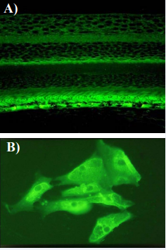
Figure 2: The abnormal antibody activity of the 45 kDa CAR patient predominated within the outer segments of the photoreceptor layer, and the cytoplasm of the RPE.
This protein has extensive influence in maintaining homeostasis, any antibody mediated interference with its activity could prove to have far reaching effects, and could optimize the molecule for immunologic removal by phagocytosis.
The extensive anatomical distribution of PEDF might result in it being involved in other autoimmune reactions if a loss of tolerance develops as a result of uncharacteristic expression.
![Controls: It is understood that normal serum from healthy donors will react with the antigens of every normal tissue component, including retina, a natural process involved in homeostasis that does not include activity with key ‘disease-associated’ antigens [12]. Normal serum reacts with low affinity upon sections of rhesus monkey retina, and in vitro cultivated RPE, but lacks the impressive focus of activity found in the sera of 45 kDa CAR patients when evaluated at the same dilution of 1:20.](https://www.jscimedcentral.com/public/assets/images/uploads/image-1703153967-1.png)
Figure 3: Controls: It is understood that normal serum from healthy donors will react with the antigens of every normal tissue component, including retina, a natural process involved in homeostasis that does not include activity with key ‘disease-associated’ antigens [12]. Normal serum reacts with low affinity upon sections of rhesus monkey retina, and in vitro cultivated RPE, but lacks the impressive focus of activity found in the sera of 45 kDa CAR patients when evaluated at the same dilution of 1:20.
It is known to be involved in the neuronal differentiation of retinoblastoma cells, and is over expressed in other cancer cells such as Junket T-leukemia lymphocytes, and HeLa cells. The common immunologic response to PEDF in the 45 kDa CAR syndrome might therefore result from the patient’s neoplasm expressing this factor in excess, inciting the observed immunological response, comparable to that described in the aberrant expression of ‘recoverin’ in small cell carcinoma associated CAR [1]. If this proves to be the case, it would contribute to our understanding of how some cancers derive the nutrition required for both growth and metastasis. The recruitment of nurturing blood vessels could result from the inadvertent blocking of the modulating activity of PEDF. Specific neutralizing antibodies developing from the excessive production of this ubiquitous protein [13-16].
The possibility of an antibody-mediated interference in the biological activity of PEDF leads to a testable hypothesis: “Immunologic inhibition of the modulating anti-vascular proliferation properties of pigment epithelium-derived factor results in a loss of homeostasis leading to the excessive production of blood vessels that typifies some forms of cancer, and retinopathies that involve uncontrolled vascular proliferations”. If this hypothesis proves correct it will introduce the prospect of ameliorating the pathological process of unwanted vascular spread through the infusion of specific bioactive PEDF peptides [17], and/or appropriate targeted immune modulation therapy [18].
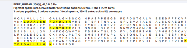
Figure 4: A 45 kDa protein band excised from the 10% acrylamide Coomassie stained gel of an extract of in vitro cultivated RPE proved to react with the cancer patient’s serum antibodies. Proteomic analysis of this band revealed four regions of amino acid sequence compatibility with the complete amino acid sequence of PEDF, implicating pigment epithelium-derived factor as the 45 kDa antigen of interest.


