The Changes of Circulating Follicular T- Helper Cells in Relation to Different Therapy in Adult Idiopathic Thrombocytopenia Patients
- 1. Department of Anesthesia and Intensive Care, Assiut University, Egypt
Abstract
Adult idiopathic thrombocytopenic purpura (ITP) is an acquired organ-specific autoimmune disorder characterized by accelerated platelet destruction. This is due to the production of autoantibodies against platelets membrane glycoproteins which results in the destruction of platelets via the reticuloendothelial system. It can be also caused by platelet production problems. The primary immunologic defect in ITP is having autoreactive B-lymphocytes secreting antiplatelet antibodies. The dysfunctional cellular immunity is also very important in the pathophysiology of ITP.
Citation
Hussein SS (2026) The Changes of Circulating Follicular T- Helper Cells in Relation to Different Therapy in Adult Idiopathic Thrombocytope nia Patients. J Autoimmun Res 7(1): 1034.
INTRODUCTION
Adult idiopathic thrombocytopenic purpura (ITP) is an acquired organ-specific autoimmune disorder characterized by accelerated platelet destruction. This is due to the production of autoantibodies against platelets membrane glycoproteins which results in the destruction of platelets via the reticuloendothelial system. It can be also caused by platelet production problems. The primary immunologic defect in ITP is having autoreactive B-lymphocytes secreting antiplatelet antibodies. The dysfunctional cellular immunity is also very important in the pathophysiology of ITP. Several studies have shown the presence of activated platelet-specific autoreactive T cells that recognize and respond to autologous platelet antigens and drive the generation of platelet reactive autoantibodies by B cells [1].
T follicular helper (TFH) cells may play a critical role in regulating the humoral immune response that occurs with autoimmune diseases, infectious diseases, and tumors. As a new subset, TFH cells regulate the immune process of antigen-specific B-cells and are characterized by the expression of molecules such as chemokine (C-X-C motif) receptor 5 (CXCR5) [2]. CXCR5 is the landmark surface marker of TFH cells. It can bind to CXCL13 and then assist B cells in the migration to the lymphatic follicular area for homing. CXCL13 is the only ligand of CXCR5 [3].
The interaction between CXCL13 and CXCR5 play critical part in the process of lymphocyte migration and formation of the germinal center (GC). It has reported that CXCL13-CXCR5 axis is closely associated with the occurrence of dozen of diseases [2,3]. TFH cells contribute to the generation and maintenance of GC, support Ig class switching and regulate B-cell responses that could produce appropriate specific antibodies and assist in differentiation of GCs into memory B cells and long – lived plasma cells [2,3].
Some studies have defined Inducible cell costimulatory (ICOS) high CXCR5+ CD4+ and/or Programmed cell death protein -1 (PD-1) high CXCR5+ CD4+ T cells as markers of circulating TFH cells that are closely correlated with the expression of antibodies in systemic lupus erythematosus (SLE) and Sjögren’s syndrome [4]. PD-1 and ICOS, which are in the CD28 family, are two distinguishing molecules that have closely related functions in TFH cells.
B cell lymphoma 6 (Bcl-6) and c-Maf are considered two important transcriptional factors of TFH cells that guide differentiation and controls the transcriptional signature of TFH cells [5].
After activation, both of these markers are upregulated on T cells. While ICOS is a co-stimulatory molecule, PD-1 (CD279) sends a signal to the T cell that inhibit it [6].
Regulatory pathways need to be engaged in the control of the development and maturation of TFH cells, as well as their interactions with B cells [5]. it is of particular importance to mention the role of another specific subset of CD4+CD25+Foxp3+ regulatory T cells, called regulatory T cells (Treg cells), that have the immune-regulatory properties and are capable of inhibiting development of autoimmune disease, preventing activation of autoreactive Tcells and inhibiting CD8+ T cell responses [3].
The role of Treg cells in the pathogenesis of human autoimmune disease has been demonstrated in several studies. Tregs, via interacting with Antigen presenting cell (APC) and decreasing CD19+ B cell and CD8+ T cell responses, appear to be the key cell types that may be responsible for the initiation of ITP. There is peripheral deficiency of Tregs which may be due to the sequestration of this cell type. On the other hand, Tregs are also modulated by CD16+ monocytes that were shown to be increased in patients with active ITP. Also, Treg cells have major roles in self-tolerance. In the case of Treg deficiency, peripheral tolerance can fail, leading to the development of autoimmunity diseases.
T helper (Th) subset balance in Peripheral blood (PB) of ITP patients is disrupted, and increased numbers of circulating Th1, Th17, Th22 cells, as well as reduced number or function of Tregs has been reported. In addition, cytotoxic T lymphocyte (CTL)-mediated platelet lysis also contributes to thrombocytopenia in ITP.
The main mechanism for autoimmune diseases is that immune tolerance has been broken down, generation of high-affinity autoantibodies results from proliferation of self-reactive B cells that participate in the pathophysiology of these disorders, so, TFH cells are considered to have an important role in their pathogenesies [5].
So further research on the role of TFH cells in the development of ITP are still required.
MATERIALS AND METHODS
Study design
A prospective randomized controlled clinical trial was conducted from October 2023 to March 2025.
Study setting
The study carried out in Medical Microbiology and Immunology Department. Patients recruited from the unit of Clinical Hematology, Internal Medicine department, and its outpatient clinic at Assiut University Hospitals.
Ethical approval
The study was approved by the Research Ethics Committee at the Faculty of Medicine of Assiut University . Informed consent was obtained from all patients or their relatives before the enrollment in the study.
Study subjects
Inclusion criteria: All ITP patients who were diagnosed as primary newly diagnosed or chronic ITP according to the latest guidelines (American Society Of Hematology (ASH)).The age was more than 18 years up to 50 and informed consent was taken from all participants. The control group was selected from age and sex matched population with normal platelets count.
Exclusion criteria: Patients with secondary causes for ITP (systemic lupus erythematosus, lymphoproliferative disorders, evidence of HIV or hepatitis B or C infections). Also, underlying solid or hematological malignancies and known primary or secondary immune deficiency syndromes are excluded. Concurrent liver disease or renal disease (serum creatinine > 1.5 mg/dl), congenital thrombocytopenia and pregnancy also excluded. None of the ITP patients had undergone a splenectomy and did not have any other relevant diseases during the recent 3 months.
Total number of 60 patients divided as: Twenty healthy controls with no history of hematologic or autoimmune diseases.Twenty newly diagnosed ITP patients (treated with first line of treatment (steroid only) and second line of treatment (steroid and iv immunoglobulin, rituximab or triple therapy).Twenty chronic ITP patients (treated with third line of treatment (Eltromopag 50 mg).
Data collection
Patient’s data were collected in predesigned data sheet. The data were obtained by reviewing patient’s medical records. The patient’s data included the following details:
• Demographic details (age, gender).
• Clinical presentation of the patients
• Comorbid conditions and CBC.
Laboratory investigations
Routinely laboratory tests including blood sample upon admission for hemoglobin concentration, white blood, platelet counts and lymphocytes were measured.
Sample collection
Five ml whole blood samples were drawn under aseptic conditions from all participants including newly diagnosed ITP patients, chronic ITP patients, and healthy control. Blood samples were collected from patients at the time of hospitalization. five milliliters of polystyrene round bottom falcon EDTA tubes were labelled with patient’s name, the date of collection and staining antibody , including controls.
Flow cytometric analysis of circulating TFH cells and its phenotypic marker
Procedure: The following antibodies used for flow cytometry analysis: CD4-PerCP (Cluster of differentiation conjugated to Peridinin-chlorophyll-protein) (BD Biosciences, USA) to stain surface CD4 , CXCR5-PE cy7 ( C-X-C chemokine receptor type5 conjugated to phycoerythrin )(BD Biosciences,USA) to stain surface of CXCR5 and PD1-Apc (Programmed Cell Death-1 conjugated to Allophycocyanin) (BD Biosciences, USA) to stain surface of PD-1.
Wash whole blood sample with 3 ml phosphate-buffered saline (PBS) and remove the supernatant.Dispense Fluorochrome conjugated monoclonal antibodies according titration into all appropriately labeled tubes.
A negative control tube contain cell suspension without CD will be necessary.Add 100 ul of cell suspension to the monoclonal antibodies mixture in each tube. Agitate the tubes gently to mix and incubate in the dark for 15 minutes at room temperature.
After incubation red blood cells (RBCs) were lysed with 3 ml of lysing solution, invert once, keep for 0.5:1 minute,Centrifuge at (2500 rpm) for 1 minute , the supernatant was discarded. The cell mixture was Washed with 3 ml of PBS and centrifuged at 1500xg (2500 rpm) for 1 minute, remove supernatant and add 300mic Dullbecco’s phosphate-buffered saline (DPBS) to remove unbound antibodies.
Sample is now ready for acquisition by multicolor flow cytometry .once stained with appropriate antibodies , sanples were acquired using a flow cytometer ( BD FACS Canto II ) , with a minimum of 50,000–100,000 events recorded per sample, and analyzed with Facs Diva software (BD Biosciences, San Jose , CA , USA ; serial no V33896201978; 3 lasers , 8 colors).
Instrument settings and fluorescence compensation were established using single-stained controls.For data analysis, lymphocytes were initially gated based on forward scatter (FSC) and side scatter (SSC) properties. T lymphocytes were identified followed by gating of CD4? helper T cells. Within the CD4? population, CXCR5? cells were identified, and the expression level of PD-1 was assessed. Circulating T follicular helper (cTfh) cells were defined as CD4? CXCR5? PD-1^high cells.
Results were expressed as the percentage of cTfh cells within the CD4? T-cell population, and the mean fluorescence intensity (MFI) of PD-1 expression was calculated. Comparative analyses were performed among newly diagnosed ITP patients, chronic ITP patients, and healthy controls.
Correlation analyses were conducted to evaluate the association between circulating Tfh cell frequencies and clinical parameters, including platelet count, bleeding severity, disease duration, and response to different lines of treatment.
Statistical analysis of data
Data was collected and analyzed using SPSS (Statistical Package for the Social Science, version 20, IBM, and Armonk, New York). Continuous data was expressed in form of mean ± standard deviation (SD), median and range while nominal data was expressed in form of frequency (percentage).
Chi²-test was used to compare the nominal data of both studied groups while Mann Whitney-U test, independent sample t-test were used to compare mean of continuous variables between both study and control groups. Spearman’s correlation coefficient, Fischer exact test were used to measure the statistical association between the continuous variables. A probability (P-value) of < 0.05 was considered statistically significant.
RESULTS
A total of 60 participants who met the predefined inclusion and exclusion criteria were enrolled in the study, including 20 newly diagnosed ITP patients, 20 chronic ITP patients, and 20 age- and sex-matched healthy controls. All participants were adults (>18 years), and secondary causes of thrombocytopenia, autoimmune diseases, malignancies, chronic liver or kidney disease, pregnancy, congenital thrombocytopenia, and prior splenectomy were excluded to ensure sample homogeneity. All patients underwent detailed clinical evaluation and routine laboratory investigations, including complete blood count and ANA testing, in addition to full assessment of disease duration and previous treatment history. The results presented below describe the clinical and hematological characteristics of the studied groups and the expression levels of circulating PD1^high CXCR5? CD4? T cells, representing circulating TFH cells, and their relationship with disease, treatment, clinical manifestations, and response to therapy.
Demographic data of the studied groups
Regarding sex distribution, females represented the majority in all studied groups. In the newly diagnosed ITP group, females constituted 80.0% of patients, while males accounted for 20.0%. Similarly, females represented 95.0% of the chronic ITP group compared with 5.0% males, and 90.0% of the control group compared with 10.0% males.There was no statistically significant difference in sex distribution among the studied groups (p = 0.635).
The mean age of patients with newly diagnosed ITP was 32.00 ± 11.31 years, whereas patients with chronic ITP had a significantly higher mean age of 41.95 ± 8.18 years, compared with 36.48 ± 4.74 years in the control group. A statistically significant difference in age was observed among the studied groups (p = 0.002) (Table 1).
Table 1: The newly diagnosed ITP group, chronic ITP group, and healthy controls.
|
1. Demographic data |
|
|
|
8. P-value |
|||
|
9. No. |
10. % |
11. No. |
12. % |
13. No. |
14. % |
||
|
15. Sex: |
|
|
|
|
|
|
|
|
16. Male |
17. 4 |
18. 20.0% |
19. 1 |
20. 5% |
21. 2 |
22. 10% |
23. 0.635 |
|
24. Female |
25. 16 |
26. 80.0% |
27. 19 |
28. 95% |
29. 18 |
30. 90% |
|
|
31. Age: (years) |
|
|
|
|
|||
|
32. Mean ± SD |
33. 32.00 ± 11.31 |
34. 41.95 ± 8.18 |
35. 36.48 ± 4.74 |
36. 0.002* |
|||
|
37. Residence: |
|
|
|
|
|
|
|
|
38. Rural |
39. 15 |
40. 75.0% |
41. 12 |
42. 60% |
43. 2 |
44. 10% |
45. 0.000* |
|
46. Urban |
47. 5 |
48. 25.0% |
49. 8 |
50. 40% |
51. 18 |
52. 90% |
|
P value: p values were generated using Mannwhitny test, using Chi square test, T test. P value less than 0.05 was considered significant. SD: standard deviation.
With respect to residence, most patients in the newly diagnosed ITP group were from rural areas (75.0%), while 25.0% were from urban areas. In the chronic ITP group, 60.0% of patients were from rural areas and 40.0% were from urban areas. In contrast, the majority of the healthy control group were urban residents (90.0%), while only 10.0% were from rural areas. A highly statistically significant difference was detected regarding residence among the studied groups (p < 0.001).
Overall, while sex distribution was comparable across groups, significant differences were observed in both age and residence between patients with immune Thrombocytopenia and healthy controls (Figure 1).
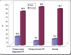
Figure 1: Sex distribution among the studied groups.
Laboratory data for the studied groups:
In patients with newly diagnosed Immune Thrombocytopenia, the mean pre-treatment white blood cell (WBC) count was 7.71 ± 2.14 ×10³/µL, with a median value of 7.2 (range: 3.4–12.4). After treatment, the mean WBC count increased to 13.30 ± 6.55 ×10³/µL, compared with 7.65 ± 6.10 ×10³/µL in chronic ITP patients and 6.03 ± 0.73 ×10³/µL in the control group. A highly statistically significant difference was observed among the groups regarding post-treatment WBC counts (p < 0.001). Additionally, comparison between pre- and post treatment values in newly diagnosed patients showed a significant increase (p = 0.004) (Table 2).
Table 2: The complete blood count (CBC) parameters of the studied groups.
|
|
CBC |
newly diagnosed ITP |
chronic ITP |
Control |
P-value1 |
P-value2 |
|
WBC |
Pre-treatment |
|
|
|
|
|
|
Mean ± SD |
7.71 ± 2.14 |
-- |
-- |
-- |
|
|
|
Median (Range) |
7.2 (3.4-12.4) |
-- |
-- |
|
0.004* |
|
|
Post-treatment |
|
|
|
|
||
|
Mean ± SD |
13.30 ± 6.55 |
7.65 ± 6.10 |
6.03 ± 0.73 |
0.000* |
||
|
Median (Range) |
10.3 (3.1-25.0) |
7.0 (2.4-32.4) |
5.8 (5.1-7.2) |
|
|
|
|
HB |
Pre-treatment |
|
|
|
|
|
|
Mean ± SD |
10.07 ± 2.89 |
-- |
-- |
-- |
|
|
|
Median (Range) |
10.8 (4.5-15.0) |
-- |
-- |
|
0.130 |
|
|
Post-treatment |
|
|
|
|
||
|
Mean ± SD |
10.52 ± 2.57 |
10.24 ± 1.83 |
12.10 ± 1.39 |
0.010* |
||
|
Median (Range) |
10.8 (6.1-16.0) |
10.9 (6.0-13.3) |
12.1 (10.0-14.2) |
|
|
|
|
Platelet count |
Pre-treatment |
|
|
|
|
|
|
Mean ± SD |
7.25 ± 4.09 |
-- |
-- |
-- |
|
|
|
Median (Range) |
7.0 (2.0-17.0) |
-- |
-- |
|
0.000* |
|
|
Post-treatment |
|
|
|
|
||
|
Mean ± SD |
102.15 ± 82.28 |
90.00 ± 79.17 |
327.67 ± 58.80 |
0.000* |
||
|
Median (Range) |
116.0 (3.0-266.0) |
43.0 (10.0-283.0) |
340.0 (231.0-448.0) |
|
|
|
|
Lymphocyte count % |
Pre-treatment |
|
|
|
|
|
|
Mean ± SD |
2.56 ± 2.80 |
-- |
-- |
-- |
|
|
|
Median (Range) |
1.5 (0.2-10.8) |
-- |
-- |
|
|
|
|
Post-treatment |
|
|
|
|
|
|
|
Mean ± SD |
4.36 ± 3.49 |
6.80 ± 4.09 |
13.91 ± 4.16 |
0.000* |
0.05* |
|
|
Median (Range) |
3.3 (0.2-13.5) |
7.6 (1.0-15.5) |
12.8 (5.6-21.4) |
|
P value: p values were generated using Mannwhitny test, using Chi square test, T test.
P value less than 0.05 was considered significant.
P value 1: Compare between the 3 groups, P value 2: Compare before and after treatment in newly diagnosed ITP SD: standard deviation, CBC: complete blood picture, WBC; white blood cells,
Hg: hemoglobin.
Regarding hemoglobin (Hb) levels, the mean pretreatment Hb concentration in newly diagnosed ITP patients was 10.07 ± 2.89 g/dL, with a median value of 10.8 (range: 4.5–15.0). Following treatment, the mean Hb level slightly increased to 10.52 ± 2.57 g/dL in newly diagnosed patients and 10.24 ± 1.83 g/dL in chronic ITP patients, while the control group demonstrated a significantly higher mean Hb level of 12.10 ± 1.39 g/dL. A statistically significant difference was observed among the groups post-treatment (p = 0.010), whereas the difference between pre- and post-treatment Hb levels in newly diagnosed patients was not statistically significant (p = 0.130).
The platelet count showed marked changes following treatment. The mean pre-treatment platelet count in newly diagnosed ITP patients was 7.25 ± 4.09 ×10³/µL, with a median value of 7.0 (range: 2.0–17.0). After treatment, platelet counts increased substantially to 102.15 ± 82.28 ×10³/µL in newly diagnosed patients and 90.00 ± 79.17 ×10³/µL in chronic ITP patients, although these values remained significantly lower than those observed in the control group (327.67 ± 58.80 ×10³/µL). Both the comparison between pre- and post-treatment values in newly diagnosed patients and the comparison among the three groups demonstrated highly statistically significant differences (p < 0.001).
Regarding lymphocyte count percentage, the mean pre-treatment value in newly diagnosed ITP patients was 2.56 ± 2.80, with a median of 1.5 (range: 0.2–10.8). Post-treatment values increased to 4.36 ± 3.49 in newly diagnosed patients and 6.80 ± 4.09 in chronic ITP patients, while the control group showed the highest mean value (13.91 ± 4.16). A highly statistically significant difference was observed among the three groups (p < 0.001). Moreover, comparison between pre- and post-treatment values in newly diagnosed patients revealed a statistically significant increase (p = 0.05).
The expression of TFH cells phenotypic markers in newly diagnosed ITP
The proportion of CD4? CXCR5? cells showed no statistically significant difference following treatment. The mean ± SD decreased slightly from 13.4 ± 5.6 in the pre treatment group to 12.6 ± 6.8 post-treatment (P = 0.146). Similarly, the median values were comparable between the two measurements, indicating that treatment did not significantly affect the frequency of CD4? CXCR5? cells (Table 3).
Table 3: The expression of TFH cells phenotypic markers in patients with primary newly diagnosed ITP was evaluated before and after treatment.
|
|
Pre-treatment |
Post-treatment |
P-value |
|
CD+ CXCR5+ Mean ± SD Median (Range) |
13.4±5.6 12.4 |
12.6±6.8 13.1 |
0.146 |
|
CD4+ CXCR5+ PD1+: |
|
|
|
|
Mean ± SD |
34.3 ± 14.1 |
29.4 ± 15.2 |
0.002* |
|
Median (Range) |
35.2 |
31.3 |
|
|
CD4+ PD1+: Mean ± SD Median |
29.4 ± 15.2 25.8 |
30.2±16.7 24.5 |
0.004* |
In contrast, a significant reduction was observed in the expression of CD4? CXCR5? PD-1? cells after treatment. The mean ± SD declined from 34.3 ± 14.1 before treatment to 29.4 ± 15.2 after treatment, with a statistically significant difference (P = 0.002). The median values also decreased from 35.2 pre-treatment to 31.3 post-treatment, suggesting that therapy was associated with a reduction in this cell subset.
Regarding CD4? PD-1? cells, a statistically significant difference was also detected between the two measurements (P = 0.004). The mean ± SD increased slightly from 29.4 ± 15.2 before treatment to 30.2 ± 16.7 after treatment, while the median values showed a slight decrease from 25.8 to 24.5. These findings indicate that treatment had a measurable effect on PD-1–expressing CD4? T cells in patients with primary newly diagnosed ITP
The expression of TFH cell phenotypic markers post treatment in the studied groups :
Patients with newly diagnosed ITP showed the highest levels, with a mean ± SD of 12.6 ± 6.8 and a median of 13.1, followed by patients with chronic ITP (11.53 ± 4.5; median 11.8). The lowest values were recorded in the normal control group (9.4 ± 3; median 9.6). These findings indicate an increased frequency of circulating TFH cells in ITP patients compared with healthy individuals (Table 4).
Table 4: Regarding CD4? CXCR5? cells, a statistically significant difference was observed among the three studied groups (P < 0.001).
|
|
newly diagnosed ITP |
chronic ITP |
Control |
P-value |
|
CD4+ CXCR5+: Mean ± SD Median |
12.6±6.8 13.1 |
11.53 ± 4.5 11.8 |
9.4 ± 3 9.6 |
0.000* |
|
CD4+ CXCR5+ PD1+%: Mean ± SD Median |
29.4 ± 15.2 31.3 |
27.9 ± 8.8 27.4 |
22.8 ± 6.3 22.1 |
0.001* |
|
CD4+ PD1+%: Mean ± SD Median |
30.2±16.7 24.5 |
23.6 ± 7.5 22.2 |
15.3 ± 5.2 14.2 |
1.000 |
P value: p values were generated using Mannwhitny test, using Chi square test, T test.
P value less than 0.05 was considered significant SD: standard deviation
Similarly, CD4? CXCR5? PD-1? cells demonstrated a statistically significant difference between the groups (P= 0.001). The highest expression was observed in newly diagnosed ITP patients (mean ± SD: 29.4 ± 15.2; median: 31.3), followed by chronic ITP patients (27.9 ± 8.8; median: 27.4), while the control group showed the lowest levels (22.8 ± 6.3; median: 22.1). This suggests an increased activation of TFH cells in ITP patients.
In contrast, the comparison of CD4? PD-1? cells among the three groups did not show a statistically significant difference (P = 1.000), despite higher mean values in newly diagnosed ITP patients (30.2 ± 16.7; median 24.5) compared with chronic ITP patients (23.6 ± 7.5; median 22.2) and the control group (15.3 ± 5.2; median 14.2). These results indicate that although TFH-associated markers are elevated in ITP patients, PD-1 expression on CD4? T cells alone did not differ significantly among the studied groups.
The expression of TFH cells phenotypic markers in studied groups:
Peripheral blood from newly diagnosed ITP patients (n = 20) and chronic ITP patients (n=20) and healthy controls (n = 20) were stained with labeled antibodies as described in methods and meterials. Lymphocytes were gated by forward and side scatters in the inter box represent as p1. CD4+T cell were gated by flow cytometry Plots in the inter box represent as p2. CD4+CXCR5+T subsets were gated by flow cytometry Plots in the inter box represent as p3.CD4+CXCR5PD-1hi T cells gated by flow cytometry plots in the inter box represent as p4.
Flow cytometric analysis of proportion of circulating TFH cells in patients with nITP and cITP compared with Hcs
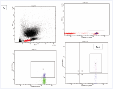
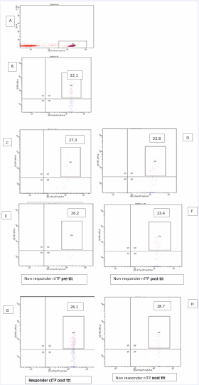
A) CD4+lymphocytes were gated and CD4+CXCR5 T subsets were gated using flow cytometry. plots in the inter box of p4 represent circulating TFH cells ( CD4+CXCR5+PD 1high+)
B) Circulating TFH cells in healthy controls.
C) Circulating TFH cells in nITP patient responders at pre-treatment.
D) Circulating TFH cells in nITP patient responders at post-treatment.
E) Circulating TFH cells in nITP patien non responders at pre-treatment.
F) Circulating TFH cells in nITP patient non responders at post-treatment
G) Circulating TFH cells in cITP patient responders at post-treatment.
H) Circulating TFH cells in cITP patient non responders at post-treatment.
Increased frequencies of circulating TFH cells in peripheral blood from ITP patients. Percentages of CD4+CXCR5PD-1hi T cells was significantly higher in the nITP, cITP than control groups which reduced after treatment in responded groups when compered with control while in non-responder ITP patients percentages of CD4+CXCR5PD-1hi T cell resist reduction in percentage.
Flow cytometric analysis of circulating TFH cells in three groups from A to G; P1 is TFH cells, P2 is CD4+PD1+, P3 is CD4+ CXCR5+, P4 is CD4+ CXCR5+PD1+, H is bar graph quantification of circulating TFH cell phynotypic markers in three groups (Figure 2).
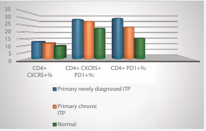
Figure 2: The expression of TFH cell phenotypic markers post treatment in the studied groups
Correlation between TFH cell phenotypic markers and Platelet count, lymphocytes and Hemoglobin in pre treatment in patients with newly diagnosed ITP
Regarding CD4?CXCR5?PD1? cells, negative correlations were found with platelet count (r = −0.046, p = 0.846), lymphocyte count (r = −0.321, p = 0.168), and hemoglobin (r = −0.373, p = 0.106); however, these correlations were not statistically significant. Similarly, CD4?PD1? cells showed weak negative correlations with platelet count (r = −0.070, p = 0.768), lymphocyte count (r = −0.304, p = 0.193), and hemoglobin levels (r = −0.231, p = 0.328), with no statistically significant associations detected (Table 5 and Figure 3).
Table 5: Showed a weak negative correlation with platelet count (r = −0.125, p = 0.599) and lymphocyte count (r = −0.061, p = 0.797), while a moderate negative correlation with hemoglobin levels was observed (r = −0.505), which reached statistical significance (p = 0.023).
|
|
|
Platelet count |
Lymphocytes |
Hb |
|
CD4+ CXCR5+ |
R value |
-0.125 |
-0.061 |
-0.505 |
|
P value |
0.599 |
0.797 |
0.023 |
|
|
CD4+ CXCR5+ PD1+%: |
R value |
-0.046 |
-0.321 |
-0.373 |
|
P value |
0.846 |
0.168 |
0.106 |
|
|
CD4+ PD1+%: |
R value |
-0.070 |
-0.304 |
-0.231 |
|
P value |
0.768 |
0.193 |
0.328 |
|
|
P value: p values were generated using Pearson test. p value less than 0.05 was considered significant. R value = +1 prefect positive correlation, -1 prefect negtive correlation, 0 no correlation. |
||||
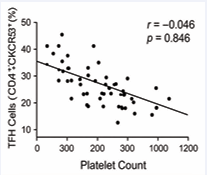
Figure 3: Correlation between markers and Platelet count. Possible association between percentage of circulating TFH cells and platelet count were analyzed. Negative correlation between percentage of circulating TFH cells and platelet count was found.
Bleeding manifestations in the studied patients were classified according to their clinical severity. Mild bleeding manifestations such as petechiae and ecchymotic patches were categorized as Grade 1. Moderate bleeding manifestations, including gum bleeding, epistaxis, and subconjunctival hemorrhage, were classified as Grade 2. More severe bleeding manifestations, such as menorrhagia, were considered Grade 3. This grading system was used to standardize the assessment of bleeding severity among patients with ITP and to facilitate the analysis of its relationship with immunological markers (Table 6).
Table 6: Bleeding grading according to WHO 2020.
|
Bleeding pattern |
Suggested grade |
Newly diagnosed ITP |
Chronic ITP |
|
Petechiae |
Grade 1 |
0 |
10 (47.6%) |
|
Ecchymotic patches |
Grade 1 |
8 (40%) |
7 (33.5%) |
|
Gum bleeding |
Grade 2 |
6 (30%) |
2 (9.5%) |
|
Epistaxis |
Grade 2 |
2 (10%) |
4 (19%) |
|
Subconjunctival bleeding |
Grade 2 |
2 (10%) |
0 |
|
Menorrhagia |
Grade 3 |
4(20%) |
2 (9.5%) |
DISCUSSION
ITP is an acquired autoimmune disorder characterized by isolated thrombocytopenia resulting from immune mediated platelet destruction and impaired platelet production. The pathogenesis of ITP involves a complex dysregulation of both humoral and cellular immunity, including the production of autoantibodies against platelet surface glycoproteins and abnormal T-cell–mediated immune responses. Increasing evidence indicates that aberrant CD4? T-cell activation plays a central role in disease initiation and persistence, particularly through altered T-cell help to autoreactive B cells.
cTfh cells, defined by the expression of CD4, CXCR5,and high levels of programmed cell death protein-1 (PD 1), represent a specialized subset of CD4? T cells that promote B-cell differentiation and antibody production. The CXCR5 chemokine receptor enables Tfh cell migration toward B-cell follicles, while PD-1 expression reflects functional activation and regulatory balance within germinal center reactions. Dysregulation of the Tfh–B cell axis has been implicated in several autoimmune diseases and is increasingly recognized as a key contributor to autoantibody-driven conditions such as ITP.
Clinically, ITP exhibits distinct immunological characteristics across its disease stages. Newly diagnosed (acute) ITP is often associated with heightened immune activation and inflammatory responses, whereas chronic ITP reflects sustained immune dysregulation, failure of immune tolerance, and ongoing autoreactive lymphocyte activity. The persistence of activated cTfh cells and increased PD-1 expression may therefore contribute to chronic disease evolution by maintaining continuous B-cell stimulation and pathogenic autoantibody production. Accordingly, evaluating the differential expression of PD-1^high CXCR5? CD4? T cells across acute and chronic ITP provides valuable insight into disease progression, immune imbalance, and potential prognostic biomarkers.
The present study was designed to investigate the immunological role of circulating T follicular helper (cTfh) cells in immune thrombocytopenic purpura (ITP). Specifically, the study aimed to evaluate differences in the expression of PD-1^high CXCR5? CD4? T cells among newly diagnosed ITP patients, chronic ITP patients, and healthy individuals. In addition, the study sought to assess the potential of circulating Tfh cells as predictive biomarkers for therapeutic response across different stages of ITP. Furthermore, the relationship between circulating Tfh cell frequencies and clinical manifestations of the disease, including platelet count, bleeding severity, and disease duration, was examined to elucidate their possible role in disease activity and progression.
Sixty (60) eligible participants who fulfilled the predefined inclusion and exclusion criteria were enrolled in the present study and were equally divided into three groups. Group I included twenty (20) newly diagnosed immune thrombocytopenic purpura (ITP) patients, Group II comprised twenty (20) patients with chronic ITP, and Group III consisted of twenty (20) age- and sex-matched apparently healthy individuals who served as the control group. All participants were adults aged over 18 years, and all enrolled subjects completed the study protocol and were included in the final analysis without any loss to follow-up. Secondary causes of thrombocytopenia, other autoimmune diseases, malignancies, chronic liver or kidney diseases, pregnancy, congenital thrombocytopenia, and previous splenectomy were excluded to avoid confounding immunological factors and to ensure homogeneity of the studied population.
In the present study, a total of 60 participants were enrolled, including newly diagnosed primary ITP patients, chronic primary ITP patients, and healthy controls. The mean age differed significantly among the studied groups (p = 0.002), with newly diagnosed ITP patients showing a mean age of 32.00 ± 11.31 years, chronic ITP patients 41.95 ± 8.18 years, and healthy controls 36.48 ± 4.74 years. The relatively higher mean age observed in chronic ITP patients may reflect disease persistence and progression over time, which is consistent with previous reports indicating that chronic ITP is more frequently observed in older adult populations [7].
Regarding sex distribution, females predominated across all study groups, accounting for 80% of newly diagnosed ITP patients, 90% of chronic ITP patients, and 90% of the control group, with no statistically significant difference between groups (p = 0.635). This female predominance aligns with the well-established autoimmune nature of ITP and is consistent with multiple studies demonstrating a higher prevalence of ITP among adult females [8] while in children the predominance aligns show higher prevalences of ITP among male children
A statistically significant difference was observed in residence distribution among the studied groups (p < 0.001). Most newly diagnosed (75%) and chronic ITP patients (60%) were from rural areas, whereas the majority of healthy controls (90%) resided in urban areas. Although residence is not considered a direct etiological factor in ITP, this finding may reflect differences in healthcare access, environmental exposures, or referral patterns, as previously suggested in epidemiological studies of autoimmune diseases in developing countries [7].
The clinical profile observed in the present study is largely consistent with previously published data on adult primary immune thrombocytopenia. The low prevalence of hypertension and diabetes mellitus among both newly diagnosed and chronic ITP patients is in agreement with recent reports indicating that metabolic comorbidities do not represent core features of primary ITP and are not considered major contributors to disease pathogenesis or clinical course [8].
The significant difference in abortion history observed between newly diagnosed and chronic ITP patients aligns with recent evidence suggesting that immune dysregulation during early or active phases of ITP may adversely affect pregnancy outcomes. Similar associations between ITP activity and obstetric complications have been reported in contemporary cohort studies, supporting the concept that immune-mediated thrombocytopenia may influence reproductive outcomes particularly during active disease stages.
With regard to bleeding manifestations, the observed bleeding patterns are in agreement with recent clinical studies demonstrating that bleeding phenotype in ITP varies according to disease duration rather than following a uniform pattern. The predominance of cutaneous bleeding manifestations in chronic ITP and the variability of mucosal bleeding across disease stages are consistent with findings reported in recent observational and real-world studies of adult ITP patients [9]. These studies emphasize that bleeding severity and pattern in ITP are influenced by chronic immune activation and platelet dysfunction rather than platelet count alone [10].
The absence of antinuclear antibody positivity in all studied patients is consistent with current diagnostic recommendations and recent clinical studies confirming that primary ITP should be clearly differentiated from secondary autoimmune thrombocytopenia. This finding supports appropriate patient selection and aligns with contemporary international guidelines and cohort studies published after 2020 [11-67].
In this study, we noticed that the frequencies of TFH cells were markedly increased in peripheral blood of newly diagnosed and chronic ITP, and there was a strong negative correlation between the proportion of TFH cells and platelet count which suggested that aberrant increased TFH cells may involve in the pathogenesis of ITP . This was agreement with Xie et al. and Simpson et al. who demonstrated a higher frequency of TFH cells in the chronic ITP patients than in healthy control group. The function of TFH cells is closely related to the expression of PD-1. Thus, it was important to determine the expression of TFH phenotype to show if typically associated with TFH cells altering in ITP cases.
Our study revealed that, expressions of CXCR5+CD4+/ PD-1+ TFH cells were significantly increased in ITP patients compared to control group. In addition, to further analyze whether the increased TFH cells had also high expression of PD-1 molecules, the correlation study revealed a strongly positive correlation between the frequency of the CD4+CXCR5+PD-1TFH cells and PD-1+ on CD4+ cells in ITP patients, but not in control group. These results are in accordance of Xie et al.
Moreover, we divided patients into nITP and cITP groups in this study, and found that the frequencies of TFH cells in the nITP group recovered to normal levels after therapy in respect with response. However, the persistent increase in frequencies of TFH cells was noticed in cITP non-responder patients . This result is in agreement with Feng et al. and Qian et al. ,also Li et al.
The frequency of TFH cells was not significantly influenced by the treatment , otherwise cause shifts in the frequency of PD-1 high on CD4+ T cells that observed between early and later follow up and treating with different lines of therapy .
In summary , our study demonstratesthat the expansion of TFH cells and its phenotypic markers may be involve in the immunopathogenesis in adult ITP , furthermore, the immunotherapy resulted in reduction of TFH cells and its phenotypic markers .
CONCLUSIONS
Finally we conclude that, the increasing frequencies of circulating TFH cells with associated phenotype PD 1+ expressions of TFH cell might play a critical role in the pathogenesis of ITP via associated markers . Steroid treatment in ITP may significantly reduce the frequencies of TFH cells and its phenotypic markers which will reduce illness severity. Further studies are still needed to explore the exact pathogenesis of ITP. This will help identify disease features and predictors of treatment response, thus facilitating the decision-making process for ITP management in the future.
ACKNOWLEDGEMENTS
Dr /Asmaa Mohamed Zahran
Professor and Head of Clinical Pathology Department, South Egypt Cancer Institute, Assiut University
Ethics approval and consent to participate
Approval for the present study was obtained by the Ethics Committee of The First Affiliated Hospital of assuit University.
Patient consent for publication
All patients admitted to the study provided informed consent for their participation of the present study and publication of the data.
REFERENCES
- Karnell JL, Albulescu M, Drabic S, Wang L, Moate R, Baca M, et al. A CD40L-targeting protein reduces autoantibodies and improves disease activity in patients with autoimmunity. Sci Transl Med. 2019; 11: eaar6584.
- Christodoulou M, Moysidou E, Lioulios G, Stai S, Lazarou C, Xochelli A, et al. T-Follicular Helper Cells and Their Role in Autoimmune Diseases. Life (Basel). 2025; 15: 666.
- Gensous N, Charrier M, Duluc D, Contin-Bordes C, Truchetet ME, Lazaro E, et al. T Follicular Helper Cells in Autoimmune Disorders. Front Immunol. 2018; 9: 1637.
- Ahmadivand S, Fux R, Pali? D. Role of T Follicular Helper Cells in Viral Infections and Vaccine Design. Cells. 2025; 14: 508.
- Garcia-Espinoza JA, López-Villalobos EF, García-Chagollán M, Uribe-Martínez JF, Torres-Lizárraga S, Muñoz-Valle JF, et al. Analysis of CCR9, CXCR5 and ICOS in Circulating Follicular Helper T Cell-like Populations in Sjögren’s Disease. Int J Mol Sci. 2026; 27: 1765.
- Del Carmen Crespo Oliva C, Labrie M, Allard-Chamard H. T peripheral helper (Tph) cells, a marker of immune activation in cancer and autoimmune disorders. Clin Immunol. 2024; 266: 110325.
- Cooper N, Ghanima W. Immune Thrombocytopenia. N Engl J Med. 2019; 381: 945-955.
- Audia S, Mahévas M, Nivet M, Ouandji S, Ciudad M, Bonnotte B. Immune Thrombocytopenia: Recent Advances in Pathogenesis and Treatments. Hemasphere. 2021; 5: e574.
- Kuter DJ, Efraim M, Mayer J, Trn?ný M, McDonald V, Bird R, et al. Rilzabrutinib, an Oral BTK Inhibitor, in Immune Thrombocytopenia. N Engl J Med. 2022; 386: 1421-1431.
- Lambert MP, Gernsheimer TB. Clinical updates in adult immune thrombocytopenia. Blood. 2017; 129: 2829-2835.
- KUTER, D. J. The treatment of immune thrombocytopenia (ITP)—focus on thrombopoietin receptor agonists. Annals of Blood. 2021; 6.
- Georgi JA, Middeke JM, Bornhäuser M, Matzdorff A, Trautmann-Grill K. Deciphering the genetic basis of immune thrombocytopenia: current evidence for genetic predisposition in adult ITP. Blood Adv. 2023; 7: 3710-3724.
- Al-Samkari H, Kuter DJ. Immune Thrombocytopenia in Adults: Modern Approaches to Diagnosis and Treatment. Semin Thromb Hemost. 2020; 46: 275-288.
- Almendros A, Choi YR, B?czkowski PM, Baiker K, Barrs VR, Beatty JA. Babesia gibsoni Infection in a Cat with Immune-Mediated Haemolytic Anaemia and Thrombocytopenia. Animals (Basel). 2023; 13: 2128.
- ?Altahan RM. Acquired platelet disorders. Front Med (Lausanne). 2025; 12: 1638994.
- Audia S, Bonnotte B. Emerging Therapies in Immune Thrombocytopenia. J Clin Med. 2021; 10: 1004.
- Audia S, Mahévas M, Samson M, Godeau B, Bonnotte B. Pathogenesis of immune thrombocytopenia. Autoimmun Rev. 2017; 16: 620-632.
- Audia S, Rossato M, Trad M, Samson M, Santegoets K, Gautheron A, et al. B cell depleting therapy regulates splenic and circulating T follicular helper cells in immune thrombocytopenia. J Autoimmun. 2017; 77: 89-95.
- Bariana TK, Ouwehand WH, Guerrero JA, Gomez K; BRIDGE Bleeding, Thrombotic and Platelet Disorders and ThromboGenomics Consortia. Dawning of the age of genomics for platelet granule disorders: improving insight, diagnosis and management. Br J Haematol. 2017; 176: 705-720.
- Beckman JD, Rollins-Raval MA, Raval JS, Park YA, Mazepa M, Ma A. Bortezomib for Refractory Immune-Mediated Thrombocytopenia Purpura. Am J Ther. 2018; 25: e270-e272.
-
Bhasin TS, Sharma S, Manjari M, Mannan R, Kansal V, Chandey M, et al. Changes in megakaryocytes in cases of thrombocytopenia: bone marrow aspiration and biopsy analysis. J Clin Diagn Res. 2013; 7: 473-479.
- Bosma DMT, Busselaar J, Staal MD, de Koning M, Relji? M, Lei X, et al. Requirements for development of T helper 1 and T follicular helper cells from a common precursor. Cell Rep. 2026; 45: 117296.
- Bradbury CA, Pell J, Hill Q, Bagot C, Cooper N, Ingram J, et al. Mycophenolate Mofetil for First-Line Treatment of Immune Thrombocytopenia. N Engl J Med. 2021; 385: 885-895.
- Broome CM, Röth A, Kuter DJ, Scully M, Smith R, Wang J, Reuter C, Hobbs W, Daak A. Safety and efficacy of classical complement pathway inhibition with sutimlimab in chronic immune thrombocytopenia. Blood Adv. 2023; 7: 987-996.
- Bu S, Liu M, Yang L, Lee P, Miller H, Park CS, et al. The function of T cells in immune thrombocytopenia. Front Immunol. 2025; 16: 1499014.
- Bussel J, Arnold DM, Grossbard E, Mayer J, Treli?ski J, Homenda W, et al. Fostamatinib for the treatment of adult persistent and chronic immune thrombocytopenia: Results of two phase 3, randomized, placebo-controlled trials. Am J Hematol. 2018; 93: 921-930.
- Bussel JB, Garcia CA. Diagnosis of immune thrombocytopenia, including secondary forms, and selection of second-line treatment. Haematologica. 2022; 107: 2018-2036.
- Caserta S, Zaccuri AM, Innao V, Musolino C, Allegra A. Immune thrombocytopenia: options and new perspectives. Blood Coagul Fibrinolysis. 2021; 32: 427-433.
- Chaudhary PK, Kim S, Kim S. An Insight into Recent Advances on Platelet Function in Health and Disease. Int J Mol Sci. 2022; 23: 6022.
- Chen M, Shortt J. Plasma Cell Directed Therapy for Immune Thrombotic Thrombocytopenic Purpura (iTTP). Transfus Med Rev. 2022; 36: 204-214.
- Chen Y, Hu J, Chen Y. Platelet desialylation and TFH cells-the novel pathway of immune thrombocytopenia. Exp Hematol Oncol. 2021; 10: 21.
- Chen Y, Luo L, Zheng Y, Zheng Q, Zhang N, Gan D, et al. Association of Platelet Desialylation and Circulating Follicular Helper T Cells in Patients With Thrombocytopenia. Front Immunol. 2022; 13: 810620.
- Corsiero E, Nerviani A, Bombardieri M, Pitzalis C. Ectopic Lymphoid Structures: Powerhouse of Autoimmunity. Front Immunol. 2016; 7: 430.
- Cuker A, Buckley B, Mousseau MC, Barve AA, Haenig J, Bussel JB. Early initiation of second-line therapy in primary immune thrombocytopenia: insights from real-world evidence. Ann Hematol. 2023; 102: 2051-2058.
- Davoren A, Aster RH. Heparin-induced thrombocytopenia and thrombosis. Am J Hematol. 2006; 81: 36-44.
- DELVES, P. J. Innate and adaptive systems of immunity. The autoimmune diseases. Elsevier. 2020; 45-61.
- Devine DV, Serrano K. The platelet storage lesion. Clin Lab Med. 2010; 30: 475-87.
- Dimitriu T, Szczelkun MD, Westra ER. Evolutionary Ecology and Interplay of Prokaryotic Innate and Adaptive Immune Systems. Curr Biol. 2020; 30: R1189-R1202.
- Eyerich K, Dimartino V, Cavani A. IL-17 and IL-22 in immunity: Driving protection and pathology. Eur J Immunol. 2017; 47: 607-614.
- Fiyouzi T, Pelaez-Prestel HF, Reyes-Manzanas R, Lafuente EM,Reche PA. Enhancing Regulatory T Cells to Treat Inflammatory and Autoimmune Diseases. Int J Mol Sci. 2023; 24: 7797.
- French SL, Butov KR, Allaeys I, Canas J, Morad G, Davenport P, et al. Platelet-derived extracellular vesicles infiltrate and modify the bone marrow during inflammation. Blood Adv. 2020; 4: 3011-3023.
- Fu W, Ishikawa-Ankerhold H, Gaertner F. Homeostasis of megakaryocytes: balancing tissue residency and consumptive platelet production. Trends Cell Biol. 2026; 36: 453-470.
- Giordano P, Delvecchio M, Lassandro G, Valente F, Palladino V, Chiarito M, et al. Can Anti-Thyroid Antibodies Influence the Outcome of Primary Chronic Immune Thrombocytopenia in Children? Endocr Metab Immune Disord Drug Targets. 2020; 20: 351-355.
- Grodzielski M, Goette NP, Glembotsky AC, Constanza Baroni Pietto M, Méndez-Huergo SP, Pierdominici MS, et al. Multiple concomitant mechanisms contribute to low platelet count in patients with immune thrombocytopenia. Sci Rep. 2019; 9: 2208.
- Grozovsky R, Giannini S, Falet H, Hoffmeister KM. Regulating billions of blood platelets: glycans and beyond. Blood. 2015; 126: 1877-1884.
- Hassan M, Elzallat M, Mohammed DM, Balata M, El-Maadawy WH. Exploiting regulatory T cells (Tregs): Cutting-edge therapy for autoimmune diseases. Int Immunopharmacol. 2025; 155: 114624.
- Hillion S, Arleevskaya MI, Blanco P, Bordron A, Brooks WH, Cesbron JY, Kaveri S, Vivier E, Renaudineau Y. The Innate Part of the Adaptive Immune System. Clin Rev Allergy Immunol. 2020; 58: 151-154.
- Jurczak W, Chojnowski K, Mayer J, Krawczyk K, Jamieson BD, Tian W, et al. Phase 3 randomised study of avatrombopag, a novel thrombopoietin receptor agonist for the treatment of chronic immune thrombocytopenia. Br J Haematol. 2018; 183: 479-490.
- Kailashiya J. Platelet-derived microparticles analysis: Techniques, challenges and recommendations. Anal Biochem. 2018; 546: 78-85.
- Kapur R, Zufferey A, Boilard E, Semple JW. Nouvelle cuisine: platelets served with inflammation. J Immunol. 2015; 194: 5579-5587.
- Karnell JL, Rieder SA, Ettinger R, Kolbeck R. Targeting the CD40-CD40L pathway in autoimmune diseases: Humoral immunity and beyond. Adv Drug Deliv Rev. 2019; 141: 92-103.
- Kashiwagi H, Kuwana M, Murata M, Shimada N, Takafuta T, Yamanouchi J, et al. Reference guide for the diagnosis of adult primary immune thrombocytopenia, 2023 edition. Int J Hematol. 2024; 119: 1-13.
- Khan AM, Mydra H, Nevarez A. Clinical Practice Updates in the Management Of Immune Thrombocytopenia. P T. 2017; 42: 756-763.
- Khellaf M, Viallard JF, Hamidou M, Cheze S, Roudot-Thoraval F, Lefrere F, et al. A retrospective pilot evaluation of switching thrombopoietic receptor-agonists in immune thrombocytopenia. Haematologica. 2013; 98: 881-7.
- Kiessling P, Lledo-Garcia R, Watanabe S, Langdon G, Tran D, Bari M, et al. The FcRn inhibitor rozanolixizumab reduces human serum IgG concentration: A randomized phase 1 study. Sci Transl Med. 2017; 9: eaan1208.
- Kim DS. Recent advances in treatments of adult immune thrombocytopenia. Blood Res. 2022 Apr 30; 57: 112-119.
- Kim S, Lee K. Emerging Roles of Megakaryocytes in Immune Regulation and Potential Therapeutic Prospects. Cells. 2025; 14: 1677.
- Koupenova M, Livada AC, Morrell CN. Platelet and Megakaryocyte Roles in Innate and Adaptive Immunity. Circ Res. 2022; 130: 288-308.
- Kühne T, Berchtold W, Michaels LA, Wu R, Donato H, Espina B, et al. Newly diagnosed immune thrombocytopenia in children and adults: a comparative prospective observational registry of the Intercontinental Cooperative Immune Thrombocytopenia Study Group. Haematologica. 2011; 96: 1831-1837.
- Lee JY, Lee JH, Lee H, Kang B, Kim JW, Kim SH, et al. Epidemiology and management of primary immune thrombocytopenia: A nationwide population-based study in Korea. Thromb Res. 2017; 155: 86-91.
- Li H, Guan Y, Sun B, Dou X, Liu X, Xue F, et al. Role of bone marrow-derived mesenchymal stem cell defects in CD8+ CD28- suppressor T-lymphocyte induction in patients with immune thrombocytopenia and associated mechanisms. Br J Haematol. 2020; 191: 852-862.
- Li J, Sullivan JA, Ni H. Pathophysiology of immune thrombocytopenia. Curr Opin Hematol. 2018; 25: 373-381.
- Li X, Sheng Z, Sun Y, Wang Y, Xu M, Zhang Z, et al. Human leukocyte antigen-G upregulates immunoglobulin-like transcripts and corrects dysfunction of immune cells in immune thrombocytopenia. Haematologica. 2021; 106: 770-781.
- Liebman HA, Saleh MN, Bussel JB, Negrea OG, Horne H, Wegener WA, et al. Comparison of two dosing schedules for subcutaneous injections of low-dose anti-CD20 veltuzumab in relapsed immune thrombocytopenia. Haematologica. 2016; 101: 1327-1332.
- Ling LE, Hillson JL, Tiessen RG, Bosje T, van Iersel MP, Nix DJ, et al. M281, an Anti-FcRn Antibody: Pharmacodynamics, Pharmacokinetics, and Safety Across the Full Range of IgG Reduction in a First-in-Human Study. Clin Pharmacol Ther. 2019; 105: 1031-1039.
- Liu XG, Hou Y, Hou M. How we treat primary immune thrombocytopenia in adults. J Hematol Oncol. 2023; 16: 4.
- Loriamini M, Cserti-Gazdewich C, Branch DR. Autoimmune Hemolytic Anemias: Classifications, Pathophysiology, Diagnoses and Management. Int J Mol Sci. 2024; 25: 4296.