Significance of Comprehensive Dermatological Examination in Diagnosing Atypical Superficial Fungal Infections
- 1. Department of Dermatology, School of Medicine, Ankara Yildirim Beyazit University, Turkey
- 2. Department of Dermatology, Ankara City Hospital, Turkey
Abstract
Superficial fungal infections often exhibit atypical presentations in routine dermatological practice, posing diagnostic challenges as they imitate diverse dermatoses with challenging and confusing features. Distinguishing these infections is crucial for accurate diagnosis and implementing an appropriate treatment strategy. This case report details a 35-year-old female patient with pruritic erythematous scaly lesions, notably on her upper extremities and face. The superficial fungal infection was initially overlooked due to its atypical clinical features and an insufficient primary dermatological examination. This case highlights the importance of a detailed and complete dermatological examination in making a correct diagnosis and planning treatment. The report also serves to raise awareness about the diverse clinical manifestations and varied presentations of superficial fungal infections, aiming to prevent diagnostic delays in clinical practice
Citation
Kalkan G, Albayrak ID (2025) Significance of Comprehensive Dermatological Examination in Diagnosing Atypical Superficial Fungal Infections. J Dermatolog Clin Res 13(1): 1168.
INTRODUCTION
Superficial fungal infections frequently manifest with atypical presentations in routine dermatological practice, often resembling diverse dermatoses and posing diagnostic challenges. The accurate identification of these infections is essential for prescribing appropriate treatments. The literature extensively discusses these atypical appearances [1-6], highlighting the complexity of diagnosis in various clinical forms.
A comprehensive examination of the entire body is imperative for precise diagnoses. In this context, we present a case involving a 35-year-old female patient who presented with pruritic erythematous scaly lesions, notably concentrated on her upper extremities and face. The initial oversight of considering superficial fungal infection, attributed to its atypical clinical features and an insufficient dermatological examination, underscores the necessity for a thorough evaluation. Through this case, we underscore the significance of conducting comprehensive dermatological examinations to ensure accurate diagnoses. This serves as a reminder and raises awareness regarding the diverse and atypical clinical manifestations of superficial fungal infections, aiming to mitigate delays in diagnosis.
CASE REPORT
A 35-year-old woman presented to our outpatient clinic with complaints of yellowing and thickening in her fingernails that had appeared three months ago, along with red, itchy lesions on both dorsal surfaces of her hands and forearms a few days later (Figures 1,2).

Figure 1:Erythematous plaques on bilateral hand dorsum, yellow discoloration and thickening of nails
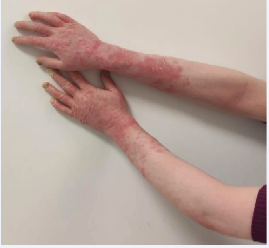
Figure 2:Sharply demarcated erythematous plaques starting from the fingers and extending to the forearms
Fifteen days subsequently, she observed a sharply circumscribed plaque with erythema and scaling around the left eye, followed by a similar erythematous plaque on the right cheek.
When the patient removed the clothing covering her arms, sharply circumscribed erythematous plaque like lesions became visible, allowing for a detailed dermatological examination. Afterwards, when she removed the scarf from her head and the mask from her face, we noticed a sharply defined erythematous plaques on the face. A mycological microscopic examination with 20% potassium hydroxide (KOH) was performed on samples from the fingernail, dorsum of the hand, around the eyelid, and cheek, suspecting a fungal infection, and fungal hyphae were observed intensively in all samples. Treatment was initiated with oral terbinafine at a dose of 250 mg/day.
At the one-month follow-up, an excellent response to the treatment was observed, with the lesions showing signs of resolution (Figures 3,4).

Figure 3:One month after the treatment, redness on the dorsum of the hand has decreased, and there is partial improvement in the nails

Figure 4:One month after the treatment, the redness on the forearms decreased.
We continued the oral terbinafine treatment at the same dose, and the patient expressed satisfaction with the improvement
DISCUSSION
Dermatophyte infections, the predominant cause of superficial fungal infections worldwide, are typically identified by their characteristic clinical presentations. However, contemporary dermatology encounters cases with uncommon and atypical clinical appearances. The standard clinical images of these infections involve scaly erythematous patches with an annular configuration that expands towards the periphery with central healing. Nevertheless, the emergence of cutaneous dermatophyte infections with challenging atypical clinical presentations complicates the distinction from other dermatological diseases, leading to the adoption of the term “tinea atypica” in current literature. Various factors, including the use of topical or systemic immunosuppressants and corticosteroids, the invasion power and location of the dermatophyte, sun exposure, climatic conditions, excessive washing of the affected area, and the individual’s immune status, are considered probable causes of this atypical appearance [1-6].
The atypical clinical appearance of superficial fungal infections can mimic various dermatoses with unusual clinical manifestations, such as psoriasis, impetigo, eczematous dermatitis, seborrheic dermatitis, lupus erythematosus, polymorphous light eruption, erythema multiforme, lichen planus, keratoderma, pyoderma gangrenosum, subcorneal pustular dermatosis, and rosacea. A recent study by Sharquie et al. extensively covered a large series of cases with atypical superficial dermatophyte infections presenting as major imitators, with psoriasis being the most common clinical presentation (50.2%). Additionally, photosensitive dermatoses, keratoderma, seborrheic dermatitis/napkin dermatitis,folliculitis, rosacea, alopecia, lupus erythematosus, discoid lupus erythematosus, granuloma annulare, and pityriasis alba-like lesions were observed. While similar mimicry clinical findings have been reported in other studies, this study emphasizes the need for superficial fungal infections to be recognized as major imitators in dermatological diseases to avoid diagnostic delays with these atypical clinical manifestations [1-5]. In such atypical cases, histopathological examination, with its emphasized sensitivity in various studies, emerges as a valuable method for diagnosis compared to direct microscopic examination and culture [6]
In our case, the initial consideration was the diagnosis of psoriasis due to the erythematous scaly plaque-like appearance and nail thickening, as consulted with our colleagues. However, after the patient took off all her clothes, the distinct border line of the fungal disease, especially noticeable on the arms, became apparent. Similarly, after she removed the mask and scarf, facial lesions indicative of fungal infection became evident. This case report underscores the imperative of conducting a thorough dermatological examination,particularly for clinicians navigating time constraints in their daily practice. Moreover, it underscores the significance of recognizing superficial fungal infections with diverse atypical clinical manifestations, increasingly observed in contemporary practice, as significant imitators in the realm of dermatology


