Hydatidosis among Imported Animals in Jeddah, Saudi Arabia
- 1. Department of Biology, King Abdulaziz University, Saudi Arabia
- 2. Department of Medical Laboratory Technology, King Abdulaziz University, Saudi Arabia
Abstract
Background: This epidemiological study was conducted over two years in one of the main governmental abattoir in Jeddah Province in West Region of Saudi Arabia to evaluate the condition of hydatidosis caused by Echinococcus granulosus in imported slaughtered animals.
Methods: We examined a total of (132858) imported internationally animals including camels, cattle, sheep and goats for infection rate, organs affected, size and fertility of cysts.
Results: The infection rate of hydatidosis in imported internationally animals was 0.42%. We also find that the highest prevalence of infection was in cattle representing 0.60%. Summer revealed the highest prevalence in sheep and goats, while cattle showed higher prevalence in autumn. The most affected organs were found to be the liver, and secondly the lung. The cysts of small size had the highest percentage in sheep and goats (25.76% and 20.20%) respectively. Sheep show the highest percentage of fertile cysts (42.26%), followed by cattle (31.58%).
Conclusions: Our study offers insights into hydatidosis hosts infection, the need for intensive controlling programs and effective treatments.
Keywords
Echinococcus granulosus ; Hydatidosis ; Protoscoleces ; Intermediate host ; Fertility.
CITATION
Toulah FH, El Shafi AA, Alsolami MN, Wakid MH (2017) Hydatidosis among Imported Animals in Jeddah, Saudi Arabia. J Liver Clin Res 4(1): 1031.
INTRODUCTION
Hydatidosis or echinococcosis is an infection caused by the larva or adult of Echinococcus spp., which belong to the family Taeniidae [1]. Echinococcosis is known as one of the major zoonoses affecting domestic animals and humans in different parts of the world [2]. Echinococcus includes four species named E. granulosus, E. multilocularis, E. vogeli and E. oligarthrus. However, E. granulosus is the most widely important agent of the disease [3,4].
The rate of infection in human reaches 1 million worldwide [5].The life cycles of the parasites are maintained with carnivores such as dogs and foxes, which serve as final hosts carrying the adult worms in their intestine. Herbivorous animals such as cattle, goats, sheep, camels, and pigs serve as intermediate hosts in which the metacestode stage (hydatid cysts) develops in tissues after ingestion of eggs. Humans are infected with hydatid cyst after ingestion of eggs [6,7].
Echinococcosis has a major public health importance and a great impact on the economy of countries where livestock production is a main part of the agriculture and when manufacture of livestock is mainly based on expanded pasture system. The intimate relationship between man, dogs and sheep makes the life cycle of the parasite more likely to be completed [8].
Echinococcosis has a global distribution; it is endemic or hyperendemic in the Middle East and neighboring countries, such as Iraq [9], Yemen [10], Libya [11], Oman [12], Iran [13], Tunisia [14], Ethiopia [15,16] Turkey [17], and Egypt [18]. In Saudi Arabia, echinococcosis is a significant endemic disease in various provinces, particularly in the Western Region [19-27].
The high prevalence of echinococcosis is enhanced by many factors related to economic and social status of the population. This includes pastoralism practice, the high dog population, the role of dogs in guarding livestock, inadequate medical/ veterinary services and low hygiene/education standards [8,28]. Furthermore, importing of live animals from endemic areas is a continuous risk of re-introducing E. granulosus and maintaining its zoonotic life cycle [29].
It is so difficult to estimate a precise prevalence status of hydatidosis in intermediate hosts in any continent. This may be explained by poor accuracy and costly diagnostic tests. Currently, most of the prevalence status studies have based on slaughter data [10-12,18,25,27,29,30]. Gathering information on livestock diseases, especially subclinical conditions is frugal and economical. Additionally no reliable test exists for hydatidosis in living ruminants [31].
Her we evaluate the condition of hydatidosis in imported slaughtered animals in Jeddah Province in West Region of Saudi Arabia, based on infection rate, affected organs, together with status of cysts in regard to size and fertility.
This study took place during two years in one of the main governmental abattoirs in Jeddah Province in west of Saudi Arabia. The nature of its climate is temperate in winter (20°C) and very hot in summer (48°C). We visited the abattoir periodically to examine the slaughtered imported internationally animals.
MATERIALS AND METHODS
Examination of slaughtered animals
Imported internationally animals including camels, cattle, sheep and goats were examined in the present study. Thorough examination of slaughtered animals was carried out with the aid of veterinarians by visual inspection, palpation and systemic incision of all visceral organs according to the recommended procedures [32].
Examination of cysts size, fertility and viability of protoscoleces
Cysts were released from the organs and accurately examined to detect their count, size, fertility status and viability of protoscoleces.
Examinations of infected organs were carried out in the laboratory of Faculty of Science for Girls, King Abdul-Aziz University. To check the presence of any calcification and degeneration, we examined the cysts macroscopically. Then we withdrew the content of each cyst and (i) determined the size of the cyst through measuring the hydatid fluid volume according to Berhe [15] (small if <6 ml, medium if 6-20 ml and large if > 20 ml); (ii) assessed fertility and infertility by examining a drop of the hydatid fluid sediment microscopically for protoscoleces. Infertile cysts were divided as sterile (contain no protoscoleces and slightly turbid fluid) or calcified (a gritty sound feeling up is produced on incision); (iii) evaluated the protoscoleces viability by staining with 0.1% eosine stain and assessing flame cells motility. Only dead protoscoleces take up the stain [33].
Statistical analysis
To evaluate differences between the rate of infection relative to host species, organ distribution, cyst count, cyst fertility and the mean of protoscoleces viability we used student’s test and Chi-square. The statistical tests were conducted using the software packages SPSS version 19.
RESULTS AND DISCUSSION
Results
We examined a total of 132858 imported internationally animals including camels, cattle, sheep and goats (2361, 17796, 89787, 22914) represented as (1.78%, 13.39%, 67.58%, 17.25%) for the hydatidosis in Jeddah city, Saudi Arabia. Sheep were the most abundant slaughtered animals in the studied area. There was a statistically significant difference of hydatidosis infection among the studied animals (P< 0.001) (Table 1).
|
Table 1: Number and infection rate of hydatidosis among slaughtered animals. |
|||
|
Animals |
No. of slaughtered |
No. of infected animals |
Infection rates* (%) |
|
Camels |
2361 |
12 |
0.51 |
|
Cattle |
17796 |
106 |
0.60 |
|
Sheep |
89787 |
337 |
0.38 |
|
Goats |
22914 |
108 |
0.47 |
|
Total |
132858 |
563 |
0.42 |
|
*Number of infected animal/ total number of the same animal. |
|||
The prevalence of cystic echinococcosis among the same species showed that cattle (0.60%) and camels (0.51%) have higher infection rate than goats (0.47%) and sheep (0.38%).
Comparing seasonal variations and prevalence of infection (Table 2),
|
Table 2: Seasonal prevalence of hydatidosis among slaughtered animals. |
||||
|
Animals
Season |
Camels |
Cattle |
Sheep |
Goats |
|
No. (%) |
No. (%) |
No. (%) |
No. (%) |
|
|
Winter |
1 (8.33) |
24 (22.64) |
106 (31.45) |
31 (28.70) |
|
Spring |
5 (41.67) |
28 (26.42) |
76 (22.55) |
30 (27.78) |
|
Summer |
1 (8.33) |
21 (19.81) |
122 (36.20) |
33 (30.56) |
|
Autumn |
5 (41.67) |
33 (31.13) |
33 (9.80) |
14 (12.96) |
|
Total |
12 (100 ) |
106 (100) |
337 (100) |
108 (100 ) |
we found that sheep and goats revealed higher infection prevalence in summer and winter (36.20%, 31.45%) (30.56%, 28.70%) respectively. Autumn showed higher prevalence of infection in slaughtered cattle (31.13%). On the other hand, camels revealed higher infection prevalence in both spring and autumn (41.67%).
Liver and lung were clearly the most predominantly infected visceral organs in all examined animals (Table 3).
|
Table 3: Prevalence of infection of hydatidosis in the affected organs of slaughtered animals. |
||||
|
|
Organs |
|||
|
Animals |
Liver No. (%)* |
Lung No. (%)* |
Intestine No. (%)* |
Muscle No. (%)* |
|
Camels (n = 12) |
12 (100) |
0 (0.0) |
0 (0.0) |
0 (0.0) |
|
|
|
|
|
|
|
Cattle (n = 106) |
91 (85.85) |
14 (13.21 ) |
1 (0.94) |
0 (0.0) |
|
|
|
|
|
|
|
Sheep (n = 337) |
249 (73.89) |
55 (16.32) |
13 (3.86) |
20 (5.93) |
|
|
|
|
|
|
|
Goats (n = 108) |
88 (81.48) |
7 (6.48) |
12 (11.11) |
1 (0.93) |
|
*Total number of infected animals |
||||
The infection prevalence in liver was 100%, 85.85%, 73.89% and 81.48% in camels, cattle, sheep and goats respectively. Few animals had cysts in other organs such as intestine and muscles.
The total number of small sized cysts among all animals was 149 (75.25%), followed by medium sized cysts of 41 (20.71%). Only eight large sized cysts (4.04%) were detected. There was a highly statistically significant difference in cyst size and distribution among the investigated animals (P<0.001). Liver harbor most of small sized cysts (81.63%, 75.00%, 67.61%, and 63.15%) of goats, camels, sheep and cattle respectively. Lung from sheep showed highest percentage of large sized cysts (4.23%). There was a highly significant difference in the size of the recovered cysts between liver and lung (P<0.001) (Table 4).
|
Table 4: Size distribution among recovered liver and lung cysts. |
|||||||||
|
|
Animal (cyst number) |
||||||||
|
|
Camels (40) |
Cattle (38) |
Sheep (71) |
Goats (49) |
|||||
|
Organ
Cystsize |
Liver |
Lung |
Liver |
Lung |
Liver |
Lung |
Liver |
Lung |
|
|
|
Small No. (%)*
|
30 (75) |
0 (0) |
24 (63.15) |
4 (10.53) |
48 (67.61) |
3 (4.22) |
40 (81.63) |
0 (0) |
|
|
Medium No. (%)*
|
7 (17.5) |
0 (0) |
5 (13.16) |
4 (10.53) |
10 (14.08)) |
7 (9.86) |
7 (14.29) |
1 (2.04( |
|
|
Large No. (%)*
|
3 (7.5) |
0 (0) |
0 (0) |
1 (2.63) |
0 (0) |
3 (4.23) |
0 (0) |
1 (2.04) |
|
Total No. (%)* |
40 (100) |
0 (0) |
29 (76.32) |
9 (23.68) |
58 (81.69) |
13 (18.31) |
47 (95.92) |
2 (4.08) |
|
|
*represented among the same animals |
|||||||||
The highest rate of fertile cysts was in the sheep (42.26%) followed by cattle (31.58%). Camels revealed the highest percentage of calcified cysts (70.00%), while cattle showed the lowest (42.10%).
|
Table 5: Frequency of hydatid cyst forms recovered from studied animals. |
||||
|
Animals |
All cysts No. |
Fertile No. (%)* |
Sterile No. (%)* |
Calcified No. (%)* |
|
Camels |
40 |
4 (10.00) |
8 (20.00) |
28 (70.00) |
|
Cattle |
38 |
12 (31.58) |
10 (26.32) |
16 (42.10) |
|
Sheep |
71 |
30 (42.26) |
10 (14.08) |
31 (43.66) |
|
Goats |
49 |
15 (30.61) |
9 (18.37) |
25 (51.02) |
|
*represented among the same animals |
||||
There was a highly statistically significant difference in the number of fertile cysts among the examined animals the viability of protoscoleces among the investigated animals (P<0.001) and the highest viable protoscoleces were recorded in sheep. In addition we observed a significant difference in the number of viable protoscoleces between lung and liver (P<0.001).
Discussion
Echinococcosis is among the most neglected public health problems in humans and causes serious socio-economic effects throughout the world2.
The work presented here provides assessment to the magnitude of the disease in the imported internationally animals slaughtered in Jeddah.
In our study, we revealed that the highest infection rate was in cattle followed by camels, goats and sheep. A previous study in Jeddah investigated local slaughtered animals stated that camels were the highest followed by cattle, sheep then goats [27]. Prior studies have reported similar findings where camels have the highest infection rates [25,26,30]. However, other investigators recorded highest infection rate in sheep [29,34,35], or in cattle [16,36,37]. The difference in prevalence rates in different animal species and in different localities could be attributed to the variations in the parasite strain that exists in different geographical areas [38], in addition to attitudes to dogs in different region and difference in culture [16,26,36,39].
We identified that the most frequently infected visceral organs were liver and lung in all examined animal species. Intestinal affections were limited among goats, sheep and cattle. Muscles affections were observed only in sheep and goats. Liver and lung represent the primary sites for migrating oncosp here may clarify the frequency of hydatidosis in these organs [16,18,37,40-42].
Our observations suggest seasonal variations in the prevalence of hydatidosis among the studied animals. Cystic echinococcosis (hydatidosis) in sheep and goats was prevalent in the summer. Previous study found that the highest prevalence of hydatidosis among sheep was in spring and autumn [25], while others recorded the highest prevalence in autumn and winter [29]. Similarly, autumn showed higher prevalence of infection in cattle, while camels were in highest prevalence in spring and autumn. Together these data indicate that the difference in prevalence rates is associated to several factors including livestock stocking intensity, cross–border migration of livestock, differences in environmental conditions and age variations during seasons [29,30,43]. In addition, a recent study attributed the highest prevalence of echinococcosis in local slaughtered animals to religious occasions as Muslims slaughter enormous numbers of livestock during the period of pilgrimage [26].
We recorded limited number of large cysts (Figure 1).
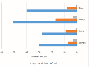
Figure 1 Distributions of cysts in investigated animals according to the size.
Small sized cysts exhibited a high proportion in all examined hosts (15.15%, 14.14%, 25.76% and 20.20%) in comparison to medium sized cysts (3.54%, 4.50%, 8.59% and 4.04%) in camels, cattle, sheep and goats respectively (Figure 2).
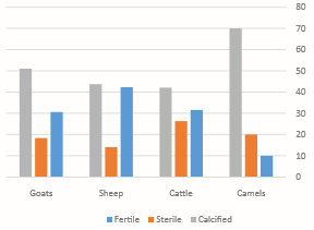
Figure 2 Distributions of cysts in investigated animals according to the fertility.
The higher frequencies of small and medium sized cysts are in liver than in lung, which mainly harbored the large cysts. Host immunological response that could prevent cyst expansion would explain the high proportion of small sized cysts, whereas the soft consistency of cells may explain the occurrence of large sized cysts in lung [16].
In the present study, we observed that fertility rates of hydatid cysts are higher in sheep, cattle and goats than in camels, while previous studies obtained slightly different findings [25, 44-46].
Our observation pinpoints that sheep, cattle and goats are the most important reservoir of infection, which support maintaining the zoonotic life cycle of E. granulosus in the studied region.
CONCLUSION
In conclusion, the infection rates of hydatidosis among slaughtered animals in Jeddah province, is enough to cause serious economic losses and human affections. Prompt measures should be applied in the region for infection eradication. Health education should be implemented in controlling programs, and beneficial contribution of the population is indicated in order to change improper behaviors related to man-dog relationships and home slaughtering practices. Great efforts must be undertaken to inhibit incorrect disposal of parasite-infected organs from slaughterhouses in addition to meat inspection and medical treatment. Stray dogs should be strictly controlled and illegal slaughter of animals must be prohibited. It seems very necessary to intensify collaboration effects between scientists and the concerned authorities in the region to support further investigations and increase public awareness of the disease. These efforts must take place before this neglected zoonotic disease turns into a serious epidemic issue.
ACKNOWLEDGEMENTS
This study was funded (Grant Number RS-18-42) by King Abdulaziz City for Science and Technology, Saudi Arabia. The authors gratefully thanks the managers and workers of North Jeddah abattoir’ and the veterinarians, technicians for their beneficial help and cooperation.


