Innovative Use of Magnetic Crowns for Pulp Vitality and Regeneration
- 1. Imam Abdulrahman Alfaisal Hospital, Saudi Arabia
Abstract
Preserving the vitality of the dental pulp is critical in maintaining tooth longevity, yet conventional restorations often lead to pulp shrinkage or necrosis, ultimately requiring endodontic therapy. We propose a novel dental crown design incorporating magnetic elements to achieve two core objectives: [1] maintain pulp vitality by stimulating local circulation and cell viability, and [2], promote controlled ossification within the pulp chamber using repelling magnetic fields, potentially obviating the need for root canal treatment. This concept leverages existing literature on magnetic biomodulation, pulpal regeneration, and mineralization to introduce a non-invasive therapeutic pathway. We present the scientific rationale, conceptual design, and a proposed mechanism of action. The concept is expected to initiate further laboratory research and clinical feasibility testing in collaboration with academic or biomaterial development laboratories.
Keywords: Magnetic Crowns; Pulp Vitality; Dental pulp; Regeneration
Introduction
The preservation of dental pulp vitality is central to modern conservative dentistry. Vital pulp tissue ensures continued nourishment, innervation, and immune defense of the tooth, directly influencing long-term tooth survival and resistance to reinfection. However, deep carious lesions, trauma, or repeated restorative procedures often compromise pulpal health, leading to irreversible pulpitis or necrosis. Conventional endodontic therapy, though effective in many cases, is invasive, removes natural tissue, and is often followed by structural weakening of the tooth. In recent years, regenerative endodontics and biomodulation techniques have gained attention for their potential to revive compromised pulp tissue or stimulate natural healing responses. Among these, magnetic field stimulation has demonstrated promising effects in various biological contexts, including enhanced cellular proliferation, improved microcirculation, and bone mineralization. Yet, its application within the dental pulp chamber remains largely unexplored. This paper introduces a novel approach: the integration of miniaturized permanent magnets within dental crowns as a non-invasive therapeutic tool. The concept includes two main objectives: [1], to maintain pulp vitality by generating localized magnetic fields that promote cellular activity and vascular stability, and [2], to create controlled magnetic repulsion within the pulp space to stimulate ossification or mineral deposition, potentially eliminating the need for conventional root canal treatment. This hypothesis aims to initiate scientific discussion and spark preclinical research on the integration of magnetic fields in restorative dentistry. By leveraging existing evidence in bioelectromagnetics and tissue engineering, this concept could pave the way for a new class of biologically active dental restorations.
Literature Review and Scientific Background
Pulp Vitality and Its Clinical Importance
The dental pulp is a dynamic connective tissue responsible for dentin formation, immune defense, and mechanosensation. Loss of pulp vitality compromises the structural integrity of the tooth, increases susceptibility to bacterial invasion, and often necessitates endodontic therapy. While root canal treatment is effective in removing infection, it eliminates the natural defense and regenerative capacity of the tooth, often weakening it structurally over time [3].
Magnetic Fields in Biology and Regeneration
Static and pulsed magnetic fields have been widely studied for their bio-modulatory effects. Research has shown that magnetic fields can:
- Stimulate angiogenesis (formation of new blood vessels)
- Enhance cellular proliferation and differentiation, especially in stem cells
- Increase mineral deposition and bone formation
- Reduce inflammation and oxidative stress
Magnetism and Dental Applications
Studies have shown that magnets in dental applications are generally biocompatible when coated (e.g., with titanium or stainless steel) and properly isolated from saliva. The use of neodymium magnets, due to their high energy density, allows for miniaturization while maintaining functional magnetic strength [4].
Ossification and Regeneration without Root Canal Therapy
Attempts to regenerate the pulp-dentin complex have led to techniques like revascularization and the use of growth factors or scaffold materials. However, these require invasive access and are unpredictable. Magnetism-based mechanical stimulation has shown potential in inducing osteogenesis in craniofacial bones and mineralization in experimental models. Translating this to the dental pulp, a repelling magnetic force may exert enough physical influence to trigger cellular reorganization and calcific bridge formation, essentially filling the pulp chamber with mineralized tissue—an outcome that could serve as an alternative to root canal therapy.
Proposed Concept and Design
This paper proposes the development of a magnet-integrated dental crown designed to serve both restorative and therapeutic functions. Unlike conventional crowns that passively restore function and esthetics, this crown introduces an active biological influence on the underlying dental pulp through controlled magnetic fields.
Concept Overview
The innovation involves embedding one or more miniaturized permanent magnets within the structure of a dental crown. These magnets are strategically positioned to deliver magnetic influence directly toward the pulp chamber, either through attraction or repulsion, depending on the desired biological outcome [5].
A Pulp Vitality Mode (Attracting Magnetic Field)
A single centrally placed magnet is embedded within the occlusal or axial portion of the crown, oriented such that it emits a static magnetic field inward toward the pulp chamber.
B Ossification Mode (Repelling Magnetic Field)
A repelling magnetic system is proposed by embedding two magnets in the crown: one in the occlusal surface and another in the axial wall, both with like poles facing each other. This creates a repelling force, hypothesized to stimulate mechanotransduction pathways and mineralized tissue deposition [6,7].
Structural Design and Materials
- Crown Material: Zirconia, Emax, or PFM crown with internal magnet compartment
- Magnet Type: Biocompatible coated neodymium (NdFeB)
- Orientation: Fixed magnetic polarity directed toward pulp chamber axis
- Safety: Shielding from saliva, removable or MRI-compatible options
Figure 1
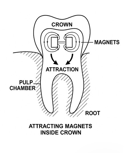
Figure 1: Diagram showing attracting magnets placed to maintain pulp vitality.
Figure 2
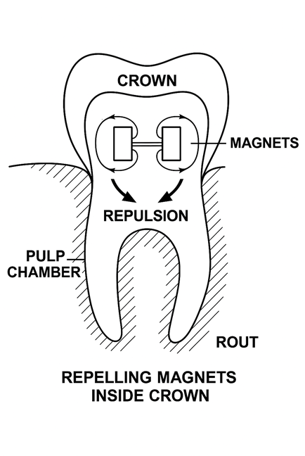
Figure 2: Diagram showing repelling magnets placed to maintain pulp vitality.
Hypothesized Mechanism of Action
Pulp Vitality Preservation via Static Magnetic Stimulation
Permanent magnets in the crown emit a static magnetic field directed toward the pulp, promoting angiogenesis, reducing inflammation, and enhancing stem cell activity.
Pulp Ossification Induction via Internal Repelling Magnetic Field
Repelling magnets within the crown create a mechanical force field that stimulates differentiation of pulp stem cells and deposition of mineralized tissue, possibly replacing root canal therapy.
Supporting Evidence from Literature:
- SMF and PEMF shown to enhance cell proliferation and bone healing
- Stem cell behavior influenced positively by low-intensity magnetic fields [8-10].
Discussion
Clinical Implications and Advantages
- Preserves pulp vitality in high-risk teeth
- Potential non-invasive alternative to RCT
- Introduces a new category of biologically active crowns
Technical and Biological Challenges
- Magnetic field strength calibration
- MRI compatibility and magnet shielding
- Patient-specific anatomy considerations
Path Forward
- Finite element modeling and in vitro testing
- Collaboration with biomedical engineering and dental research labs
- Animal model experiments to validate safety and efficacy
Conclusion
This paper presents a novel biologically active dental crown that integrates magnetic fields to preserve pulp vitality or induce ossification. This innovation holds promise as a new direction in minimally invasive endodontics and restorative dentistry. Scientific validation is essential, and the authors call upon research institutions and labs to collaborate in developing and testing this concept.
References
- Bassett CAL. Beneficial effects of electromagnetic fields. J Cellular Biochem. 1993; 51: 387-393.
- Yamaguchi DT. Pulsed electromagnetic fields increase osteoblast migration through a calcium/calmodulin pathway. Bioelectromagnetics. 2006; 27: 519-530.
- Markov MS. Pulsed electromagnetic field therapy: History, state of the art and future. The Environmentalist. 2007; 27: 465-475.
- Buzalaf MAR, Kato MT, Hannas AR. The role of matrix metalloproteinases in dental wear and erosion. Journal of Dental Research.2010; 89: 292-303.
- Diniz IMA. Restoring the pulp-dentin complex using tissue engineering strategies. Regenerative Biomaterials. 2020; 7: 331-349.
- Nakao Y. Effect of a static magnetic field on bone formation in rat bone marrow stromal cell cultures. Int J Oral Maxillofacial Implants. 2002; 17: 231-236.
- Iseri U. Effects of pulsed electromagnetic fields on tooth movement and root resorption: A histological study in rats. Am J Orthodontics Dentofacial Orthopedics. 2006; 130: 636
- D'Angelo C. Effects of electromagnetic fields on human stem cells for regenerative medicine: A review. Electromagnetic Biol Med. 2015; 34: 146-155.
- Choi BK. Orthodontic magnets and corrosion: A literature review. Am J Orthodontics Dentofacial Orthopedics. 2007. 131: 501-510.
- Kakehashi S, Stanley HR, Fitzgerald RJ. The Effects of Surgical Exposures of Dental Pulps in Germ-Free and Conventional Laboratory Rats. Oral Surg Oral Med Oral Pathol. 1965; 20: 340-349.


