CT-MRI Image Fusion Effect on Dose Delivered and Distribution for Astrocytoma Radiation Therapy
- 1. Department of Clinical Oncology and Nuclear Medicine, Mansoura University, Egypt
- 2. Department of Physics, Mansoura University, Egypt
- 3. Department of Science, Mansoura University, Egypt
Abstract
Aim: This study described the benefits of CT-MRI fusion to consolidate the accuracy of conformal radiation therapy for Egyptian astrocytoma patients.
Materials and methods: Thirty four patients with astrocytoma tumor who underwent 3D conformal radiotherapy were included in this study. CT and MRI imaging was done for all the patients after surgery. The PTVCT and PTVMRI tumor were measured and compared with each other. The center of mass (CM) of the PTV delineated on CT and MRI were computed and the shift between the two CMs was determined.
Results and Discussion: The mean and median for PTV measured by CT-MRI was 66.77cm3 ±48.77 and 43.65 (range 8.6 to 162.2) respectively. For CT the mean and median for PTV was 53.82cm3 ± 45.66 and 31.7 (range 12.4 to 180.1) respectively. The differences between the centers of mass (CM) of the two PTV’s detected spatial 3D shifts ranging from (-4mm to 5mm) with an average of 0.3 mm for x direction, (-2mm to 5mm) with an average of 1.3 mm for y, and (-2mm to 4mm) with an average of 0.7 mm for z direction respectively. The prescribed dose was fitted on PTVCT. The V95-PTVMRI was lower than the V95-PTVCT by 22.5%.
Conclusion: The result showed that MRI is an essential modality in radiotherapy for planning astrocytoma tumors.
Keywords
• Astrocytoma
• CT
• MRI
• Image fusion
Citation
Dawod T, Abdelrazek EM, El-Hadaad HA, Isam M (2015) CT-MRI Image Fusion Effect on Dose Delivered and Distribution for Astrocytoma Radiation Therapy. J Radiol Radiat Ther 3(1): 1054.
ABBREVIATIONS
CM: Center Mass; 3D: Three Dimension; CT: Computed Tomography; MRI: Magnetic Resonance Imaging; PET: Positron Emission Tomography; PTV: Planning Target Volume; RT: Radiotherapy; GTV: Gross Target Volume; CTV: Clinical Target Volume
INTRODUCTION
Medical imaging techniques in the past decades became crucial for medical diagnosis or analyses. Techniques such as X-rays, computed tomography (CT), magnetic resonance imaging (MRI), and positron emission tomography (PET) enabled different approaches and insights of human body, important for analysis and diagnosis. For example, CT scans provide high resolution information on bony structure while MRI scans provide detailed information on tissue types within the body [1,2].
In recent years, external beam radiotherapy, such as intensitymodulated radiotherapy and 3D conformal radiotherapy have been employed in many institutions. Precise determination of target volumes is the most crucial and difficult part of radiotherapy (RT) planning process, especially for the treatment of astrocytoma to avoid a geographical miss [3].
The most suitable sources of detailed 3D anatomic information for treatment planning are the CT and MRI devices [4]. In CT aspects, the data set of target lesion and OARs were provided but there was a lack of tissue discrimination. MRI showed good tissue discrimination but the use of MRI in radiotherapy was limited in many centers. In fact, the CT simulator was the primary equipment in many radiotherapy units [5].
A powerful technique used in medical imaging analysis is medical image fusion, where streams of information from medical images of different modalities are combined into a single fused image. The effectiveness of medical image fusion can be illustrated by the fused image of an axial slice through the head using MRI scan and a CT scan [6].
To perform fusion between medical images, it is first necessary to align the images with each other. A number of techniques have been proposed for the purpose of image alignment. These methods are often categorized based on the similarity metric used to perform the comparison between images [6], such as Cross-correlation [7], Sum of squared differences (SSD) [8], Mutual information [9], and Phase correlation [10]. In this work we described the benefits of CT-MRI fusion to consolidate the accuracy of conformal radiation therapy for Egyptian astrocytoma patients.
MATERIALS AND METHODS
This work was carried out at clinical oncology and nuclear medicine in Mansoura University, Mansoura Scan Center, and AyadiAlmostkbal Oncology Center in Alexandria. A total of 34 patients with astrocytoma tumors (grade II, grade III and glioblastoma multiform) were prepared for standard RT treatment. The CT scanner used was GE Healthcare, Bright Speed. Each patient was scanned in treatment position, with an immobilization applied. CT data were then exported from CT and imported to the RT treatment planning system. The MR data sets were obtained by 0.2 Tesla signal profile. Patients were immobilized in supine position with the same thermoplastic mask as that used during CT scan, where, the patients were scanned according to the standard diagnostic protocol, and images were transferred to the RTP workstation for treatment planning.
The first step correlating CT and MR images is image registration. Image registration is the process in which two image data sets are put into the common coordinate system. The most often is a combination of CT and MR images, where CT set is used as a reference, and MR set is reoriented and registered to the CT coordinate system. Finally, two images can be fused; it is done with the fusion license of a treatment planning software (Prowess Panther treatment planning).When the results were not considered satisfactory, manual correction of image registration was necessary in some cases, depending on the matching result, by matching points on anatomical landmarks.
The following step was the target delineation. A radiation oncologist contoured GTV (surgical bed plus residual mass if present and visible) on CT (GTVCT) and subsequently on CT/ MRI (GTVMRI). CTVs (CTVCT and CTVMRI) were obtained adding 0.5 cm automatic margin on the corresponding GTV. The volume of contoured structures (PTVct and PTVMRI (cm3 )) was directly calculated using a software tool within the treatment planning system. The shift in the position of the isocenter of the two PTV’s was also extracted through the use of a software tool that calculates the three dimensional coordinates of the center of mass CMs any structure. Therefore, the shift between the two centers of mass (planning isocenter shift) is found by calculating the difference between these two points. Postoperative target volumes GTV, CTV, and PTV were drawn on CT and MRI images according to the recommendations in ICRU 50.
For each patient, the treatment plan based on PTVCT was carried out with Prowess Panther TPS in terms of percentage of PTVCT and PTVMRI that received 95 % of the prescribed dose (V95-PTVCT, V95-PTVMRI), PTVCT total dose was 6500 cGy. A comparison between values of target volumes PTVCT and PTVMRI were demonstrated and volume differences were calculated for each patient ( MRI volume-CT volume) .Also the values of dose delivered obtained by both CT and CT-MRI fusion were calculated (V95-PTVCT, V95-PTVMRI). Shifts between the image fusion and CT target’s isocenter were analyzed. Figure 1 showed the performance of medical image fusion by the fused image of an axial slice through the brain using a CT scan and a MRI scan.
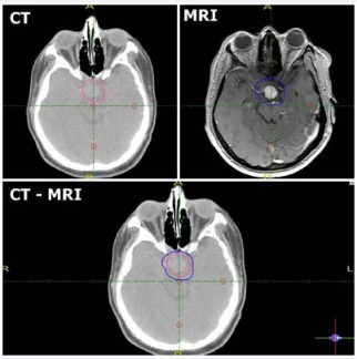
Figure 1: An axial cranial slice: Top-left image: CT scan, Top-right image: MRI scans, and Bottom image: CT-MRI fusion.
The statistical analysis of data was done by using excel program for graph and SPSS software (version 17.0). One-Way ANOVA was used to analyze the difference. A two-tailed p value of <0.05 was considered statistically significant in all cases.
RESULTS AND DISCUSSION
In this study, we found that male to female ratio was (1.6:1), 21 were male and 13 were female. Most of the patients had glioblastoma multiform GBM (56%) and had undergone subtotal surgical resection, followed by grade three astrocytoma (23%) and grade two astrocytoma (21%).Regarding the Location of tumor, we found that, 13 cases frontal, 6 cases fronto-parietal , 9 tempro parietal cases, 5 cases occipital, and one patient are brain stem.Two cases underwent complete resection, 18 cases underwent subtotal resection and 13 cases underwent a biopsy .The demographic details are listed in (Table 1).
Table 1: Patient charateristics, There a total of 34 study sets. Each study set contained the contours for PTV structure via both CT and fusion techniques.
|
Characteristics |
Distribution |
|
|
Age (in years) |
(range 21-68) |
|
|
Sex |
||
|
Male |
21(62%) |
|
|
Female |
13(38%) |
|
|
Ratio |
1.6:1 |
|
|
Location of the tumor |
||
|
Frontal |
13 |
|
|
Fronto - pariatial |
6 |
|
|
Tempro - pariatal |
9 |
|
|
Occipital |
5 |
|
|
Brain stem |
1 |
|
|
Tumor Grade |
||
|
Grade II |
7(21%) |
|
|
Grade III |
8(23%) |
|
|
Grade four(GBM) |
19(56%) |
|
|
Type of surgery |
||
|
Biopsy |
13 |
|
|
subtotal resection |
18 |
|
|
Total |
2 |
|
The target volumes contoured on fusion CT/MRI were significantly larger than those delineated on CT (PTV), the change in the planning target volumes for CT and MRI fusion for 34 patients is shown in Figure 2, the data showed that the target volumes contoured on fusion CT/MRI were larger than those delineated on CT in 31 of the 34 patients; it was the same for one patient and marginally smaller for two patient. The mean and median for PTV measured by CT-MRI was 66.77cm3 ±48.77 and 43.65 (range 8.6 to 162.2) respectively. For CT the mean and median for PTV was 53.62cm3 ± 45.52 and 31.7 (range 12.4 to 180.1) respectively. The results showed that PTVMRI larger than PTVCT by 1.24 times, which indicated a 24.06% increase in the mean PTV, these data showed in table 2.
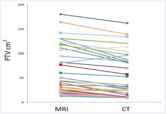
Figure 2: The change in planning target volume (PTV) for both CT-MRI fusion and CT.
Table 2: Volume analysis.
|
Item |
PTVct |
PTVMRI |
|
Mean ±SD |
53.62±45.52 |
66.77±48.77 |
|
Median |
31.7 |
43.65 |
|
Min-Max |
8.6-162.2 |
12.4-180.1 |
|
P value |
0.001 |
|
The average volume difference for PTVs was < 15cm3 . The volumes of the PTVs, also exhibit a wide range of differences (-15.5, 42 cm3 ).The result for the PTV comparison indicates that, in most cases, the volume of PTVMRI was larger than PTVCT except in three cases where the opposite occurs. Statistical analysis for the difference in CT and MRI tumor volumes was highly significant p < 0.001.
Examining the differences between the centers of mass (CM) of the two PTV’s detected spatial 3D shifts ranging from (-4mm to 5mm) with an average of 0.3 mm for x direction, (-2mm to 5mm) with an average of 1.3 mm for y, and (-2mm to 4mm) with an average of 0.7 mm for z direction respectively (refer to fig.3 and table 3).
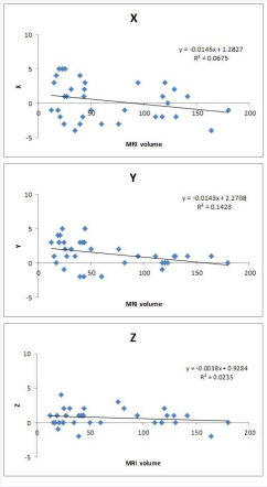
Figure 3: The relationship between 3D isocenter shift and MRI volume.
Table 3: 3D shift between planning target volume PTV marked in CT and MRI.
|
Patient No. |
CT |
MRI |
3D shift (mm) |
||
|
X |
Y |
Z |
|||
|
1 |
162.2 |
180.1 |
-1 |
0 |
0 |
|
2 |
57.2 |
76.3 |
-3 |
2 |
3 |
|
3 |
83.2 |
120.09 |
3 |
0 |
2 |
|
4 |
70.1 |
82.2 |
-1 |
0 |
2 |
|
5 |
8.8 |
12.4 |
-1 |
3 |
1 |
|
6 |
32.9 |
34.7 |
-4 |
1 |
0 |
|
7 |
27.2 |
43.4 |
1 |
-2 |
1 |
|
8 |
10.2 |
24.6 |
-3 |
3 |
0 |
|
9 |
120.3 |
130.5 |
-2 |
1 |
0 |
|
10 |
14.9 |
39.4 |
4 |
3 |
-2 |
|
11 |
52.4 |
60 |
-3 |
-2 |
0 |
|
12 |
18.9 |
31.08 |
2 |
2 |
2 |
|
13 |
80.9 |
111.3 |
-2 |
1 |
0 |
|
14 |
11.4 |
25.4 |
5 |
2 |
2 |
|
15 |
97.5 |
117.7 |
-2 |
-1 |
0 |
|
16 |
11.3 |
18.8 |
-1 |
4 |
1 |
|
17 |
87.2 |
129.2 |
2 |
1 |
1 |
|
18 |
24.1 |
24.7 |
1 |
-1 |
0 |
|
19 |
30.5 |
50.2 |
-1 |
2 |
0 |
|
20 |
9.6 |
19.9 |
5 |
3 |
-1 |
|
21 |
36.2 |
43.9 |
2 |
3 |
1 |
|
22 |
9.9 |
15.2 |
3 |
1 |
0 |
|
23 |
83.2 |
94.4 |
3 |
1 |
1 |
|
24 |
138.9 |
164.2 |
-4 |
1 |
-2 |
|
25 |
90 |
80.5 |
3 |
5 |
2 |
|
26 |
18.4 |
39.5 |
-3 |
-2 |
1 |
|
27 |
26.4 |
27.2 |
1 |
1 |
1 |
|
28 |
12.3 |
20.9 |
-2 |
4 |
0 |
|
29 |
133.8 |
141.6 |
1 |
1 |
1 |
|
30 |
8.6 |
22.9 |
5 |
5 |
4 |
|
31 |
29.4 |
42.02 |
-2 |
3 |
1 |
|
32 |
8.9 |
17.2 |
4 |
0 |
0 |
|
33 |
113 |
106 |
1 |
0 |
0 |
|
34 |
104.3 |
122.8 |
0 |
0 |
1 |
These shifts are significant when we consider that treatment field geometry. Figure 2 showed the relationship between CM shift and MRI volume which indicated that CM shift decreased by increasing in tumor volume, this result indicated also that a possible tumor underdosage occurred when CT alone was used for planning which may lead to geometric miss especially in small tumors.
The prescribed dose was fitted on PTVCT. The V95-PTVMRI was lower than the V95-PTVCT, the dose was (72.5% versus 95%, %, p = 0.001; Table 4), thus 72.5 % of the PTVMRI receives 95 % of the prescribed dose which mean the MRI target volume received an insufficient dose.
Table 4: Dose analysis.
|
Item |
V95 CT |
V95 MRI |
|
Mean ±SD |
95.20 ± 1.36 |
72.52±18.86 |
|
Median |
95 |
80 |
|
Min-Max |
90-97 |
33-92 |
|
P value |
0.001 |
|
Prior to the era of routine availability of CT and MRI, postoperative management of malignant glioma involved whole brain irradiation. Although, various clinical trials showed a positive gain with postoperative radiotherapy in these patients, the need for accurate target localization was perhaps not felt since the radiation portals were reasonably generous in covering the entire cranial contents. However, with the gradual acceptance of PBI for malignant glioma, accuracy in target delineation needs to be ensured. The target should include the residual postoperative tumor and a margin of usually 2-3 cm around the tumor to take care of the possibility of microscopic disease infiltration into the adjoining brain tumors [11].
An obvious limitation to designing precise high dose radiation treatment volumes is the ability of a physician to delineate target volume from normal surrounding tissues [12]. MRI is considered the diagnostic study of choice for assessing brain lesions.For GBM radiotherapy plan, the accurate delineation of PTV is an unsolved issue.The planning target volume (PTV), as defined in Report 62 of the International Commission on Radiation Units and Measurements [12], is a geometrical concept used to define beam apertures thatensure the prescribed dose is delivered to the CTV. As with all other external beam radiation therapy treatments, inter and intra-fractional set-up errors must be accounted for by assigning a margin to the CTV [13,14].
The PTVs in this study from both contrast-enhanced CT and MRI were drawn out by the radiation oncologist and then we determined the differences between PTV delineated on CT and on CT/MRI for astrocytoma patients. To evaluate the differences in volume, PTVCT and PTVMRI (cm3 ), the shift of CM, the agreement and disagreement indexes were measured. Moreover, since the low accuracy in the target definition had implication on the radiotherapy plan, the PTVs dose delivered was evaluated.
Our data showed that MRI showed more than 24% increase in the tumor volume, the statistics clearly show that MRI plays a very important role for target delineation for brain tumors and MRI should be incorporated for radiotherapy treatment planning. In the CM shift indicated a possible tumor under-dosage when CT alone is used for planning, these results agreed with Prabhakar et al [15]. The difference in terms of dose when radiotherapy plans based on PTVCT and PTVMRI were compared. Our result showed that that the ICRU recommendation was not respected for MRI target volumes (V95-PTVMRI 72.5%, V95-PTV CT 95 %), receiving a lower dose with respect to those based on CT (p = 0.001).Due to the GBM aggression, our result should be not neglected because it described the risk of target missing using the delineation based on CT alone.
CONCLUSION
CT and MRI image fusion technique is more precise than either by CT alone in defining the PTV of 3-dimensional conformal radiotherapy for astrocytoma. It is necessary that a proper delineation of targets based on the MRI images should be carried out to minimize the risks of geographical misses and, thereby, enable delivery of the intended doses to the target volumes. Also MRI-CT fusion can reduce the uncertainty of PTV delineation in radiation treatment planning for glioma patients after operation. Fusion allows to defining smaller more accurate volumes which may decrease dose to normal tissues and improve tumor coverage. Results show the optimal modality for planning astrocytoma is CT-MRI Fusion.
REFERENCES
- DDjan I, Petrovi? B, Erak M, Nikoli? I, Luci? S. Radiotherapy treatment planning: benefits of CT-MR image registration and fusion in tumor volume delineation. Vojnosanit Pregl. 2013; 70: 735-739.
- Mazzara GP, Velthuizen RP, Pearlman JL, Greenberg HM, Wagner H. Brain tumor target volume determination for radiation treatment planning through automated segmentation. Int J Radiation Oncology Biol Phys. 2004; 59: 300–312.
- Khoo VS. MRI--"magic radiotherapy imaging" for treatment planning? Br J Radiol. 2000; 73: 229-233.
- Prabhakar R, Julka PK, Ganesh T, Munshi A, Joshi RC, Rath GK. Feasibility of using MRI alone for 3D radiation treatment planning in brain tumors. Jpn J Clin Oncol. 2007; 37: 405-411.
- Tharavichitkul E, Samorn N, Wanwilairat S, Lookaew S, Nopnop W, Wannasri J, et al. The comparison of high-risk clinical target volumes between computed tomography and magnetic resonance imaging in cervical cancer. Biomed Imaging Interv J. 2013; 9: e4.
- Wong A, Bishop W. Efficient least squares fusion of MRI and CT images using a phase congruency model. Pattern Recogn Lett. 2008; 29: 173–180.
- Solaiyappan M, Gupta S. Predictive registration of cardiac MR perfusion images using geometric invariants. In: Proc Internat Soc. Magnetic Resonance in Medicine 2000; 1-37.
- Fitch AJ, Kadyrov A, Christmas WJ, Kittler J. Fast robust correlation. IEEE Trans Image Process. 2005; 14: 1063-1073.
- Shekhar R, Zagrodsky V. Mutual information-based rigid and nonrigid registration of ultrasound volumes. IEEE Trans Med Imaging. 2002; 21: 9-22.
- Averbuch A, Keller Y. FFT based image registration. In: Proc. IEEE Internat Conf on Acoustics, Speech, and Signal Processing. 2002; 4: 3608–3611.
- Datta NR, David R, Gupta RK. Implications of contrast-enhanced CT-based and MRI-based target volume delineations in radiotherapy treatment planning for brain tumors. J Can Res Ther. 2008; 4: 9-13.
- Lattanzi JP, Fein DA, McNeeley SW, Shaer AH , Movsas B, Hanks GE. Computed Tomography-Magnetic Resonance Image Fusion: A Clinical Evaluation of an Innovative Approach for Improved Tumor Localization in Primary Central Nervous System Lesions. Radiation Oncology Investigations. 1997; 5: 195–205.
- Prescribing, Recording and Reporting Photon Beam Therapy. Supplement to ICRU Report 50. ICRU Report 62, 1999.
- Fiorentino A, Caivano R, Pedicini P, Fusco V. Clinical target volume definition for glioblastoma radiotherapy planning: magnetic resonance imaging and computed tomography. Clin Transl Oncol. 2013; 15: 754-758.
- Prabhakar R, Haresh KP, Ganesh T, Joshi RC, Julka PK, Rath GK. Comparison of computed tomography and magnetic resonance based target volume in brain tumors. J Cancer Res Ther. 2007; 3: 121-123.


