Penile Mondor’s Disease Due to Local Trauma: A Case Report
- 1. Urology B Department, University Hospital Center IBN SINA, University Mohammed V, Morocco.
ABBREVIATIONS
PMD: Penile Mondor’s Disease; MRI: Magnetic Resonance Imaging.
Keywords
• Mondor’s Disease; Penile Trauma; Superficial Vein Thrombophlebitis; Diagnosis; Treatments
Citation
Amine EM, Anass R, Amine S, Tarik K, Abdellatif K, et al. (2025) Penile Mondor’s Disease Due to Local Trauma: A Case Report. J Urol Res 12(2): 1166.c
INTRODUCTION
The rare superficial thrombophlebitis known as “Mondor Disease” (MD) can affect the veins of the penile, abdomen, or chest. She is frequently benign and has an unknown provenance. In a small number of reported cases, MD is linked to a sub-jacent etiology such as vascularitis or prostate carcinoma. She may also be the consequence of a local trauma. we report today on a case of Mondor’s disease involving the penile superficial dorsal vein. When to think about it? What to do?
CASE PRESENTATION
E.H.Z, a 44-years-old male patient with no notable medical or surgical history, came to our clinic with penile pain and edema.
The examination found and that the edema was secondary to local trauma due to a sexual intercourse. There was no notion of immediate detumescence, creak or hematoma (Figure 1).

Figure 1 The clinical aspect of the PMD.3
The patient underwent penile doppler ultrasound, which revealed
-
- A partial thrombosis of the penile dorsal vein and collateral veins in the middle third of the penis
- A diffuse, hyperechoic thickening of the albuginea without visible focal rupture
- A soft tissue infiltration
We prescribed oral anticoagulants (rivaroxaban) and anti-edema treatment associated with pain killers.The follow-up was unremarkable, with improvement of symptoms after 2 weeks (resumption of the sexual activity, disappearance of the oedema and pain) (Figure 2).
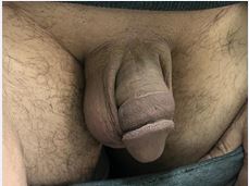
Figure 2 The follow up 2 weeks later.
DISCUSSION
Penile Mondor’s Disease (PMD) is a benign genital ailment that is uncommon and poorly understood. Men of any age who engage in sexual activity are susceptible to be affected by this disease. The patients ages range from 18 to 70 years old. In the cases that have been recorded thus far, no particular etiology has been identified [1]. PMD symptoms lack distinguishing characteristics. Some others have no symptoms at all. Patients typically arrive with a rope-like stiffness at the dorsum of the penis. They complain of throbbing and intermittent pain. The penile skin may exhibit edema and erythema. Some patients experience distention at the thrombosis site. Pain frequently gets worse when you get an erection. Symptoms of irritative voiding may appear in certain patients [2,3].
A medical history and physical examination can be used to identify Penil Mondor’s disease. In differential diagnosis, a color Doppler ultrasound examination is crucial. There have also been reports of patients who were diagnosed by Magnetic Resonance Imaging (MRI) [4].
The most crucial differential diagnosis in pathological examination is penile sclerosing lymphangitis. Microvascular structures can easily be mistaken for the lymphatic system. It is possible to differentiate them using immunohistochemical examinations. Histopathological analysis may show thrombus, increased connective tissue in the vessel wall, swelling of the endothelial cells, and infiltration of lymphocytes, histiocytes, and plasma cells in the perivascular region. The endothelium is stained with monoclonal antibodies to CD31 and CD34 in order to make a differential diagnosis of sclerosing lymphangitis. Sclerotic lymphangitis does not stain with these antibodies [6].
The use of color Doppler ultrasound examination is crucial. Dorsal vein thrombosis and the resulting hemodynamic alterations are seen during a Doppler Ultrasound (US) test. While color Doppler ultrasound data are thought to be adequate for diagnosing Mondor’s illness when superficial dorsal vein thrombosis appears without flow signals in this area, some research has indicated that this is not the case. Apart from the traditional results previously documented, a cavernous arterial flow-signal pattern has been established for Mondor’s disease [5].
Peyronie’s disease and sclerosing lymphangitis are the two most crucial disorders in the differential diagnosis. The tunica albuginea is not firm, according to both physical examination and color Doppler US [6].Medical treatment may include using anticoagulant medications, sexual activity should be limited during the acute phase. In the subacute and chronic phases, heparin- containing creams and anti-inflammatory medications are administered. The patient should be encouraged to limit his sexual activity throughout these phases until any infection-related symptoms and excruciating pain go away. No long-term, lasting sequel has been identified [6].
Surgical treatment may be indicated in individuals who are not responding to medicinal treatment, thrombectomy and superficial penile vein excision are performed surgically.
Patients who exhibit symptoms but do not exhibit flow in the color Doppler US after six weeks should be deemed resistant to treatment. There should be a surgical therapy option available [6].
CONCLUSION
To sum up, every urologist should be aware of Penil Mondor’s Disease, an uncommon clinical illness. Early and precise diagnosis reduces the need for surgery and improves the effectiveness of medical care. In this regard, a thorough physical examination and knowledge of color Doppler US exam results are crucial for early diagnosis and therapy. Patients with Penil Mondor’s disease may have issues like anxiety and sexual dysfunction. Therefore, prompt and precise diagnosis and treatment are crucial.
DECLARATION
Ethical Approval
Ethics approval has been obtained to proceed with the current study. Ethical approval for this study (Ethical Committee N009-24) was provided by the Ethical Committee Ibn University Hospitals, Rabat Morocco on 22 January 2024
Consent
Written informed consent was obtained from the patient for publication of this case report and any accompanying images. A copy of the written consent is available for review by the Editor-in-Chief of the journal.
Scare Guidelines
The work has been reported in line with the SCARE criteria [7].
REFERENCES
- Hamilton J, Mossanen M, Strote J. Mondor’s Disease of the Penis. West J Emerg Med. 2013; 14: 180.
- Thomazeau H, Alno L, Lobel B. Thrombose de la veine dorsale de la verge. A propos de 2 observations [Thrombosis of the dorsal vein of the penis. Apropos of 2 cases]. J Urol. 1983; 89: 691-692.
- Sasso F, Gulino G, Basar M, Carbone A, Torricelli P, Alcini E. Penile Mondors’ disease: an underestimated pathology. Br J Urol. 1996; 77: 729-732.
- Boscolo-Berto R, Iafrate M, Casarrubea G, Ficarra V. Magnetic resonance angiography findings of penile Mondor’s disease. J Magn Reson Imaging. 2009; 30: 407-410.
- Han HY, Chung DJ, Kim KW, Hwang CM. Pulsed and color Doppler sonographic findings of penile Mondor’s disease. Korean J Radiol. 2008; 9: 179-181.
- Öztürk H. Penile Mondor’s disease. Basic Clin Androl. 2014; 24: 5.
- Sohrabi C, Mathew G, Maria N, Kerwan A, Franchi T, Agha RA; Collaborators. The SCARE 2023 guideline: updating consensus Surgical CAse REport (SCARE) guidelines. Int J Surg. 2023; 109: 1136- 1140.


