Management of the Complications Due to Renal Injury: Case Report and Review of the Literature
- 1. Urology B Department, IBN SINA Hospital, University Hospital Center IBN SINA, University Mohammed V, Morocco
Abstract
Renal trauma accounts for approximately 1 to 5% of abdominal trauma. While the majority of cases can be treated conservatively, certain complications may require specific management.
Keywords
• Renal injury • Pseudoaneurysm • Hematuria • Embolization • Conservative treatment
Citation
ELAFARI MA, RHAYOUR A, MAMAD A, BAKKALI BE, SLAOUI A, et al. (2026) Management of the Complications Due to Renal Injury: Case Report and Review of the Literature. J Urol Res 13(1): 1169.
ABBREVIATIONS
CTA: Computed Tomography Angiography; MRA: Magnetic Resonance Angiography
INTRODUCTION
Renal trauma accounts for approximately 1 to 5% of abdominal trauma. While the majority of cases can be treated conservatively, certain complications may require specific management. Among these, urinary extravasation and renal pseudoaneurysm are described separately, but their association remains rare.
Urinary extravasation indicates damage to the collecting system and may require diversion via a double J stent to promote healing.
Pseudo-renal aneurysm, on the other hand, is a vascular complication that can manifest as delayed hematuria, sometimes severe, and its standard treatment is selective embolization. We report a case combining these two complications, treated sequentially with an internal urinary diversion and selective embolization, and discuss the lessons learned from this observation.
CASE PRESENTATION
M.M, a 33 years old male patient with no notable medical or surgical history, come by an ambulance to the emergency department, The patient claims to have been the victim of a traffic accident (motorcyclist struck by a car).
The examination finds a hemodynamically unstable patient with bruising on the right side and fever 38,8 C.
The resuscitation measures were implemented, and the patient underwent a Body-TDM which reveals: (Figure 1A-1B)
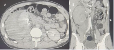
Figure 1 A: Axial cut-CT showing the pseudoaneuvrysm. B: frontal cut-CT showing the renal injury with the pseudoaneuvrysm
- Kidney with multiple lacerations in its middle and upper poles, with the presence at the same level of several hypodense triangular areas related to areas of infarction.
- Individualization of a rounded formation in contact with the segmental artery measuring 10X09 mm, initially suggesting a pseudoaneurysm of the superior segmental artery.
- Late individualization of a discreet extravasation of contrast medium into the retroperitoneal cavity through the upper calyceal system
- Significant retroperitoneal collection with spontaneously hyperdense content (68 HU) associated with a perirenal hematoma measuring approximately 126x85x91 mm. - Renal injury scale AAST IV.
The blood reports Hg: 7g/dl, the rest of the balance sheet without any particular features. The patient underwent blood transfusion, an ureteral stent double antibiotic and watchful waiting.
The consequences were minor, and the patient was discharged in stable condition in all respects.
1 month later, the patient came back to our clinic with a severe anemia Hg 5g/dl and hematuria
An CT-Uroscan was done showing: (Figure 2A-2B)
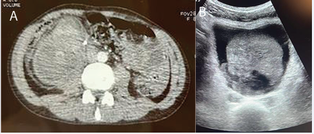
Figure 2 A: Axial Cut-CT showing the double J stent. B: Ultrasounds image, showing a massive bladder clot.
- Destroyed kidney and partially thrombosed hematoma fed by the superior segmental artery, measuring 116x110 mm
- End of the drain at the hematoma (Double J stent)
- Large hypodense intravesical collection
The patient received a blood transfusion and then embolization was indicated (Figure 3A-3C). Rupture of pseudoaneurysm at the expense of the superior segmental artery and its embolization - Seal checks Ok 3 Days after the patient underwent endoscopic clot removal. The patient was discharged after 10 days of hospitalization.
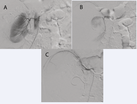
Figure 3 A: Pre-embolization arteriography showing the pseudoaneurysm. B: During the embolization (reduced the extravasation). C: After the embolization – Seal check Ok
DISCUSSION
Blunt trauma linked to high-velocity deceleration accounts for 90% of kidney injury cases, while penetrating trauma (gunshot and stab wounds) accounts for 1.4% to 3.3% However, the geographical region of the world affects these incidents.
The kidney is well protected in the retroperitoneum, but because it is solely fixed by the vascular pedicle and the renal pelvis in the uretero-pelvic junction, it is especially susceptible to forceful trauma accompanied by fast deceleration. The most common blunt mechanisms among adults include assault, falls from a height, skiing accidents, and injuries sustained in traffic accidents. The kidneys may be impacted by penetrating trauma, particularly if the superior abdomen is affected. Compared to blunt trauma, renal vascular injuries are more common while isolated penetrating kidney injuries are uncommon. Up to 90% of kidney injuries in both adults and children are mild and affect the segmental vessels or parenchyma. Isolated renal artery transection or intimal disruption is a rare and distinct kind of damage that mainly happens during fast deceleration [1,2].
A real aneurysm is a ball-shaped growth of the vascular wall, whereas a pseudoaneurysm is brought on by the tissues surrounding the arteries [3]. Aneurysms that develop at the site of artery injury but are not actual aneurysms are known as pseudoaneurysms. This results in the creation of a localized hematoma that is characterized by turbulent blood flow. The vessel wall cannot be penetrated by pseudoaneurysms, in contrast to aneurysms. Since pseudoaneurysms do not pierce any layer of the vessel wall, they differ from genuine aneurysms. A wall made of fibrin is eventually weaker than a real aneurysm [1-4].
The majority of renal artery pseudoaneurysms are associated with medicinal treatment. A kidney biopsy, percutaneous renal surgery, and open and endoscopic surgery are the most frequent reasons. The development of a renal artery pseudoaneurysm as a result of traumatic events is rare. The effect of deceleration forces on the vascular system is thought to be the cause of pseudoaneurysms resulting from renal damage [1-5].
Pseudoaneurysms are commonly diagnosed using the widely recognized standard angiography procedure. However, non-invasive imaging techniques such as computed tomography angiography (CTA), magnetic resonance angiography (MRA), and ultrasound are effective means of detecting pseudoaneurysms. Percutaneous endovascular and surgical techniques have been used more frequently in the modern era to treat arterial diseases. Surgical procedures have been replaced by interventional radiology, which has advanced significantly in recent years [6].
It has been demonstrated that embolization works well for treating pseudoaneurysm treatments. Stent deployment and embolization are the two main methods that are frequently used in clinical practice. The neck region’s measurements and the terminal organ’s additional provisions determine the best course of treatment [6].
Following the embolization process, use coil sizes of 3 x 5 cm and 2 x 3 cm to embolize the right upper pole. The-pseudoaneurysm was completely occluded in the post embolization run. The patient was monitored following the embolization treatment; within a day, there were no more hematuria complaints, and the pain on the right side had decreased. According to the research, percutaneous embolization is a safe and economical substitute for surgical exploration in the management of traumatic renal pseudoaneurysms [7]. Moreover, it is seen as a less complicated process. Postembolization syndromes, which include nausea, vomiting, fever, leukocytosis, and abdominal pain due to injury to the renal parenchyma, are among the possible side effects of renal artery embolization. Furthermore, there is a chance of renal failure, transient arterial hypertension, and hematoma at the catheter insertion site [8].
CONCLUSION
Although renal artery pseudoaneurysms are rare, they are a serious and severe consequence of renal trauma, as in this instance. A minimally invasive technique called renal artery embolization is used to diagnose and treat renal artery pseudoaneurysms. While a CT scan is only used to diagnose pseudoaneurysms, an embolization technique guided by an angiographic catheterization can also be used to treat them directly. An interventional radiologist can promptly diagnose and initiate treatment using an angiographic catheter to prevent potential issues such as hemorrhage, anemia or hemodynamic instability, and renal failure.
DECLARATION
Ethical Approval
Ethics approval has been obtained to proceed with the current study
Ethical approval for this study (Ethical Committee N009-24) was provided by the Ethical Committee Ibn University Hospitals, Rabat Morocco on 22 January 2024
Consent
Written informed consent was obtained from the patient for publication of this case report and any accompanying images. A copy of the written consent is available for review by the Editor-in-Chief of the journal.
Guarantor of Submission
The corresponding author is the guarantor of submission.
Availability of Data and Materials
Supporting material is available if further analysis is needed.
Scare Guidelines
The work has been reported in line with the SCARE criteria.
REFERENCES
- Ginanda Nabilla Hardiyanti, Ahmad Kabir AK, Mustafa NLM, Tharek A, AlKhafiz Kamis MF, Nasir Aslam, et al. Embolization for the Treatment of Delayed Hematuria Renal Artery Pseudoaneurysm Following Blunt Trauma. The Interventionalist J. 2023; 3: 1-9.
- Coccolini F, Moore EE, Kluger Y, Biffl W, Leppaniemi A, Matsumura Y, et al. WSES-AAST Expert Panel. Kidney and uro-trauma: WSES-AAST guidelines. World J Emerg Surg. 2019; 14.
- Dan AM, Azli S, Khairul-Asri MG, Jagwani AV. A rare case of renal artery aneurysm -dawning of new treatment approach. Urol Ann. 2022; 14: 99-101.
- Rivera PA, Dattilo JB. Pseudoaneurysm. 2022 Mar 9. In: StatPearls. Treasure Island (FL): StatPearls Publishing; 2023
- Yamaçake KG, Lucon AM, Mesquita JL, Srougi M. Renal artery pseudoaneurysm after blunt renal trauma: report on three cases and review of the literature. Sao Paulo Med J. 2013; 131: 356-362.
- Sarioglu O, Capar AE, Belet U. Interventional treatment options in pseudoaneurysms: different techniques in different localizations. Pol J Radiol. 2019; 84: 319-327.
- Boutakioute B, Chehboun A, Ouali M, Ganouni NCIE. Embolization for the treatment of renal artery pseudoaneurysm following surgical nephrolithotomy: A case report. Radiol Case Rep. 2022; 17: 4021-4024.
- Irwine C, Kay D, Kirsch D, Milburn JM. Renal artery embolization for the treatment of renal artery pseudoaneurysm following partial nephrectomy. Ochsner J. 2013; 13: 259-263.