Review of the Epidemiology of Camel Brucellosis and Prospects for Control in Africa
- 1. Abdou Moumouni University (UAM) of Niamey, Faculty of Agronomy, Regional Centre of Excellence on Pastoral Production (CERPP) Niger, Niger
- 2. Inter-State School of Veterinary Science and Medicine of Dakar (EISMV), Senegal
Abstract
Brucellosis is an infectious, contagious and zoonotic disease affecting humans and many animal species, including camels. In Africa, studies on camel brucellosis are mainly based on serological surveys, while studies based on bacterial isolation or molecular biology remain rare. The reported prevalence rates vary widely: from 0.5 to 51.2% in East Africa, from 1 to 28.5% in North Africa, and from 0.4 to 11.4% in West and Central Africa, reflecting the methodological differences and epidemiological contexts specific to each region. Molecular analyses have confirmed the presence of Brucella abortus and Brucella melitensis in milk, aborted fetuses and vaginal swabs from infected camels. Extensive farming practices, combined with the dietary habits of pastoral populations, particularly the consumption of raw milk, and poor control of risk factors, constitute a significant zoonotic risk to animal and human health. The disease remains largely underdiagnosed, particularly due to its insidious clinical progression and the lack of validated serological tests for camelids, as highlighted by the World Organization for Animal Health (WOAH). The combination of different methods (serology, culture isolation, PCR) is strongly recommended for reliable diagnosis and to support brucellosis control and eradication strategies in camelids. This review provides an updated summary of knowledge on camel brucellosis in Africa, focusing on its socio-economic impact, epidemiology, and available diagnostic and preventive approaches.
Keywords
• Camel Brucellosis • Epidemiology • Diagnosis • Prevention • Africa
Citation
HAROUNA MT, NDOUR APN, HAMANI M, BADA-ALAMBEDJI R, AKAKPO AJ (2026) Review of the Epidemiology of Camel Brucellosis and Prospects for Control in Africa. J Vet Med Res 13(1): 1295
INTRODUCTION
The global camel population is estimated at around 42.5 million head [1], with more than 80% found in Africa. Camels play an important socio-economic role in the pastoral and agricultural systems of arid and semi-arid areas of Asia and Africa [2]. Camels have unique qualities that make them particularly well suited to desert ecosystems. They can withstand heat, drought and malnutrition, and are resistant to disease and dehydration. These abilities make them superior to other domesticated species in hot, arid desert areas, where they play an important role in combating desertification and contributing to food security [2,3]. Camels are an important source of milk, meat, leather and wool in many parts of the world [2]. In addition, they have been used as a source of long term investment and savings, in sport, transport and tourism [4,5]. For a long-time, dromedaries were considered resistant to many pathogens [6,7]. However, it has been shown that they are susceptible, like other livestock, to common pathogens that affect other animal species, including brucellosis [2-9]. Brucellosis is a globally distributed anthropozoonosis, recognized as a notifiable infectious and contagious disease caused by bacteria of the genus Brucella [10,11]. This zoonosis can affect virtually all domestic species, with the possibility of cross-transmission between cattle, sheep, goats, camelids and other susceptible species [12,13]. In livestock farming, brucellosis causes significant economic losses, particularly through abortions, infertility, neonatal mortality, reduced milk and meat production, and the costs associated with replacing infected animals. Furthermore, due to its transmissibility to humans, it poses a real public health challenge [9-15].
In humans, contamination occurs mainly through the consumption of infected raw milk, unpasteurized and contaminated dairy products, or through direct contact with infected animals, particularly when handling abortion products, thus posing a significant risk to the health of consumers and farmers [16-18]. Although brucellosis has been extensively researched in cattle and small ruminants, its impact on camelids remains largely underestimated. However, these animals play an important role in pastoral systems in arid and semi-arid areas of Africa. Camel brucellosis has been reported in all countries where camels are raised, with sometimes high prevalence rates in certain regions (Figure 1).
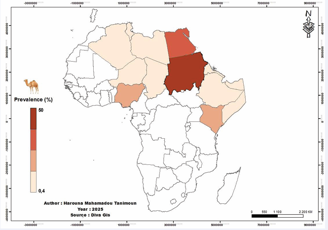
Figure 1 Map of African countries that have reported cases of brucellosis in camelids up to 2025
It therefore represents a real animal health challenge, particularly in African countries with a high density of camelids [19-21]. Despite this worrying situation, data on the origin of the infection remain limited. Inter-camel transmission and the persistence of the disease within herds are still poorly documented [2-20]. Brucellosis in camelids remains an insidious disease, rarely manifesting itself through obvious clinical signs [22]. The prevalence of subclinical infections makes diagnosis particularly difficult, especially since, unlike in cattle, the disease may manifest very late, particularly in latent carriers, and clinical signs are often subtle in camelids [2]. Furthermore, none of the available serological tests have yet been validated specifically for camelids, as recognized by the World Organization for Animal Health (WOAH) [20]. In this context, it seems necessary to take stock of the current state of knowledge on camel brucellosis. This review provides an up-to-date summary of the epidemiology of the disease in Africa, its socio-economic impact, and the diagnostic methods and preventive measures currently available. It is based on documentary research conducted using the following search engines: Google Scholar, PubMed and African Journals Online, in order to obtain relevant data and recent scientific articles.
Socio-Economic and Ecological Importance of Camels
Camels play a key role in the economic, social and environmental life of many arid regions. Their meat, which is low in fat, and their milk, which is rich in insulin and specific proteins, are major nutritional resources [23,24]. Camel milk, in particular, is a staple food for nomadic populations and helps to improve herders’ incomes [2].
Beyond their nutritional value, camels are also used for wool production, racing, transport, tourism and competitions [25]. In some cultures, such as in Niger, the dromedary is much more than just a farm animal: it represents capital, a means of transport, a source of food and leather, but also a symbol of social success and a central element of community life [3-27]. Camels also perform agricultural functions. They are used to draw water, plough fields, operate mills, transport loads, and even crush grain or sugar cane [28]. Furthermore, their presence is essential to maintaining ecological balance in pastoral areas. Thanks to their morphology (wide, flexible soles) and feeding habits, they have a minimal impact on soil and vegetation, unlike ruminants with hard hooves. They thus help to limit overgrazing and preserve the fragile ecosystems of desert regions [29,30].
In the face of global challenges such as drought, climate change and resource scarcity, camels appear to be a strategic asset for the future. Their robustness, adaptability and diversity of uses make them particularly valuable animals for meeting the challenges of tomorrow [2-31].
Impact of Brucellosis on the Economy and Public Health
Brucellosis is a zoonotic disease found in many parts of the world, but its perception varies depending on the context. While its impact on human health is well recognized, its economic consequences are often underestimated, particularly in low-income countries. In intensive livestock farming systems, it causes significant losses due to abortions, infertility, reduced milk production and stillbirths. It also hinders trade, particularly for livestock-exporting countries [15-32]. In addition, the measures required to eradicate it are costly. In sub-Saharan Africa, brucellosis remains a disease with a significant socio-economic impact. In regulated livestock farms in some African countries, economic losses in sedentary herds can be estimated at 150 million CFA francs per year, or 10% of the owners’ income [32]. In terms of health, human brucellosis is a major public health risk that affects social and economic development in various countries due to acute and chronic illness, physical disability and loss of labor [10].
Occupational brucellosis is a real public health problem due to the high risk of transmission between infected animals and people who work in contact with them. The professionals most at risk are slaughterhouse workers responsible for slaughtering animals, milkers, shepherds and veterinarians [2]. The latter are particularly at risk when working with infected animals, for example during placental retention or vaccination campaigns, often without adequate hand protection, i.e. wearing gloves [33]. The most frequent and serious human cases are observed among shepherds and their families, slaughterhouse employees and veterinarians. For this reason, brucellosis is considered both an accidental zoonosis and an occupational disease [34]. The isolation of B. melitensis and B. abortus from camel milk [35,36], and seropositivity to B. abortus and B. melitensis indicate that brucellosis is a public health hazard. Human infection with Brucella occurs mainly through the consumption of raw milk [17,18]. However, camel milk is becoming increasingly popular with consumers, hence the need to pasteurize it in order to protect public health, especially since pasteurization does not affect the chemical composition of camel milk [2-37].
In 2010, the World Health Organization (WHO) estimated that there were 0.83 million cases of human brucellosis worldwide, 47% of which were foodborne. However, this f igure is likely to be an underestimate due to significant under reporting and frequent misdiagnosis [38]. More recent data indicate that there are now more than 1 million cases per year worldwide, while the actual number of undiagnosed cases is likely to be much higher than official statistics suggest [39]. This development highlights that human brucellosis remains a major and under-recognized public health problem, with foodborne transmission still active in many regions. In view of this situation, enhanced cooperation between public health services and veterinarians appears essential to limit the spread of human brucellosis in endemic areas [40].
Clinical Manifestations of Brucellosis in Camelids
Clinical signs play an important role in predicting and diagnosing brucellosis in cattle [36]. In camelids, brucellosis may be asymptomatic or manifest with various clinical signs, including abortion, infertility, retained placenta, endometritis, orchitis, epididymitis, arthritis, and hygroma [16-41]. According to several authors, the clinical signs of brucellosis in breeding camelids are the same as those in cattle and small ruminants. However, infection in breeding camelids causes fewer abortions than in cattle and small ruminants [2-16].
It should be noted that brucellosis in camels rarely causes clinical signs [42], and the disease may manifest itself in a subclinical form or with only mild signs while producing positive serological or molecular results [2-22]. This lack of symptoms makes diagnosis particularly difficult, especially since, unlike cattle, clinical signs are often subtle in camelids [2-43]. Furthermore, the asymptomatic form of the infection plays an important role in the transmission of the disease to humans, particularly through the consumption of contaminated, unpasteurised milk or through direct contact with infected animals [2,16].
Epidemiology of Camel Brucellosis
First reported in 1931 [44], camel brucellosis has been reported in all camel-breeding countries except Australia. Its incidence appears to be closely linked to breeding practices and local climatic conditions [2-20]. According to Ekereet al. [45], the disease has a cosmopolitan distribution and affects economically important domestic animals, such as dromedaries, as well as wildlife. In sub-Saharan Africa, the available data on camel brucellosis remain limited. Reports submitted to the World Organization for Animal Health (WOAH) are mainly based on serological surveys conducted in cattle, sheep and goats, while studies on dromedaries are much less frequent. However, in a context where pastoral systems are becoming increasingly important and where demand for meat and livestock products is expected to double by 2050, brucellosis represents a significant health threat to this region [46]. Camels can contract brucellosis through interspecies transmission, particularly from other domestic animals such as cattle, sheep or goats [13]. The emergence and spread of the disease are closely linked to the presence of Brucella in these species sharing the same environment, as well as the type of farming system [16]. Camelids are often raised in close proximity to other ruminants, sharing the same water sources and pastures, which promotes the circulation of the pathogen between species. This situation could explain the sometimes high infection rates observed in camelids in certain regions [47]. Transmission can occur in several ways: through direct contact with animals that have recently aborted, via feed or an environment contaminated with faeces or abortion products, or sexually. In males, excretion of the pathogen in semen may play an important role in spreading the infection within the herd [10]. Despite advances in surveillance and control, the prevalence of brucellosis is increasing in many developing countries due to various health, socio-economic and political factors [48].
High prevalence rates of brucellosis in camels have been reported in several African countries, with considerable regional variability, posing a major risk to both human health and livestock. In East Africa, camel brucellosis is considered endemic, causing significant economic losses in most member states of the Intergovernmental Authority on Development (IGAD), including Ethiopia, Kenya, Sudan, South Sudan, Uganda, Somalia, Eritrea and Djibouti [49]. In these countries, some pastoral areas have seroprevalence rates of up to 51%. These high levels could be attributed to high animal population density, cross-border mobility, and the absence or ineffectiveness of health control measures. In North Africa, reported prevalence rates are generally more moderate, ranging from 1 to 28.5%. This variation can be explained in part by differences in the screening methods used, but also by more structured health policies in certain countries such as Algeria and Egypt. In West and Central Africa, data indicate generally lower prevalence rates, ranging from 0.4% to 11.4% according to available studies (Table I).
Table I: Review of the literature on the prevalence of brucellosis in camels in selected African dromedary-raising countries from 2000 to 2025
|
Country |
Sample size |
Type of test |
Prevalence (%) |
References |
|
Ethiopia |
1442 |
RBT |
5.7 |
[47] |
|
CFT |
4.2 |
|||
|
756 |
RBPT/CFT |
2.2 |
[50] |
|
|
1073 |
RBPT |
2.1 |
[50] |
|
|
CFT |
1.8 |
|||
|
646 |
RBPT |
2 |
[50] |
|
|
CFT |
1.5 |
|||
|
768 |
RBPT |
11.9 |
[51] |
|
|
CFT |
7.6 |
|||
|
461 |
mRBPT/CFT |
5.4 |
[52] |
|
|
458 |
RBT/ELISA |
0.9 |
[53] |
|
|
1152 |
RBPT |
5 |
[54] |
|
|
CFT |
4.1 |
|||
|
822 |
RBPT/CFT |
2.43 |
[55] |
|
|
1500 |
RBPT/CFT |
0.53 |
[56] |
|
|
415 |
RBPT |
5.8 |
[57] |
|
|
CFT |
3.37 |
|||
|
813 |
RBPT/CFT |
2.09 |
[58] |
|
|
261 |
RBPT |
2.3 |
[59] |
|
|
CFT |
1.53 |
|||
|
384 |
RBPT |
3.6 |
[60] |
|
|
CFT |
3.1 |
|||
|
201 |
RBPT |
6.5 |
[61] |
|
|
CFT |
4.5 |
|||
|
384 |
CFT |
1.04 |
[62] |
|
|
350 |
mRBPT |
8.3 |
[63] |
|
|
CFT |
2 |
|||
|
450 |
RBPT and cELISA |
2.9 |
[64] |
|
|
450 |
RBPT et CFT |
4.8 |
[65] |
|
|
460 |
mRBPT et iELISA |
7.83 et 4.78 |
[66] |
|
|
Somalia |
1246 |
RBT |
3.9 |
[13] |
|
iELISA |
3.1 |
|||
|
180 |
RBPT, m RBPT, mSAT, C-ELISA |
4.4 |
[67] |
|
|
RBPT |
1.7 |
|||
|
mRBPT, mSAT, C-ELISA |
3.9 |
|||
|
Sudan |
3274 |
RBT, SAT and CFT |
7.82 |
[22] |
|
29 |
cELISA |
13.8 |
||
|
RBT |
10.3 |
|||
|
110 |
MRT |
2.7 |
||
|
4791 |
RBT |
12.3 |
[68] |
|
|
6032 |
RBT |
15.5 |
[68] |
|
|
3549 |
RBT |
30.5 |
[68] |
|
|
14372 |
RBPT |
18.13 |
[68] |
|
|
21 |
RBT and SAT |
23.8 |
[16] |
|
|
62 |
RBPT |
9.67 |
[16] |
|
|
SAT |
8.06 |
|||
|
2225 |
RBPT, mRBPT, cELISA et SAT |
37.5 |
[69] |
|
|
2000 |
RBPT |
39.9 |
[69] |
|
|
cELISA |
40.5 |
|||
|
225 |
MRT |
16.9 |
||
|
383 |
RBPT, mRBPT et ELISA |
28.5 |
Musa et al., (2013) |
|
|
8220 |
RBPT |
51.2 |
[70] |
|
|
1225 |
RBPT et ELISA |
23.6 |
Zein (2015) |
|
|
400 |
mRBPT |
10.8 |
Mohammed and Abdelgadir (2016) |
|
|
200 |
RBPT et SAT |
32 et 23 |
Ahmed et al., (2017) |
|
|
343 |
RBPT, CFT, cELISA |
36.2; 42.6 et 19.8 |
Abdelnassir et al., (2020) |
|
|
743 |
RBPT |
13.5 |
Abdelrahman (2020) |
|
|
500 |
RBPT et SAT |
3.8 et 3.4 |
Abdalla et al., (2021) |
|
|
60 |
RBPT |
8.3 |
El Ayis (2022) |
|
|
400 |
RBPT and ELISA |
29.5 et 27.8 |
Bashir (2022) |
|
|
388 |
mRBPT, BPAT et SAT |
11 10.5 et 7.7 |
[71] |
|
|
Djibouti |
72 |
RBT, BWT |
1.38 |
[72] |
|
Kenya |
384 |
MRT |
15.36 |
[73] |
|
|
200 |
RBPT |
2 |
|
|
|
|
SAT |
10.5 |
|
|
Eritrea |
98 |
CFT |
3.1 |
[74] |
|
Algéria |
157 |
EAT, FCT and cELISA |
0 et 2.3 |
[33] |
|
|
|
EAT |
5 |
|
|
|
|
cELISA |
1.27 |
|
|
|
132 |
RBPT |
1.5 |
[75] |
|
|
|
ELISA |
5.3 |
|
|
Egypt |
592 |
STAT |
1 |
[76] |
|
|
|
Card test |
1.7 |
|
|
|
750 |
RBPT |
4.9 |
[77] |
|
|
|
BAPAT |
3.9 |
|
|
|
766 |
RBT |
8.7 |
[78] |
|
|
|
ELISA |
9.3 |
|
|
|
340 |
CFT |
7.4 |
[79] |
|
|
1126 |
mRBPT |
4.17 |
[35] |
|
|
|
cELISA |
3.73 |
|
|
|
801 |
RBPT |
11.60 |
[80] |
|
|
|
BAPAT |
12.90 |
|
|
|
|
CFT |
11.50 |
|
|
|
271 |
BAPAT |
9.5 |
[81] |
|
|
|
mRBPT |
8.8 |
|
|
|
|
TAT |
7.7 |
|
|
|
312 |
RBPT |
28.5 |
[82] |
|
|
|
ELISA |
27.8 |
|
|
|
|
CFT |
25.9 |
|
|
|
272 |
RBPT |
4.04 |
[83] |
|
|
|
cELISA |
3.6 |
|
|
|
618 |
RBPT et iELISA |
26.8 et 18.3 |
[36] |
|
Libya |
520 |
RBT, SAT, cELISA |
1.4; 1.2 et 3 |
[84] |
|
|
14 |
RBPT |
14.28 |
[85] |
|
|
35 |
RBPT, SAT, CFT |
5.7 |
[86] |
|
Tunisia |
500 |
ELISA |
7.4 |
[87] |
|
|
470 |
iELISA |
2.12 |
[88] |
|
Chad |
288 |
ELISA and RBT |
0.4 et 1.4 |
[89,90] |
|
Niger |
275 |
RBT and ELISA |
4 |
[91] |
|
Nigeria |
329 |
RBT, SAT, cELISA |
11.4 |
[92] |
|
|
181 |
MRT |
8.3 |
[93] |
|
|
311 |
RBPT and LFiA |
1.9 |
[94] |
|
|
180 |
RBPT |
2.2 |
|
|
|
32 |
RBPT |
6.3 |
|
|
|
980 |
RBPT |
11.2 |
[95] |
|
|
|
SAT |
10.5 |
|
SAT : Serum Agglutination Test, TAT : Tube Agglutination Test, CFT : Complement Fixation Test, RBT : Rose Bengal Test, RBPT : Rose Bengal Plate Test, ELISA : Enzyme Linked Immuno Sorbent Assay, iELISA : indirect Enzyme Linked Immunosorbent Assay, cELISA : competitive Enzyme Linked Immune Sorbent Assay ; MRT : Milk Ring Test, LFiA : Lateral Flow Immunoassay, BAPAT : Buffered Acidified Plate Antigen Test,
Variations in the prevalence of brucellosis between African countries can be explained by a combination of factors. These include local climatic conditions, husbandry and management practices, camel population density, the virulence of the Brucella strains present, the presence of infected animals in certain areas, as well as inadequate veterinary services, lack of awareness among livestock owners and herd mobility, which also plays an important role [14]. Other factors may influence these disparities, such as the sensitivity of the screening techniques used, the nutritional and immune status of the animals, and the mobility dynamics of the farmers. In addition, several risk factors are associated with the occurrence of brucellosis in camelids, including geographical location, sex and age of animals, season, level of awareness among livestock farmers, history of abortion or orchitis, herd size and cohabitation with other ruminants [36-96].
Beyond serological studies, several molecular analyses have confirmed that Brucella abortus and Brucella melitensis are the main Brucella species circulating in camel livestock in Africa. These are also the strains most frequently isolated in milk, aborted foetuses, lymph nodes and vaginal swabs from infected female camels [16-99] (Table II).
Table II: Brucella species isolated from camelids in Africa
|
Country |
Species |
Organs |
References |
|
Egypt |
B. melitensis biovar 3 |
Milk |
[76] |
|
B. abortus biovars 1 and 7 |
Organs (uterus, placenta) Aborted fetuses (stomach contents, liver, spleen, and lungs) |
[77] |
|
|
B. melitensis biovar 3 |
|||
|
B. abortus biovar 7 |
Milk |
[100] |
|
|
B. melitensis biovar 3 |
|
[35] |
|
|
B. abortus biovar 1 |
Milk |
||
|
B. suis biovar 1 |
|
||
|
B. abortus |
Milk |
[36] |
|
|
B. melitensis |
|||
|
Libya |
B. melitensis biovar 1 |
Milk, aborted fetus, vaginal swab |
[97] |
|
Sudan |
B. abortus biovar 6 |
Lymph nodes, testis |
[69] |
|
B. abortus biovar 6 |
Lymph nodes |
[16] |
Although camelids are considered secondary hosts for Brucella species, they are among the animal species most susceptible to infection [21 102]. Thus, the prevalence of the disease in camelids depends on the infection rate in the primary hosts with which they come into contact [16]. Camel brucellosis is present in most African countries where dromedaries are raised, with incidence and prevalence rates varying greatly from country to country, and even from region to region within the same country. However, in several sub-Saharan African countries, the disease remains underreported or poorly documented, often due to its discreet progression, weak screening methods and lack of organized surveillance. Overall, the epidemiological situation of brucellosis in camels in Africa remains poorly understood. The available data show great disparity, linked to the diversity of local contexts, differences in breeding practices and the limitations of current health monitoring systems.
Diagnosis of Camel Brucellosis
In the field, some clinical signs such as repeated abortions or the presence of hygromas in a herd of dromedaries can be useful indicators for clinical diagnosis. However, these signs are often subtle, non-specific and appear late, making diagnosis based solely on clinical signs unreliable. Furthermore, seropositive cases can be observed in animals that do not show any characteristic symptoms of brucellosis, which further complicates the detection of the disease [36]. Accurate screening for brucellosis is often difficult and generally requires serological tests [103].
In Africa, brucellosis screening in camels relies mainly on serological tests, while studies based on bacterial isolation or molecular biology techniques remain limited. Several serological tests have been used to detect anti-Brucella antibodies in camel blood, including: the Rose Bengal Test (RBT), the complement fixation test (CFT), the standard or rapid slide agglutination test (SAT), also known as the Sero agglutination Wright test (SAW), ELISA (enzyme-linked immunosorbent assay) tests and rapid card tests [16-91].
However, none of these tests are currently validated specifically for use in camels, as highlighted by the World Organization for Animal Health (WOAH) [2-20]. Similarly, none of these tests have been validated for the diagnosis of human brucellosis [20-104].
However, it has been found that a combination of different serological tests can increase the effectiveness of screening in camelids, although none of the serological tests can differentiate between B. abortus, B. melitensis and B. suis infections [20]. The most common combinations used in Africa for diagnosis in camelids are RBT tests with CFT or ELISA, which are recognised as being among the most sensitive and specific [16-82].
Despite this, serological tests have some limitations, including cross-reactions with other bacteria such as Yersinia enterocolitica, and the inability to differentiate between acute and chronic infections due to the prolonged persistence of antibodies. To overcome these shortcomings, it is therefore recommended that serological methods be combined with complementary approaches such as bacterial culture, PCR or other molecular biology tools [10-106].
PCR, particularly AMOS-PCR, is increasingly used as a confirmatory method, allowing the precise identification of Brucella species in infected animals. This technique has proven effective in detecting B. abortus and B. melitensis in several African countries such as Egypt, Sudan and Ethiopia [35-109]. In addition, during calving or abortions, camels excrete large amounts of Brucella through vaginal discharge, amniotic fluid, placenta, foetal stomach contents and milk. These are valuable sources for bacterial isolation or molecular identification. Finally, although costly and more time consuming, bacteriological isolation remains the gold standard method for reliably identifying Brucella species, particularly for confirming serological test results [106]. The combined use of different methods (serology, bacterial isolation, PCR) is therefore strongly recommended for reliable diagnosis and to support brucellosis control and eradication strategies in camelids.
Control and Prevention of Camel Brucellosis
Although brucellosis has been eradicated in some parts of the world, it remains widespread in others, where it continues to pose a major public health and economic problem. In camelids, this zoonotic disease has a significant impact on human health, particularly in countries where these animals are farmed on a large scale.
Several approaches can be implemented to limit the spread of brucellosis: either by reducing its prevalence to an acceptable level (control strategy) or by completely eliminating infectious foci (eradication strategy). The choice of the most appropriate strategy depends on several factors, such as the prevalence of infection in different animal species, the clinical incidence in humans, and the organizational and technical capacities of veterinary services [110]. It is therefore essential to adapt control measures to the local realities of camel farming. Furthermore, the establishment of an effective epidemiological surveillance system is a prerequisite for any control strategy. In regions where brucellosis is endemic, eradication can only be achieved through a combination of control, prevention and rigorous surveillance. Vaccination of healthy animals remains generally the most effective and economically viable method of preventing brucellosis [2 110]. However, in many developing countries, vaccination programmes focus mainly on small ruminants and cattle, with camelids often being neglected [75].
According to Abbas and Agab [2], in low-prevalence areas, widespread vaccination of livestock may be sufficient to control brucellosis. However, in high-prevalence areas, a strategy combining screening, culling of infected animals and vaccination is more effective. This strategy has been successfully applied in Egypt, where the RB51 vaccine has been used in cattle and the Rev.1 vaccine in small ruminants [99-111]. However, applying this method to camelids is particularly difficult. On the one hand, most infected camelids are often asymptomatic, making them difficult to detect. On the other hand, the culling of seropositive animals is widely rejected by farmers due to the high economic, social and cultural value of dromedaries [110]. In this context, the decision to slaughter animals that test positive can only be considered after taking into account regulatory, economic and epidemiological factors. Keeping positive animals is less dangerous if the rest of the herd is vaccinated, but it should only be considered as a last resort [49]. In the reality of developing countries, the test-and-kill strategy is often unworkable [112] due to its high cost and social constraints. Vaccination therefore appears to be the most realistic and sustainable alternative. Although no vaccine has yet been specifically approved for camelids [49], significant progress has been made in the development of a vaccine (Rev.1 or Brucella melitensis vaccine) for camelids, with promising results [112,113]. Furthermore, trials with the RB51 vaccine have shown a good immune response in camelids in Egypt [71]. However, further studies are needed to evaluate the efficacy and feasibility of large-scale vaccination in camelids [36].
CONCLUSION
Camels play an important role in the epidemiology of brucellosis, particularly in pastoral areas of African countries where camel breeding is widespread. Camel brucellosis causes significant economic losses and poses a major public health problem, especially as it develops subclinically, making it difficult to detect in the field. This situation is exacerbated by the lack of diagnostic tests validated by the World Organization for Animal Health (WOHA) for camels and by the limited use of molecular biology or bacteriological isolation tools in many regions of sub-Saharan Africa, thus limiting surveillance and control efforts. The isolation of Brucella abortus and Brucella melitensis in apparently healthy camelids, showing no clinical signs suggestive of brucellosis, poses a real risk of transmission to other domestic animal species and to humans in close contact with them. This situation highlights the need for regular screening for infection in this species. It is therefore essential to include camels in brucellosis control programmes in order to prevent transmission to susceptible hosts. An integrated approach based on awareness-raising and screening, combined with animal vaccination, is essential for the effective prevention and possible eradication of this zoonosis. The available data underscore the urgent need to strengthen research, particularly on bacterial isolation, characterization of circulating strains, reliable epidemiological studies, and the adoption of control measures adapted to the realities on the ground in Africa.
REFERENCES
- FAOSTAT. 2023: Production: Crops and livestock products.
- Gwida M, El-Gohary A, Melzer F, Khan I, Rösler U, Neubauer H. Brucellosis in camels. Res Vet Sci. 2012; 92: 351-355.
- Teko AA. Impact socio-économique du dromadaire (camelus dromedarius) au Niger. Thèse: Méd. Vét: Dakar. 1998; 11: 127.
- Wilson RT. Productivity. The Camel. Longman House Burnt Mill, Harlow, Essex, UK. 1984. 153-172
- Rollefson K. The camel and human society. In: Gahlot, T.K. (Ed.), Selected Topics on Camelids. The Camelid Publishers, Bikaner, India. 2000. 1-17.
- RAGHEB ZAKI. Brucella infection among ewes, camels and pigs in Egypt. J Comp Pathol Ther. 1948; 58: 145-151.
- Dalling T, Robertson A, Boddie G, Spruell J. Diseases of camels. In: The Inter-national Encyclopedia of Veterinary Medicine. Edinburgh, U.K.,W. Green and Son. 1988; 585.
- Abbas B, Tilley P. Pastoral management for protecting ecological balance in Halaib District, Red Sea Province, Sudan. Nomadic Peoples 1990; 29: 77-86.
- Abbas B, Agab H. A review of camel brucellosis. Prev Vet Med. 2002; 55: 47-56.
- Corbel MJ. Brucellosis in humans and animals. Geneva. WHO. 2006; 89.
- Akakpo AJ, Ndour APN. La brucellose bovine en Afrique de l’Ouest et du Centre: état des lieux. Rev. Afr. Santé Prod. Anim. 2013; 11: 23-28.
- Agab H, Abbas B, Ahmed HJ, Mamoun IE. First report on the isolation of Brucella abortus biovar 3 from camels (Camelus dromedarius) in Sudan. Rev. Elev. Méd. vét. Pays trop. 1994; 47: 361-363.
- Ghanem YM, El-Khodery SA, Saad AA, Abdelkader AH, Heybe A, Musse YA. Seroprevalence of camel brucellosis (Camelus dromedarius) in Somaliland. Trop Anim Health Prod. 2009; 41: 1779-1786.
- Radostits OM. Gay CC, Hinchcliff KW, Constable PD. Veterinary Medicine: A Textbook of the Diseases of Cattle, Horses, Sheep, Pigs and Goats. 10th Edition, Elsevier Saunders, London. 2007: 966-994.
- Seleem MN, Boyle SM, Sriranganathan N. Brucellosis: a re-emerging zoonosis. Vet Microbiol. 2010; 140: 392-398.
- Musa MT, Eisa MZ, El Sanousi EM, Abdel Wahab MB, Perrett L. Brucellosis in camels (Camelus dromedarius) in Darfur, Western Sudan. J Comp Pathol. 2008; 138: 151-155.
- Yumuk Z, O’Callaghan D. Brucellosis in Turkey -- an overview. Int J Infect Dis. 2012; 16: e228-e235.
- Dean AS, Bonfoh B, Kulo AE, Boukaya GA, Amidou M, Hattendorf J, et al. Epidemiology of brucellosis and q Fever in linked human and animal populations in northern togo. PLoS One. 2013; 8: e71501.
- Sprague LD, Al-Dahouk S, Neubauer H. A review on camel brucellosis: a zoonosis sustained by ignorance and indifference. Pathog Glob Health. 2012; 106: 144-149.
- Wernery U. Camelid brucellosis: a review. Rev Sci Tech. 2014; 33: 839-857.
- Mohammed A, Ahmed M, Osman Ahmed A, Yousof S, Hamad S, Shuaib Y, et al. Seroprevalence and risk factors of brucellosis in dromedary camels (Camelus dromedarius) in Sudan from 1980 to 2020: a systematic review and meta-analysis. Vet Q. 2023; 43: 1-15.
- Musa MT, Shigidi MTA. Brucellosis in camels in intensive animal breeding areas of Sudan. Implications in abortion and early-life infections. Rev. Elev. Méd. vét. Pays trop. 2001; 54: 11-15.
- Farah Z, Abdulkadir O, Abdurahman SH. Milk and meat from the camel: handbook on products and processing. Hochshuleverlag AG ander ETH Zürich. 2004; 15-28.
- Sallam AM. Future opportunities for genetic improvement of the Egyptian camels. Egyptian J Animal Production. 2020; 57: 39-45.
- Ashour G, Abdel-Rahman SM. Camels as a miracle key for animal production sustainability in Egypt. Egyptian J Animal Production. 2022; 59: 33-43.
- Ramet JP. La technologie des fromages au lait de dromadaire (Camelus dromeda-rius). Etude FAO production et santé animales. 1993; 113.
- Pacholek X, Vias G, Faye B, Faugère O. Élevage camelin au Niger : Référentiel zootechnique et sanitaire, 1ère Ed. Niamey, Coopération française. 2000; 93.
- Yaqoob M, Nawaz H. Potential of Pakistani camel for dairy and other uses. Animal Sci J. 2007; 78: 467-475.
- Narjisse H. Nutrition et production laitière chez les dromadaires. Options Méditerra-néennes -Série Séminaires. 1989; 2: 163-166.
- Ould AM. Caractérisation de la population des dromadaires (Camelus dromedarius) en Tunisie. Thèse : INAT : Carthage. 2009; 172.
- Wardeh FM. Classification of the Dromedary Camels. J. Camel Science.2004; 1: 1-7.
- Akakpo AJ, Teko-Agbo A, Kone P. L’impact de la brucellose sur l’économie et la santé publique en Afrique. Conf. OIE. 2009; 71-84.
- Lounes N, Yahiaoui D, Taftaf D, Zenia S. A survey on the occupational exposure of veterinarians to brucellosis in Algeria. German J Microbiol. 2022; 2: 28-35.
- Araita HH. Etude Séro-Epidemiologique de la Brucellose animale dans la République de Djibouti. Thèse, Méd. Vét., Dakar (Sénégal). 2013; 140.
- Hosein H, Rouby S, Menshawy A, Ghazy N. Seroprevalence of camel brucellosis and molecular characterization of Brucella melitensis recovered from dromedary camels in Egypt. Res J Veterinary Practice. 2016; 4: 17-24.
- Hussein M, El-Diasty M, Farag VM, Selim AM, Elbaz E. Seroprevalence and molecular investigation of camel brucellosis in Egypt with emphasis on potential risk fac-tors. Ger. J. Vet. Res. 2025; 5: 15-23.
- Farah Z. Camel milk properties and products. Published: SKAT, Swiss Centre for Developments Cooperation in Technology and Management, Vadianstrasse-42, CH-9000 St. Gallen, Switzerland. 1996; 92.
- WHO. WHO Estimates of the Global Burden of Foodborne Diseases: Foodborne Dis-ease Burden Epidemiology Reference Group 2007-2015. 2015.
- Laine CG, Johnson VE, Scott HM, Arenas-Gamboa AM. Global Estimate of Human Brucellosis Incidence. Emerging Infectious Diseases. 2023; 29: 1789-1797.
- Jelastopulu E, Bikas C, Petropoulos C, Leotsinidis M. Incidence of human brucellosis in a rural area in Western Greece after the implementation of a vaccination programme against animal brucellosis. BMC Public Health. 2008; 8: 241.
- Tibary A, Fite C, Anouassi A, Sghiri A. Infectious causes of reproductive loss in camelids. Theriogenology. 2006; 66: 633-647.
- Pappas G. The changing Brucella ecology: novel reservoirs, new threats. Int J Antimicrob Agents. 2010; 36: S8-S11.
- Legesse A, Birhanu K, Mekuriaw A, Akalu M, Abayneh T, Adamu K, et al. Molecular detection of Brucella species from apparently healthy camels slaughtered at the Akaki abattoir, Ethiopia. 2024.
- Solonitsuin MO. Brucellosis in camels. Veterinariya Moscow. 1949; 26: 16-21.
- Ekere SO, Njoga EO, Onunkwo JI, Njoga UJ. Serosurveillance of Brucella antibody in food animals and role of slaughterhouse workers in spread of Brucella infection in Southeast Nigeria. Vet World. 2018; 11: 1171-1178.
- Racloz V, Schelling E, Chitnis N, Roth F, Zinsstag J. Persistence of brucellosis in pastoral systems. Rev Sci Tech. 2013; 32: 61-70.
- Teshome H, Molla B, Tibbo M. A seroprevalence study of camel brucellosis in three camel-rearing regions of Ethiopia. Trop Anim Health Prod. 2003; 35: 381-390.
- Pappas G, Papadimitriou P, Akritidis N, Christou L, Tsianos EV. The new global map of human brucellosis. Lancet Infect Dis. 2006; 6: 91-99.
- Wakene WZ, Mamo G. Review on Epidemiology of Camel and Human Brucello-sis in East Africa, Igad Member Countries. Sci. J. Clin. Med. 2017. 2017; 6: 109-115.
- Megersa B, Biffa D, Abunna F, Regassa A, Godfroid J, Skjerve E. Seroprevalence of brucellosis and its contribution to abortion in cattle, camel, and goat kept under pastoral management in Borana, Ethiopia. Trop Anim Health Prod. 2011; 43: 651-656.
- Zewold SW, Haileselassie M. Journal of Environmental and Occupational Science. Journal of Environmental and Occupational Science. 2012; 1: 91-98.
- Bekele WA, Tessema TS, Melaku SK. Camelus dromedarius brucellosis and its public health associated risks in the Afar National Regional State in northeastern Ethiopia. Acta Vet Scand. 2013; 55: 89.
- Gumi B, Firdessa R, Yamuah L, Sori T, Tolosa T, Aseffa A, et al. Seroprevalence of Brucellosis and Q-Fever in Southeast Ethiopian Pastoral Livestock. J Vet Sci Med Diagn. 2013; 2: 1-11.
- Hadush A, Pal M, Kassa T, Zeru F. Sero-epidemiology of camel brucellosis in the Afar region of Northeast Ethiopia. J. Vet. Med. Anim. Health. 2013; 5: 269-275.
- Tilahun B, Bekana M, Belihu K, Zewdu E. Camel brucellosis and management practices in Jijiga and Babile districts, Eastern Ethiopia. Journal of Veterinary Medicine and Animal Health. 2013; 5: 81-86.
- Gessese AT, Mulate B, Nazir S, Asmare A. Seroprevalence of Brucellosis in Camels (Camelus dromedaries) in South East Ethiopia. J Vet Sci Med Diagn. 2014; 3: 1.
- Habtamu TT, Richard B, Dana H, Kassaw AT. Camel Brucellosis: Its Public Health and Economic Impact in Pastoralists, Mehoni District, Southeastern Tigray, Ethiopia. J Microbiol Res. 2015; 5: 149-156.
- Zeru F. Prevalence and Risk Factor of Brucellosis in Dromedaries in Selected Pastoral Districts of Afar, Northeastern Ethiopia. J. Nat. Sci. Res. 2016; 6: 1-7.
- Robayo Y, Esubalew S. Seroprevalence and Associated Risk Factors of Brucellosis in Camels Kept Under Pastoral Management in Fafen Zone, Somali Regional State, Ethiopia. Int J Livestock Res. 2017; 7: 49-56.
- Admasu P, Kaynata G. Seroprevalence of Camel Brucellosis in Yabello District of Borena Zone, Southern Ethiopia. J. Vet. Med. Res. 2017; 4: 1115.
- Abebe G, Worku Y, Mam, G, Nazir S. Sero-prevalence and Associated Risk Factors of Brucellosis in Camel at Akaki Abattoir, Central Ethiopia. J Animal Res. 2017; 7: 617-622.
- Wakjira BS, Dilba GM, Ogato AB, Leliso SA. Seroprevalence and Associated Risk Factors of Camel Brucellosis in Selected Pastoral Area of East Bale Zone, Oromia, Ethiopia. Global Veterinaria. 2021; 23: 196-202.
- Waktole H, Aden M, Ashenafi H. Seroepidemiology of Camel Brucellosis in and around Dire Dawa, Eastern Ethiopia. Veterinary Medicine Int. 2022; 7.
- Ahad AA, Megersa B, Edao BM. Brucellosis in camel, small ruminants, and Somali pastoralists in Eastern Ethiopia: A One Health approach. Front. Vet. Sci. 2024; 11: 1276275.
- Mohome AA, Ahmed ZA, Jabir AA, Salad AA, Sheikh Mohamed MO, Mio JB, et al. Sero Epidemiology of Camel Brucellosis and its Public Health Significances at Three Selected Districts in Erer Zone, Somali Region. Am J Aquac Anim Sci. 2024; 3: 25-36.
- Ambaw YG, Kosi DG, Mengistu S, Wubaye AM, Wassie AT, Kallu SA. Seroprevalence of camel brucellosis and its relationship to camel owners’ knowledge, attitudes, and practices in Dire District, Borana Zone, Ethiopia: A cross-sectional study. One Health. 2025; 21: 101194.
- Kadle AAH, Ibrahim AM, Mohamed SA, Yusuf AA. Comparative evaluation of serological tests for detection of Brucella infection in Somali camels. Eur Acad Res. 2018; 5: 5499-5507.
- Omer MM, Abdelaziz AA, Abusalab MAS, Ahmed MA. Survey of brucello-sis among sheep, Goats, Camels and Cattle in Kassala Area, Eastern Sudan. J Animal and Veterinary Adv. 2007; 6: 635-637.
- Omer MM, Musa MT, Bakhiet MR, Perrett L. Brucellosis in camels, cattle and humans: associations and evaluation of serological tests used for diagnosis of the disease in certain nomadic localities in Sudan. Rev Sci Tech. 2010; 29: 663-669.
- Abdel-Aziz BE, Elbasheir HM, Altigani MA, Al-Hassan SF. A survey on some dromedary camel diseases at Tumbool slaughterhouse, Gezira State, Sudan. Bull Anim Health Prod Africa. 2014; 62: 397-403.
- Ahmed ME, Mohamed EI, Ramadan KM, Elsheikh HEM, El-Said BM, Shehata AA. Evaluation of the immunization of camels with Brucella abortus vaccine (RB51) in Egypt. Open Vet J. 2024; 14: 19-24.
- Abdoul-Latif FM, Ali AM, Abdallah OA, Aden DA, Mohamed AA, Mohamed S. Assessing brucellosis incidence among Djibouti’s livestock and exploring plant remediation approaches. J Anal Sci Appl Biotechnol. 2023; 5: 103-109.
- Wanjohi M, Gitao CG, Bebora L. The prevalence of Brucella spp. in camel milk marketed from North Eastern Province, Kenya. Res Opin Anim Vet. Sci. 2012; 2: 425-434.
- Omer MK, Skjerve E, Holstad G, Woldehiwet Z, Macmillan AP. Prevalence of antibodies to Brucella spp. in cattle, sheep, goats, horses and camels in the State of Eritrea; influence of husbandry systems. Epidemiol Infect. 2000; 125: 447-453.
- Benfodil K, Cherif AM, Ansel S, Fettata S, Bounar-Kechih S, Ait-Oudhia K. Seroprevalence and Associated Risk Factors for camel brucellosis In South Algeria. Veterinaria. 2022; 71: 17-26.
- Abou-Eisha AM. Brucellosis in camels and its relation to public health. Aus. Vet. Med. J. 2000; 44: 54-64.
- El-Seedy FR, Radwan AI, El-Shabrawy MA. Serological and bacteriological investigations on Brucella infection in one humped camels, Camelus dromedarius in Egypt. Vet Med J Cairo Univ. 2000; 48: 83-89.
- Abdel Moghney A. A Preliminary study on Brucellosis on camels at Behira province. Ass Univ Bull. Environ Res 2004; 7: 39-43.
- EL-Boshy AH, EL-Khodery S, Osman S. Cytokine response and Clinicopathological findings in Brucella infected camels (Camelus dromedarius). Vet Med. 2009; 54: 25-32.
- El-Sayed AM, Sayed-Ahmed MZ, El-Diasty MM, El-Beskawy MA, Shoieb S, Younis EE. Sero-prevalence of camel brucellosis (Camelus dromedarius) and phenotypic characteristics of Brucella melitensis biovar 3 in Shalateen City, Red Sea Governorate, Egypt. Afr J Microbiol. Res. 2017; 11: 1259-1266.
- Abdel Tawab AA, Agag MA, Khoudair RM, Anwer BR. Some studies on Brucella among camels with reference to isolated strains. Benha Vet Med J. 2018; 35: 12-21.
- Abou Gazia KA, Hazem RS, Ahmed E, Hamed TE, Mohamed ME, Rezk SG. Seroprevalence of Camel Brucellosis in Egypt. VMJ-G. 2019; 65: 18-21.
- Nayel M, Ibrahim R, Zaghawa A, Abd El- Maksoud A, Elsify A, Salaman A, et al. Seroprevalence and Associated Risk Factors of Brucellosis among Sheep, Goats and Camels in North Western Coastal Area of Egypt. J Current Vet Res. 2020; 2: 25-34.
- Azwai SM, Carter SD, Woldehiwet Z, MacMillan A. Camel brucellosis: evaluation of field sera by conventional serological tests and ELISA. J Camel Practice and Res. 2001; 8: 185-193.
- Ahmed MO, Elmeshri SE, Abuzweda AR, Blauo M, Abouzeed YM, Ibrahim A, et al. Seroprevalence of brucellosis in animals and human populations in the western mountains region in Libya, December 2006-January 2008. Euro Surveill. 2010; 15: 19625.
- Al-Griw HH, Kraim ES, Farhat ME, Perrett LL, Whatmore AM. Evidence of ongoing brucellosis in livestock animals in North West Libya. J Epidemiol Glob Health. 2017; 7: 285-288.
- Eckstein S, Ehmann R, Gritli A, Ben Rhaiem M, Ben Yahia H, Diehl M, et al. Viral and Bacterial Zoonotic Agents in Dromedary Camels from Southern Tunisia: A Seroprevalence Study. Microorganisms. 2022; 10: 727.
- Selmi R, Mamlouk A, Belkahia H, Ben Yahia H, Abdelaali H, Jemli MH, et al. Serological and molecular survey of brucellosis and chlamydiosis in dromedary camels from Tunisia. Comp Immunol Microbiol Infect Dis. 2024; 104: 102098.
- Schelling E, Diguimbaye C, Daoud S, Nicolet J, Boerlin P, Tanner M, et al. Brucellosis and Q-fever seroprevalences of nomadic pastoralists and their livestock in Chad. Prev Vet Med. 2003; 61: 279-293.
- Schelling E, Diguimbaye C, Daoud S, Nicolet J, Zinsstag J. Séroprévalences des maladies zoonotiques chez les pasteurs nomades et leurs animaux dans le chari-baguirmi du Tchad. Med Trop. 2004; 64474-477.
- Harouna MT, Ndour APN, Haladou G, Akakpo AJ, Bada RA. Prevalence and risk behaviours of camel brucellosis transmission in the peri-urban dairy basin of Niamey, Niger. Int J Biol Chem Sci. 2021; 15: 379-387.
- Junaidu AU, Oboegbulem SI, Sharubutu GH, Daneji AI. Brucellosis in camels (Camelus dromedaries) slaughtered in Sokoto, northwestern Nigeria. Anim Prod Res Adv. 2006; 2: 158-160.
- Adama NB, Okoh AEJ, Azunku UJ. Prevalence of brucellosis in nomadic herds of dromedaries in Borno State, Nigeria. J Camel Pract Res. 2007; 14: 135-138.
- Madu GA, Adama OR, James BW, Hassan M, Lubabatu I, Esther M, et al. Sero-prevalence of camel brucellosis in three abbatoirs of Nothern Nigeria. J Vet Med Anim Health. 2015; 8: 15-20.
- Salisu US, Kudi CA, Bale JOO, Babashani M. Risk factors and knowledge of Brucella infection in camels, attitudes and practices of camel handlers in Katsina State, Nigeria. Nigerian Vet J. 2018; 39: 227-239.
- Fatima S, Khan I, Nasir A, Younus M, Saqib M, Melzer F, et al. Serological, molecular detection and potential risk factors associated with camel brucellosis in Pakistan. Trop Anim Health Prod. 2016; 48: 1711-1718.
- Gameel SE, Mohamed SO, Mustafa AA, Azwai SM. Prevalence of camel brucellosis in Libya. Trop Anim Health Prod. 1993; 25: 91-93.
- Hamdy ME, Amin AS. Detection of Brucella species in the milk of infected cattle, sheep, goats and camels by PCR. Vet J. 2002; 163: 299-305.
- Wareth G, Hikal A, Refai M, Melzer F, Roesler U, Neubauer H. Animal brucellosis in Egypt. J Infect Dev Ctries. 2014; 8: 1365-1373.
- Refai MK. Brucellosis in animals and man in Egypt. Egyptian J Veterinary Sci. 2003; 37: 1-31.
- Bayasgalan C, Chultemdorj T, Roth F, Zinsstag J, Hattendorf J, Badmaa B, et al. Risk factors of brucellosis seropositivity in Bactrian camels of Mongolia. BMC Vet Res. 2018; 14: 342.
- Elbehiry A, Aldubaib M, Al Rugaie O, Marzouk E, Moussa I, El-Husseiny M, et al. Brucella species-induced brucellosis: Antimicrobial effects, potential resistance and toxicity of silver and gold nanosized particles. PLoS One. 2022; 17: e0269963.
- El-Diasty M, Wareth G, Melzer F, Mustafa S, Sprague LD, Neubauer
-
H. Isolation of Brucella abortus and Brucella melitensis from Seronegative Cows is a Serious Impediment in Brucellosis Control. Vet Sci. 2018; 5: 28.Yohannes M, Gill JP, Ghatak S, Singh DK, Tolosa T. Comparative evaluation of the Rose Bengal plate test, standard tube agglutination test and complement fixation test for the diagnosis of human brucellosis. Rev Sci Tech. 2012; 31: 979-984.
- Díaz R, Casanova A, Ariza J, Moriyón I. The Rose Bengal Test in human brucellosis: a neglected test for the diagnosis of a neglected disease. PLoS Negl Trop Dis. 2011; 5: e950.
- OIE. Brucellosis (Brucella abortus, B. melitensis and B. suis) (infection with B. abortus, B.melitensis and B. suis) 2018. In Manual of Diagnostic Tests and Vaccines for Terrestrial Animals 2018, OIE, World Health Organisation for Animal. Ed., Office International des Épi-zooties, Paris, 2018. 2, 355-398.
- Abedi AS, Hashempour-Baltork F, Alizadeh AM, Beikzadeh S, Hosseini H, Bashiry M, et al. The prevalence of Brucella spp. in dairy products in the Middle East region: A systematic review and meta-analysis. Acta Trop. 2020; 202: 105241.
- Khalafalla AI. Zoonotic diseases transmitted from the camels. Front Vet Sci. 2023; 10: 1244833.
- Mousa AE, Eisa MI, Khaudair RM, Zaki HM, Elsayed AM, Gouda HF.Seroprevalence of Brucella among Camels in Upper Egypt. J Adv Vet Res. 2023; 13: 1401-1405.
- Wernery U. Camelid Brucellosis: A Review. J Bacteriol Mycol. 2016; 3: 1019.
- El-Diasty M, Salah K, El-Hofy FI, Abd El Tawab AA, Soliman EA. Inves-tigation of an outbreak of brucellosis on a mixed dairy farm and evaluation of a test and-slaughter strategy to release the herd from quarantine. German J Vet Res. 2022; 2: 1-9.
- Benkirane A, Idrissi AH, Doumbia A, de Balogh K. Innocuity and immune response to Brucella melitensis Rev.1 vaccine in camels (Camelus dromedarius). Open Vet J. 2014; 4: 96-102.
- Radwan AI, Bekairi SI, Mukayel AA, al-Bokmy AM, Prasad PV, Azar FN, et al. Control of Brucella melitensis infection in a large camel herd in Saudi Arabia using antibiotherapy and vaccination with Rev. 1 vaccine. Rev Sci Tech. 1995; 14: 719-732.