Ear Maggot Panel: Case Report
- 1. ENT - Head and Neck Surgery, CNFSA Jean Paul II, Guinea
- 2. ENT - Head and Neck Surgery, Mamou Regional Hospital, Guinea
- 3. ENT - Head and Neck Surgery, Labé Regional Hospital, Guinea
- 4. ENT - Department of Head and Neck Surgery, Donka National Hospital, University Hospital of Conakry, Guinea
- 5. ENT - Department of Head and Neck Surgery, Ignace Deen National Hospital, University Hospital of Conakry, Guinea
- 6. FSTS, Gamal Abdel Nasser University of Conakry, Guinea
Abstract
Maggots are larvae from eggs laid by flies in decomposing organic matter. Their location in the ear is rare. We share our experience in the management of a rare case of auricular maggots in two infants. They were admitted for incessant crying with otological signs. Otorhinolaryngological examination revealed live maggots in the external acoustic meatus. Lesions ranged from congestion of the external acoustic meatus to tympanic perforation. The first case was diagnosed as otitis externa and right congestive acute otitis media; the second as otitis externa associated with right perforated acute otitis media. Maggots were extracted by lavage. Follow-up was favorable. We stressed the importance of hygiene. Ear maggots are a rare etiology of otitis. It can cause serious otological lesions. Prevention is the best alternative.
Keywords
• Maggots • Ear infections • Management • Case report
Citation
Diallo I, Diallo MMR, Cissé A, Camara A, Diallo MG, et al. (2026) Ear Maggot Panel: Case Report. Ann Otolaryngol Rhinol 13(2): 1388.
INTRODUCTION
Maggots (myiasis) are associated with the parasitism of a living organism by fly larvae or maggots. Some of these larvae are obligatory parasites, while others typically live on decomposing organic matter and accidentally develop in humans. Clinically, myiasis is classified into cutaneous, subcutaneous, cavitary, and deep forms [1]. Auricular localization is exceptionally rare and can occur at any age, particularly in children. Management is primarily medical. To our knowledge, very few cases of auricular myiasis in infants have been reported in the literature [2].
We share our experience in managing cases of auricular maggots in two infants at the Otorhinolaryngology and Head and Neck Surgery Department of CNFSA (National Center for Applied Social Training) Jean Paul II.
Observation 1
This case involved a 2-month-old infant with no known medical history, residing in an urban environment under crowded conditions. He was the second child of an intrauterine sibling pair. The infant was seen in consultation on September 5, 2028, for unexplained crying and frequent hand movements toward the right ear. The symptoms began two days prior, with the mother noticing the infant repeatedly touching his right ear. She mentioned that she habitually bathed him using non-potable water. After observing persistent and unexplained crying, she brought him in for consultation. Upon admission, the general examination found a conscious but agitated infant with the following vital signs: Weight: 6 kg; Temperature: 38.2°C; Respiratory rate: 30 breaths per minute; Pulse: 120 beats per minute. Otologic examination revealed the presence of 10 maggots in the right external auditory canal, along with congestion of the external auditory canal and tympanic membrane (Figures 1 and 2).
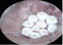
Figure 1 Otoscopy revealed the presence of 10 maggots in the right external auditory canal
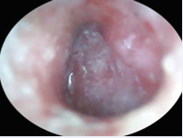
Figure 2 After extraction, congestion of the right external auditory canal and tympanic.
The rest of the physical examination was normal. A diagnosis of right acute congestive otitis externa and otitis media secondary to a maggot infestation was made. Treatment consisted of maggot extraction through irrigation (Figure 3), along with auricular care. The following medications were prescribed:
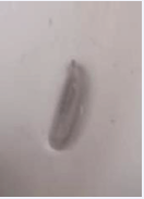
Figure 3 Identification of maggots in a kidney dish.
Paracetamol syrup (6 kg dosage, 3 times per day for 5 days); Amoxicillin syrup (200 mg, twice daily for 10 days); Polydexa ear drops (3 drops, 3 times per day for 14 days). Additionally, parents were advised to prevent water exposure in the right ear and to maintain proper hygiene for the child. The outcome was favorable, with complete recovery observed after 12 days of follow up. Parental consent was obtained before administering treatment.
Observation 2
This case involved a 3-month-old infant with no known medical history, residing in an urban environment under crowded conditions. He was the third child of an intrauterine sibling group of three. The infant was seen in consultation on April 29, 2024, due to persistent crying, frequent hand movements toward the right ear, and right-sided otorrhagia (ear bleeding). The symptoms began two days prior, with crying and repeated touching of the right ear. The parents brought him to the ENT consultation after noticing right-sided ear bleeding in a febrile context. Clinical Examination, on admission, the infant was conscious but agitated, with the following vital signs: Weight: 6.1 kg; Temperature: 38.9°C ; Respiratory rate: 32 breaths per minute ; Pulse: 125 beats per minute ; Skin and conjunctivae: Normal coloration. Otologic examination revealed maggots in the right external auditory canal, obstructing the view of the tympanic membrane (Figure 4).
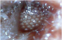
Figure 4 Otoscopy revealed multiple maggots in the right.
After extraction, findings included: Congestion of the right external auditory canal, small central tympanic perforation, inflammatory middle ear cavity (Figure 5).
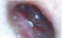
Figure 5 After extraction, congestion of the right external auditory canal was observed, along with a small central tympanic perforation and an inflamed middle ear cavity.
Additionally, rhinological examination revealed serous rhinorrhea, while oral and oropharyngeal examination showed grade I hypertrophy of the palatine tonsils without inflammation. The rest of the ENT examination was normal. Based on the clinical findings, a diagnosis of right-sided acute perforated otitis externa and otitis media secondary to maggot infestation was made. Maggot extraction via irrigation (Figure 6).

Figure 6 Identification of maggots on a compress.
Prescribed medications: Ciprofloxacin ear drops (3 drops, 3 times/day for 14 days); Amoxicillin-clavulanic acid (250 mg, twice/ day for 10 days); Betamethasone drops (60 drops/day for 5 days); Paracetamol syrup (6 kg dosage, 3 times/day for 7 days). Parents were advised to avoid water exposure in the right ear and maintain good hygiene for the child. The evolution was favorable, with complete healing confirmed by a normal follow-up otoscopy after two months. The child was subsequently declared cured. Parental consent was obtained before administering treatment.
DISCUSSION
The strength of this case series lies in highlighting the complications of a rare etiology of otitis. Indeed, auricular maggots are rare in our daily practice. Individuals at risk are mainly those living in poor sanitary conditions [3], as was the case for our two patients. The symptomatology is polymorphic and often delayed. Persistent crying is the primary warning sign for parents, who become anxious [4], and are prompted to seek medical consultation for their child. We encourage ENT specialists to conduct a thorough history-taking, particularly assessing the child’s living conditions.
Maggot larvae invade and develop within cavities or tissues, leading to pruritic, inflammatory, or destructive lesions, depending on the type of myiasis [1]. Diagnosis is straightforward, as live maggots can often be visualized in the external auditory canal, with or without tympanic membrane damage [3]. In both of our cases, live maggots were present in the external auditory canal. Inflammation was observed in both cases, with the second case also presenting a right tympanic perforation. The severity of these lesions varies from patient to patient.
Auricular care is crucial, involving maggot extraction via irrigation or suction while emphasizing the importance of preventing water entry into the affected ear. Medical treatment yields satisfactory results [3,5]. In our cases, the administration of analgesics, antibiotics, and ear drops aligns with the recommendations of other specialists [3–6]. The use of antiparasitic medications remains debatable—the optimal approach is to extract the maggots and treat the associated lesions. Close monitoring is essential to prevent lesion aggravation [3]. In our cases, otoscopic examinations were performed every three days, leading to positive outcomes, including the resolution of the tympanic perforation in the second case. Ultimately, the best approach is prevention [7,8].
CONCLUSION
Auricular maggots are rare. These parasitic infestations are typically found in immobilized individuals at extreme ages, particularly those living in poor sanitary conditions. Diagnosis is straightforward but sometimes delayed. Prevention remains the best therapeutic approach. This is an opportunity for us to raise awareness among healthcare professionals (ENT specialists, pediatricians, general practitioners, etc.) about this rare etiology of otitis.
REFERENCES
- Guiguen C, Belaz S, Chabasse D, Beaucournu JC. Apport du laboratoire pour le diagnostic des myiases. Revue Francophone des Laboratoires. 2020 ; 72-80.
- Diop K, Mbaye A, Ndiaye Diop MT, Diasse F, Diadie S, Deh A, et al. Des myiases furonculoïdes multiples chez un es myiases furonculoïdes multiples chez un nourrisson. Our Dermatology Online/Nasza Dermatologia Online. 2022 ; 13 : 15-19.
- Boumendil I, Belhaj N, Rahim H, Nitassi S, Bencheikh R, Benbouzid MA, et al. Myiase auriculaire humaine: à propos de deux cas. PAMJ-Clinical Medicine. 2021 ; 5: 1-7.
- Ouo-Ouo K. Myiase auriculaire humaine : à propos de deux cas au service de Pédiatrie de l’hôpital National de Donka: Human auricular Myiasis: about two cases in the pediatric departement of national hospital of Donka. Journal de Neurologie, Neurochirurgie et Psychiatrie. 2023 ; 2 : 64-68.
- Belchadi M, Bouzouita K, Lahmar I, Lamine H, Abdelkefi M, Ben Said M. Les myiases auriculaires : A propos de deux observations. Tunisie médicale. 2000 ; 78 : 535-537.
- Meryem A. Myiases humaines au centre hospitalier universitaire de Rabat: à propos d’un cas de myiase du cuir chevelu due à Wohlfahrtia magnifica. 2012 ; 13 : 192.
- Leclercq M. Les Myiases. Annales de la Société entomologique de France (NS). 1990 ; 26 : 335-350.
- Tligui H, Bouazzaoui A, Agoumi A. Entomologie médicale. Bull Soc Pathol Exot. 2007 ; 100 : 61-64.











































































































































