Surgical, Orthodontic, and Combined Treatment Alternatives for Mandibular Second Molar Mesio-Angular Impactions
- 1. Department of Integrated Basic Studies, College of Dentistry, University of Antioquia, Colombia
- #. These authors contributed equally to this work
Abstract
Impactions of mandibular second molars due to posterior dental crowding pose difficulties for orthodontic treatment. It is important to know aspects related to genetics and epigenetics of tooth eruption as well as orthodontic, surgical or combined treatment alternatives for these cases. The present review highlights the key role of the MSX1 gene during the intraosseous eruption process as an initiator of one of the three normal eruptive events, as well as that of other local environmental or epigenetic alterations under which this challenge in orthodontic treatment may originate. In addition, the existing surgical, non-surgical, and combined alternatives to solve these treatment difficulties will be indicated.
Keywords
• Tooth
• Impacted
• Tooth Eruption
• Orthodontics
• Surgery
• Oral
Citation
Gómez-Gil DF, Marín-Botero ML (2026) Surgical, Orthodontic, and Combined Treatment Alternatives for Mandibular Second Molar Mesio Angular Impactions. Ann Otolaryngol Rhinol 13(4): 1394.
ABBREVIATIONS
Md2B: Mandibular Second Bicuspid; Md1PM: Mandibular First Permanent Molar; Md2PM: Mandibular Second Permanent Molar; Md3PM: Mandibular Third Permanent Molar
INTRODUCTION
Tooth impaction creates a challenge to the dental multidisciplinary teams. This is particularly true with Mandibular Second Permanent Molar mesioangular impactions, one of the most challenging conditions for combined orthodontic and oral surgery treatments. This mini-review deals with its importance in diagnosis and treatment planning and will be focused on two aspects: Etiopathogenesis and clinical decisions to treat its appearance.
Molecular Etiopathogenesis of Mandibular Second Molar Mesioangular Impaction
Md2PM mesioangular impaction (ICD-11: DA07.8) is a rare but serious eruption disorder [1], with variable individual prevalence (0.4% in 6785 Colombians [2]). Md2PM impaction has several origins: a) local eruptive disturbances -like ectopic eruption pathway related with differential mesio-distal MSX1 and/or RANKL protein expression [3,4], that can be associated with mild forms of MSX1 pleiotropy; b) spatial problems -such as molar macrodontia, hyperdontia or supernumerary teeth (such as distomolars or fourth molars), loss of lower deciduous molars or first permanent molar, caries-related inter proximal dental destruction, deficient mandibular growth, or inadequate mandibular arch length- [5]; c) as the result of long-term mandibular first molars therapeutic distalization in non-extraction treatments [6,7]; d) systemic factors affecting osteoclastic activity in dental eruption [8] -or pseudoanodontia [9,10], as in cleidocranial dysplasia [11,12] or ectodermal dysplasia [13,14]; or e) pathologic conditions such as complex or compound odontomas [15], other odontogenic tumors [16], or giant cell fibromatosis [17,18] in the Md2PM eruption line.
Local eruptive disturbances in a normal preemergent -or intraosseous- dental eruption depends mainly on three different events (3): a) adaptation to eruptive movements from the periodontal ligament (mediated by MSX1) -Periodontal Ligament Traction “Theory” [19,20]; b) creation of spacing in the eruption pathway by the crown follicle –Dental Follicle “Theory” [21,22], mediated by two proteins: i) RANKL -a cytokine that upregulates osteoclastogenesis in bone resorption [1-23], whose expression in occlusal portions of the dental bony crypt is crucial to osteoclast activation during eruption pathway creation [24], under the “minimum effective strain” hypothesis that may produce time- and site-specific bone architecture changes [25,26], and ii) MMP1, a zinc-dependent endopeptidase able to degrade components of extracellular matrix that play an important role in regulating tooth development [27]; and c) eruption pressure at the apical root membrane associated to neurotrophins expression (e.g. NGF, NGFR, CGRP -calcitonin gene-related peptide- [28]) -Innervation Provoked Pressure “Theory” [3-29]- a hypothesis that have into account the neural origin of bone tissues, including dentin. These genetic alterations could affect several signaling pathways in mandibular molar eruption, following the model of spatial-temporal dynamics of gene expression [30]. Postemergent -or supraosseous- eruption take place after the crown emerges in the oral cavity until the tooth contacts the antagonist to create a functional unit of occlusion, and is mainly based on the Hydrostatic Pressure “Theory” -angiogenesis associated with VEGF- [31], which produce further vertical alveolar growth by tooth eruption even after tooth contact [32].
Osteoproliferative MSX1 gene expression to regulate dental eruption is permanent in maxilla and mandible periodontium [33]. Alveolar bone formation -a specialized form of intramembranous bone originated from dental follicle cells that gives primary support to deciduous and permanent dentition- requires the presence of not only MSX1 but also BMP4 as an early downstream target [34]. This MSX1/BMP4 signaling pathway is needed to control both DLX5 and RUNX2 expression in mandibular alveolar bone formation [34]. DLX5 gene interacts with different paralogues [35,36] to produce significant and distinct morphological mandibular differences through four grades of embryonic mandibular arch transformations [37].
These etiopathogenic/therapeutic causes of local eruptive disturbances could lead to molar closeness (in more than 95% of cases) [38,39], and/or mesialization of Md3PM over Md2PM that creates an additional distal eruption blockage [39]. Such dental spacing problems -as evidenced in 90% of 88 patients (108 molars) with impacted Md2PM [38]- give origin to severe local posterior crowding [1-43] -a malocclusion classified as having greater need of orthodontic treatment- [44]-. Such malocclusion has an altered posterior hard and soft tissue topography (gingiva, alveolar septum, vestibular and lingual alveolar bone), that create areas of biofilm retention very difficult to maintain with normal oral hygiene, requiring advanced periodontal therapy [45]. Early diagnosis of these eruption disturbances not only help to choose the optimal treatment time, but also to avoid complications [1-23].
Treatment Alternatives: Impacted Mandibular Second Permanent Molar Orthodontic Up-Righting vs. Molar Surgical Repositioning vs. Molar Extraction
Md2PM deimpaction choices depends on impaction degree, impaction depth, and associated posterior crowding (Table 1, Figure 1) [1-51]. Surgical-orthodontic uprighting felt between surgical and orthodontic deimpaction without significant differences. However, combined options allowed additional occlusal corrections compared with surgery alone [52].
Table 1: Summarized description of all treatment options for Md2PM impactions. Abbreviations: Md2B/1B: Mandibular second bicuspid/first bicuspid; Md1PM: Mandibular first permanent molar; Md2PM: Mandibular second permanent molar; Md3PM: Mandibular third permanent molar.
|
Orthodontic Options (Orthodontic up-righting) |
Mesial pushing |
Coil-spring-based mesial space opening -Rounded/Rectangular NiTi/CuNiTi archwire + NiTi open coil spring- |
|
Variable modulus/ Variable transitional-temperature based mesial space opening -Rectangular NiTi/CuNiTi occlusal arch segment- |
||
|
Distal pulling |
Cantilever-based distal traction -Squared/Rectangular S.S./ß-Ti archwire- -Rectangular ß-Ti “pole arm” up-righting spring- |
|
|
Variable-transitional-temperature-based distal molar rotation -Rounded CuNiTi piggyback overlay archwire- |
||
|
Direct/indirect skeletal anchorage-based distal traction -Mini-screw (implant) ± power chain/thread- -Indirect mini-screw (implant) alveolar bone anchorage- |
||
|
Surgical Options |
Md2PM surgical exposure |
|
|
Surgical Md2PM de-impaction |
Brass wire de-impaction |
|
|
Separation elastics de-impaction |
||
|
Surgical Md2PM up-righting (repositioning) with or without surgical fenestration (dental luxation) -rescue surgery- |
||
|
Md3PM auto-transplantation |
||
|
Dental implant replacement |
||
|
Tooth extraction |
Md2B/1B extraction |
|
|
Md1PM extraction |
||
|
Md2PM extraction with or without Md3PM auto-transplantation |
||
|
Md3PM late or early extraction with or without surgical Md2PM up-righting |
||
|
Surgical removal of pathologic lesions |
||
|
Combined Options (Surgical-orthodontic up-righting) |
Surgical Md2PM repositioning with orthodontic traction |
|
|
Surgical orthodontic button positioning + orthodontic traction |
||
|
Surgical Md2PM exposure + orthodontic traction |
||
|
Direct/indirect skeletal anchorage with Mini-screws |
||
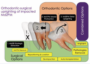
Figure 1: A summary of all strategies mentioned is presented. Combined surgical and orthodontic options for Md2PM impaction treatment are usually better than only orthodontics or surgical options. Abbreviations: Md2B: Mandibular second bicuspid; Md1PM: Mandibular first permanent molar; Md2PM: Mandibular second permanent molar.
If the etiology of delayed tooth eruption is caused by mechanical obstruction or problems of intraosseous eruption, surgical fenestration and luxation followed by the application of orthodontic traction is considered a standard procedure [53]. Correct space distribution is needed when a combination of Md2B extraction, mesial Md1PM protraction, and orthodontic Md2PM uprighting using cantilever-based distal Md2PM traction is planned (Figure 1). This strategy significantly improved Md2 and 3PM inclinations and make proper use of retromolar space [52-54]. In the developing mandible, retromolar space depends both on the resorption at the anterior border of the mandibular ramus and teeth eruptive direction during intra-osseous, supra-osseous, and post-eruptive (functional phase) tooth movements [55], modulated by orthodontic treatment [56]. Orthodontics with Md2B extractions increased eruption space in the retromolar area for Md2PM up-righting and successful eruption of Md3PM, due to mesial movement of Md1PM and Md2PM during space closure [50-62].
DISCUSSION & CONCLUSION
Vertical condylar growth together with counter-clockwise mandible growth rotation and reduced resorption at the anterior aspect of the mandibular ramus are craniofacial characteristics seen in patients with mandibular molar impaction [57-65]. Orthodontics with Md2B extractions increased eruption space in the retromolar area for Md2PM up-righting and successful eruption of Md3PM, due to mesial movement of Md1PM and Md2PM during space closure [50-62]. Successful vertical positional changes without dental angular changes with response depending on the degree of impaction could be reached with a positive mandibular counter-clockwise-forward- rotation [50].
REFERENCES
- Barone S, Antonelli A, Bocchino T, Cevidanes L, Michelotti A, Giudice A. Managing mandibular second molar impaction: A systematic review and meta-analysis. J Oral Maxillofac Surg. 2023; 81: 1403-1421.
- Ramírez Ossa D, Upegui Zea JC, Espinal Botero GE, Chaurra Marín RC. Caracterización de la impactación de segundos molares mandibulares permanentes en una población sudamericana. International Journal of Odontostomatology. 2017; 11: 165-172.
- Kjær I. Mechanism of human tooth eruption: Review article including a new theory for future studies on the eruption process. Scientifica (Cairo). 2014; 2014: 341905.
- Trybek G, Jaro? A, Grzywacz A. Association of polymorphic and haplotype variants of the MSX1 Gene and the Impacted Teeth Phenomenon. Genes (Basel). 2021; 12: 577.
- Bondemark L, Tsiopa J. Prevalence of ectopic eruption, impaction, retention and agenesis of the permanent second molar. Angle Orthod. 2007; 77: 773-778.
- Desnoës H. Abnormalities in the development of the second molars and orthodontic treatment without extraction of premolars. Management of posterior crowding. Journal of Dentofacial Anomalies and Orthodontics. 2014; 17: 406.
- Rubin RL, Baccetti T, McNamara JA. Mandibular second molar eruption difficulties related to the maintenance of arch perimeter in the mixed dentition. American Journal of Orthodontics and Dentofacial Orthopedics. 2012; 141: 146-152.
- Helfrich MH. Osteoclast diseases and dental abnormalities. Arch Oral Biol. 2005; 50: 115-122.
- Ivy RH. A case of non-eruption of entire permanent denture. Dental Cosmos. 1933; 75: 689-690.
- Hertz RS, Ward LC. Pseudoanodontia of the mandible. Oral Surgery, Oral Medicine, Oral Pathology. 1966; 22: 654-657.
- Thaweesapphithak S, Saengsin J, Kamolvisit W, Theerapanon T, Porntaveetus T, Shotelersuk V. Cleidocranial dysplasia and novel RUNX2 variants: Dental, craniofacial, and osseous manifestations. J Appl Oral Sci. 2022; 30: e20220028.
- Zeng L, He H, Sun M, Gong X, Zhou M, Hong Y, et al. Runx2 and Nell-1 in dental follicle progenitor cells regulate bone remodeling and tooth eruption. Stem Cell Res Ther. 2022; 13: 486.
- Wright JT, Fete M, Schneider H, Zinser M, Koster MI, Clarke AJ, et al. Ectodermal dysplasias: Classification and organization by phenotype, genotype and molecular pathway. Am J Med Genet A. 2019; 179: 442-447
- Zeng B, Xiao X, Li S, Lu H, Lu J, Zhu L, et al. Eight Mutations of Three Genes (EDA, EDAR, and WNT10A) identified in seven hypohidrotic ectodermal dysplasia patients. Genes (Basel). 2016; 7: 65.
- Isola G, Cicciù M, Fiorillo L, Matarese G. Association between odontoma and impacted teeth. J Craniofac Surg. 2017; 28: 755-758.
- Shoaee S, Khazaei P, Mashhadiabbas F, Varshosaz M, Sharifi F, HessarI H. Association between tooth impaction and odontogenic lesions: A matched case-control study. Med J Islam Repub Iran. 2018; 32: 57.
- Moradzadeh Khiavi M, Karimi A, Mirmohammad Sadeghi H, Derakhshan S, Tafreshi SM, Jalali S. Central odontogenic fibroma accompanied by a central giant cell granuloma-like lesion: Report of a Case and Review of Literature. Front Dent. 2021; 18: 44.
- Ramadan OR, Essawy MM. Central odontogenic fibroma with giant cell granuloma-like lesion: A Report of an additional case and review of literature. Head Neck Pathol. 2021; 15: 275-280.
- Kasugai S, Suzuki S, Shibata S, Yasui S, Amano H, Ogura H. Measurements of the isometric contractile forces generated by dog periodontal ligament fibroblasts in vitro. Arch Oral Biol. 1990; 35: 597-601.
- Olsson B, Calixto RD, da Silva Machado NC, Meger MN, Paula-Silva FWG, Rebellato NLB, et al. MSX1 is differentially expressed in the deepest impacted maxillary third molars. Br J Oral Maxillofac Surg. 2020; 58: 789-794.
- Ono T, Hayashi M, Sasaki F, Nakashima T. RANKL biology: Bone metabolism, the immune system, and beyond. Inflamm Regen. 2020; 40: 2.
- Bradaschia-Correa V, Moreira MM, Arana-Chavez VE. Reduced RANKL expression impedes osteoclast activation and tooth eruption in alendronate-treated rats. Cell Tissue Res. 2013; 353: 79-86.
- Tyrovola JB. The “Mechanostat Theory” of Frost and the OPG/ RANKL/RANK System. J Cell Biochem. 2015; 116: 2724-2729.
- Frost HM. A determinant of bone architecture. The minimum effective strain. Clin Orthop Relat Res. 1983; 286-292.
- Sternlicht MD, Werb Z. How matrix metalloproteinases regulate cell behavior. Annu Rev Cell Dev Biol. 2001; 17: 463-516.
- Brodetska L, Natrus L, Lisakovska O, Kaniura O, Iakovenko L, Skrypnyk I, et al. The regulatory role of the RANKL/RANK/OPG signaling pathway in the mechanisms of tooth eruption in patients with impacted teeth. BMC Oral Health. 2020; 20: 261.
- Kim SG, Kim MH, Chae CH, Jung YK, Choi JY. Downregulation of matrix metalloproteinases in hyperplastic dental follicles results in abnormal tooth eruption. BMB Rep. 2008; 41: 322-327.
- Mahdee A, Eastham J, Whitworth JM, Gillespie JI. Evidence for changing nerve growth factor signalling mechanisms during development, maturation and ageing in the rat molar pulp. Int Endod J. 2019; 52: 211-222.
- Rabea AA. Recent advances in understanding theories of eruption (evidence based review article). Future Dental Journal. 2018; 4: 189-196.
- Vieux-Rochas M, Mantero S, Heude E, Barbieri O, Astigiano S, Couly G, et al. Spatio-temporal dynamics of gene expression of the Edn1-Dlx5/6 pathway during development of the lower jaw. Genesis. 2010; 48: 262-373.
- Shimada A, Shibata T, Komatsu K. Relationship between the tooth eruption and regional blood flow in angiotensin II-induced hypertensive rats. Arch Oral Biol. 2004; 49: 427-433.
- Iseri H, Solow B. Continued eruption of maxillary incisors and first molars in girls from 9 to 25 years, studied by the implant method. Eur J Orthod. 1996; 18: 245-256.
- Wehrhan F, Hyckel P, Amann K, Ries J, Stockmann P, Schlegel K, et al. Msx-1 is suppressed in bisphosphonate-exposed jaw bone analysis of bone turnover-related cell signalling after bisphosphonate treatment. Oral Dis. 2011; 17: 433-442.
- Zhang Z, Song Y, Zhang X, Tang J, Chen J, Chen Y. Msx1/Bmp4 genetic pathway regulates mammalian alveolar bone formation via induction of Dlx5 and Cbfa1. Mech Dev. 2003; 120: 1469-1479.
- Kawasaki K, Buchanan AV, Weiss KM. Gene duplication and the evolution of vertebrate skeletal mineralization. Cells Tissues Organs. 2007; 186: 7-24.
- Bailon-Zambrano R, Sucharov J, Mumme-Monheit A, Murry M, Stenzel A, Pulvino AT, et al. Variable paralog expression underlies phenotype variation. Elife. 2022; 11: e79247.
- Depew MJ, Simpson CA, Morasso M, Rubenstein JL. Reassessing the Dlx code: The genetic regulation of branchial arch skeletal pattern and development. J Anat. 2005; 207: 501-561.
- Varpio M, Wellfelt B. Disturbed eruption of the lower second molar: Clinical appearance, prevalence, and etiology. ASDC J Dent Child. 1988; 55: 114-118.
- Shapira Y, Lai Y, Schonberger S, Shpack N, Finkelstein T. Mandibular Second Molar Impaction-Part II: Etiology and Role of the Third Molar. Applied Sciences (Switzerland). 2022; 12: 11520.
- Cassetta M, Altieri F, Di Mambro A, Galluccio G, Barbato E. Impaction of permanent mandibular second molar: A retrospective study. Med Oral Patol Oral Cir Bucal. 2013; 18: e564-e568.
- Turley PK. The management of mesially inclined/impacted mandibular permanent second molars. J World Fed Orthod. 2020; 9: S45-S53.
- Okihara H, Ono T. Correction of bilateral heavily impacted second molar with improved super-elastic nickel-titanium alloy wires. Am J Orthod Dentofacial Orthop. 2020; 158: 114-125.
- Cassetta M, Altieri F. The influence of mandibular third molar germectomy on the treatment time of impacted mandibular second molars using brass wire: A prospective clinical pilot study. Int J Oral Maxillofac Surg. 2017; 46: 905-911.
- Bourzgui F, Sebbar M, Hamza M, Lazrak L, Abidine Z, El Quars F. Prevalence of malocclusions and orthodontic treatment need in 8-to 12-year-old schoolchildren in Casablanca, Morocco. Prog Orthod. 2012; 13: 164-172.
- Diedrich P. Periodontal relevance of anterior crowding. J Orofac Orthop. 2000; 61: 69-79.
- la Monaca G, Cristalli MP, Pranno N, Galluccio G, Annibali S, Pippi R. First and second permanent molars with failed or delayed eruption: Clinical and statistical analyses. Am J Orthod Dentofacial Orthop. 2019; 156: 355-364.
- Allgayer S, Platcheck D, Vargas IA, Loro RC. Mini-implants: Mechanical resource for molars uprighting. Dental Press J Orthod. 2013; 18: 134-142.
- Vexiau J, Castaneda B. Retained second molars: Therapeutic approach. Journal of Dentofacial Anomalies and Orthodontics. 2015; 18: 405.
- Caminiti MF, El-Rabbany M, Lou T, Reinish EI. Surgical uprighting of mandibular second molars: A single-group retrospective cohort study. Am J Orthod Dentofacial Orthop. 2020; 158: 849-855.
- Miclotte A, Grommen B, Cadenas de Llano-Pérula M, Verdonck A, Jacobs R, Willems G. The effect of first and second premolar extractions on third molars: A retrospective longitudinal study. J Dent. 2017; 61: 55-66.
- Al Kuwari HM, Talakey AA, Al-Sahli RM, Albadr AH. Influence of orthodontic treatment with first premolar extraction on the angulation of the mandibular third molar. Saudi Med J. 2013; 34: 639-643.
- Barone S, Antonelli A, Bocchino T, Cevidanes L, Michelotti A, Giudice A. Managing mandibular second molar impaction: A Systematic Review and Meta-Analysis. J Oral Maxillofac Surg. 2023; 81: 1403-1421.
- Han T, Christensen BJ. Surgical treatment of impacted mandibular second molars: A Systematic Review. J Oral Maxillofac Surg. 2022; 80: 29-36.
- Turley PK. The management of mesially inclined/impacted mandibular permanent second molars. J World Fed Orthod. 2020; 9: S45-S53.
- Kim TW, Artun J, Behbehani F, Artese F. Prevalence of third molar impaction in orthodontic patients treated nonextraction and with extraction of 4 premolars. Am J Orthod Dentofacial Orthop. 2003; 123: 138-145.
- Kandasamy S, Woods MG. Is orthodontic treatment without premolar extractions always non-extraction treatment? Aust Dent J. 2005; 50: 146-151.
- Behbehani F, Artun J, Thalib L. Prediction of mandibular third-molar impaction in adolescent orthodontic patients. Am J Orthod Dentofacial Orthop. 2006; 130: 47-55.
- Türköz C, Ulusoy C. Effect of premolar extraction on mandibular third molar impaction in young adults. Angle Orthod. 2013; 83: 572-577.
- Patel AK, Deshmukh SV, Naik CR, Jethe S, Patel KA. Radiographic assessment for predicting the mandibular third molar eruption after orthodontic treatment in first premolar extraction group and non-extraction group: A-retrospective Study. International Journal of Dental Health Concerns. 2015; 1: 2-7.
- Elsey MJ, Rock WP. Influence of orthodontic treatment on development of third molars. Br J Oral Maxillofac Surg. 2000; 38: 350-353.
- Richardson ME. The effect of mandibular first premolar extraction on third molar space. Angle Orthod. 1989; 59: 291-294.
- Celikoglu M, Kamak H, Akkast I, Oktay H. Effects of orthodontic treatment and premolar extractions on the mandibular third molars. Aust Orthod J. 2010; 26: 160-164.