Clinical Stewardship in Action: Leveraging Hematological and Inflammatory Serological Markers to Optimize Infection Management
- 1. Professor & Head, Neuropsychopharmacology, Institute of Human Behaviour & Allied Sciences,, India
- 2. Assistant Professor, Pathology, Institute of Human Behaviour & Allied Sciences, India
- 3. Professor, Pathology, Institute of Human Behaviour & Allied Sciences, India
- 4. Lead Consultant, Infectious Diseases, Aster DM Healthcare, India
Abstract
Fever and suspected sepsis are common clinical challenges requiring rapid evaluation and evidence-based management to prevent adverse outcomes. Early identification and stratification of risk markers are essential for guiding antimicrobial therapy and escalation of care. The aim of this review is to evaluate the role of clinical stewardship in optimizing the use of inflammatory serological markers for fever and suspected sepsis. The objectives were to classify inflammatory serological markers based on diagnostic and prognostic utility; Review current evidence on marker-guided antimicrobial therapy; highlight strategies for integrating stewardship principles into clinical practice.
The review begins with an introduction on the burden of sepsis and the need for rational diagnostics, followed by the concept of clinical stewardship and its relevance in infection management. It then discusses inflammatory serological markers, their classification, diagnostic accuracy, and clinical implications. Challenges such as cost-effectiveness, accessibility, and emerging biomarkers are addressed, concluding with recommendations for practice. A structured pathway is presented for clinicians to initiate antimicrobial therapy promptly, monitor treatment response, and escalate care when indicated. By differentiating markers based on diagnostic specificity and prognostic value, the tool supports clinical stewardship and reduces unnecessary interventions. It emphasizes dynamic reassessment based on evolving clinical status and abnormal laboratory trends.
Clinical stewardship and rational use of inflammatory serological markers are essential for improving diagnostic precision, guiding timely interventions, and reducing unnecessary antibiotic exposure. A structured algorithm integrating clinical assessment with tiered marker utilization can enhance patient outcomes and support antimicrobial stewardship. Future research should validate marker combinations and develop cost-effective protocols for diverse healthcare settings.
Keywords
• Fever • Sepsis
• Clinical Algorithm
• Biomarkers
• Antimicrobial Stewardship
• Critical Care
• Procalcitonin
• Interleukin-6
• Presepsin
• Clinical Stewardship
Citation
Sharma S, Pant I, Chaturvedi S, Warrier A (2026) Clinical Stewardship in Action: Leveraging Hematological and Inflammatory Serological Markers to Optimize Infection Management. Ann Clin Pathol 13(1): 1185.
INTRODUCTION
Inflammation is a key process in many acute and chronic diseases. Although fever often suggests infection, it can also result from non-infectious inflammation such as autoimmune disorders or tissue injury, where cytokines and prostaglandins raise the body’s temperature setpoint. Inflammation typically presents with redness, swelling, and pain, while infections involve pathogen invasion and replication, producing systemic symptoms like fever, chills, and malaise. Sepsis triggers a dynamic immune response beginning with a proinflammatory phase, progressing to venostasis, and later shifting to immune suppression, which may enable viral reactivation and can sometimes lead to fatal outcomes [1].Figure 1 illustrates the temporal evolution of this dysregulated immune response, highlighting the overlap between hyperinflammation and immune suppression.

Figure 1: Immunologic response during sepsis over time
Recent studies show that sepsis triggers simultaneous pro and anti-inflammatory responses, but the early phase is largely hyperinflammatory, with severity influenced by pathogen load, virulence, and comorbidities. Accurate differentiation between bacterial, viral, and non-infectious inflammation is essential; without it, treatment becomes guesswork. Misinterpretation of clinical signs can lead to over or underdiagnosis, and difficulty distinguishing infection types often results in unnecessary antimicrobial use, driving resistance.
Treating all fever as infection undermines stewardship, delays correct diagnosis, and risks overlooking non infectious causes. A clearer distinction allows targeted therapy and better outcomes. Serological markers help differentiate inflammation from infection, but overuse of specialized tests without first using routine markers increases cost, misinterpretation, and resource strain as seen during COVID19.
Specialized inflammatory serological markers can provide condition specific insights but using them without first performing routine tests increases costs and risks misinterpretation. Relying solely on advanced markers may omit essential clinical context obtainable through basic haematology, leading to diagnostic errors and delayed treatment. This pattern became common during the COVID19 pandemic, resulting in unnecessary testing and inefficient use of resources [2-4].
A growing tendency to depend on specialized tests stems from limited awareness of marker utility, high workload in busy clinical environments, easy availability of expensive assays, fear of missing diagnoses, and misreading reports. Skipping routine tests which offer broad information such as infection clues, anaemia status, and immune changes can cause clinicians to overlook important aspects of patient health and treatment response [5-7].
Although routine haematology and serological markers are nonspecific, they help identify inflammatory states and support diagnosis and monitoring when interpreted correctly. Understanding biomarker relevance is essential for appropriate use [8-11]. Categorizing markers into diagnostic, prognostic, and predictive groups improves interpretation: diagnostic markers aid early disease detection, prognostic markers indicate disease course, and predictive markers help anticipate treatment response, enabling more individualized care [12-15].
Appropriate timing of tests is equally important. Consistent and timely testing ensures early detection of clinical changes and allows rapid treatment adjustments. Testing too early, for example, antibody testing in the first days after suspected COVID19 exposure can yield false negatives [16-18].
Using serological markers where they are not indicated, such as substituting them for cultures or PCR in bacterial infections, can also lead to inappropriate management. This also applies to fungal biomarkers like Galactomannan (GM) and βDGlucan (BDG), which must be interpreted cautiously due to assay variability, false positives, and limited species specificity, and therefore should never be used as standalone diagnostic tools [6-19]. In autoimmune diseases, poorly timed or infrequent testing can miss flareups or remission, resulting in suboptimal management. Acute infections may require frequent CBC monitoring to evaluate response and detect complications early, whereas chronic or stable inflammatory conditions generally need less frequent testing such as weekly or biweekly intervals to track disease progression and guide therapy.
Serological markers used to evaluate antibody levels or infection status are repeated at intervals ranging from days to months, depending on disease type and treatment duration. Individualizing test frequency ensures timely detection of changes and supports appropriate clinical intervention.
Although many advanced laboratory tests exist, routine, widely available investigations still play a crucial role, especially in resource limited settings. When combined with clinical and diagnostic information, these tests effectively support infectious disease management. Clinical stewardship emphasizes accurate diagnosis and targeted therapy. Correct interpretation of serological and haematological markers helps distinguish infection from sterile inflammation, reducing unnecessary antimicrobial use and ensuring patients receive appropriate treatment. Incorporating these principles into routine practice promotes evidence based, patient centred care and improves recovery outcomes.
The objective of this review is to examine how haematological and inflammatory serological markers can be used to improve the diagnosis and management of fever and suspected sepsis. It aims to classify these markers based on their diagnostic, prognostic, and predictive roles, evaluate evidence for marker guided antimicrobial therapy, and highlight strategies to integrate clinical stewardship into routine practice for more accurate, timely, and cost effective infection management.
METHODOLOGY
This narrative review adopted a structured yet flexible approach to identify, appraise, and synthesize literature relevant to infectious disease management and the stewardship oriented use of hematological and inflammatory serological markers. The review was guided by established principles for high quality narrative reviews, emphasizing transparent literature selection, balanced interpretation, and clinical applicability. Its primary objective was to provide an integrated synthesis of current evidence on the diagnostic, prognostic, and predictive utility of serological markers within antimicrobial and clinical stewardship frameworks.
The narrative design enabled inclusion of a broad range of evidence sources, including peer reviewed observational studies, clinical trials, international and national guidelines, consensus statements, and policy documents related to the evaluation of fever, sepsis, and other infectious syndromes, with particular reference to authoritative guidance from the Surviving Sepsis Campaign, the World Health Organization (WHO), and the Infectious Diseases Society of America (IDSA), ensuring that discussions on the use of serological markers and antimicrobial decision making were aligned with current evidence based standards.
Key thematic domains included appropriate marker selection, test timing and kinetics, serial monitoring, marker guided antimicrobial decision making, and integration of routine and advanced biomarkers into clinical algorithms. This approach facilitated a clinically grounded interpretation of evolving practices, highlighted gaps between evidence and real world use, and supported stewardship driven strategies to optimize diagnostics, limit unnecessary testing, and guide rational antimicrobial therapy.
CONVENTIONAL MARKERS
Erythrocyte Sedimentation Rate (ESR)
ESR is a nonspecific marker of inflammation reflecting acute phase reactants, mainly fibrinogen. It rises more slowly than CRP usually within 24–48 hours and peaks at 3–5 days, then normalizes over days to weeks due to the long half-life of acute phase proteins.
Table 1: Erythrocyte Sedimentation Rate (ESR) and C-Reactive Protein (CRP) Levels, Kinetics, and Influencing Factors
|
Parameter |
ESR (General Characteristics) |
CRP (General Characteristics) |
CRP in Bacterial Infections |
CRP in Viral Infections |
|
Normal Range |
Men: 0–15 mm/h; Women: 0–20 mm/h |
< 5–10 mg/L (lab-dependent) |
- |
- |
|
Time to Rise After Inflammation |
Begins rising in 24–48 hours |
Within 6–8 hours |
Begins rising within 6–8 hours |
Similar onset (6–8 hours) but less pronounced |
|
Peak Timing |
Peaks in 3–5 days |
Peaks at 48 hours |
Peaks 24–48 hours with rapid, high increase |
Peaks 24–48 hours, but milder |
|
Peak Levels |
- |
- |
High, robust elevation (~100–150 mg/L peaks in bacteremia/serious bacterial infection (e.g., peak ≈ 140.9 mg/L in Gram-negative bacteremia); median at ED presentation ≈133 mg/L. Values >100 mg/L in primary care strongly favor bacterial etiology. |
Lower peak levels, reflecting weaker inflammation (~15–30 mg/L typical (e.g., peak ≈ 15.4 mg/L; median presentation ≈ 23 mg/L). |
|
Severity Association |
Lower sensitivity; weaker early response |
High sensitivity for acute-phase reactions |
Magnitude correlates with severity |
Levels less dramatic even in moderate disease |
|
Specificity |
Less specific; affected by non-inflammatory factors |
More specific for acute inflammation |
Supports bacterial etiology |
Helps differentiate viral vs bacterial |
|
Influencing Conditions |
Infections, autoimmune disease, malignancy, anemia |
Acute bacterial infections, trauma, chronic inflammation |
Bacterial infections strongly elevate CRP |
Viral infections cause modest rise |
|
Physiological Influences |
Influenced by age, sex, pregnancy, anemia |
Less affected by age, sex, anemia |
- |
- |
|
Clinical Utility |
Monitoring chronic inflammation: TB, RA, vasculitis |
Monitoring acute infections, post-operative inflammation, sepsis |
Supports bacterial diagnosis; helpful in monitoring response |
Aids differentiation from bacterial infections |
|
Response to Therapy |
Slow to normalize; may take weeks |
Quick drop when inflammation resolves |
Declines rapidly with effective antibiotics |
Decline slower and less pronounced |
Table 1 summarizes key differences in ESR kinetics. Because bacterial infections trigger stronger acute phase responses than viral infections, ESR tends to rise more in bacterial illness. However, anaemia can falsely elevate ESR, and nomograms exist to correct for this. Repeating ESR daily is not useful, as rapid changes are unlikely. For chronic or subacute conditions (e.g., tuberculosis, autoimmune diseases), ESR can be repeated every 1–2 weeks, while in acute infections it is checked at diagnosis and again after several days alongside clinical findings and markers like CRP or WBC count [8-10].
ESR should not be used in isolation for acute decision making but is valuable for tracking chronic inflammation, guiding therapy in osteomyelitis, vasculitis, and rheumatologic diseases, and predicting relapse in Hodgkin’s disease. Markedly elevated ESR suggests serious underlying disease such as infection, collagen vascular disorders, or malignancy [9]. If ESR is elevated without a clear cause, repeating the test after an interval is preferred over extensive immediate investigation. Overall, ESR is useful for screening, differentiating bacterial from viral patterns, and monitoring disease progression and treatment response.
C-reactive protein (CRP)
C reactive protein (CRP) is a hepatic acute phase reactant induced by IL6/IL1/TNFα; it rises within 6–8 h after an inflammatory stimulus and peaks by 24–48 h. Elevations occur in bacterial, viral, and fungal infections, as well as autoimmune and malignant conditions. In bacterial infections (e.g., pneumonia, sepsis, meningitis) the rise is typically higher and faster, supporting both diagnosis and prognosis. Table 1 summarizes key differences in CRP kinetics and peak levels between bacterial and viral infections, highlighting the substantially higher CRP elevations seen in bacterial etiologies (median ≈ 133 mg/L vs ≈ 23 mg/L in viral infection at presentation; peaks ≈ 140 mg/L reported in Gram-negative bacteraemia). These data emphasize the diagnostic value of CRP trends in distinguishing bacterial from viral inflammatory responses. Leptospirosis shows early CRP elevation in the septicaemic phase, with higher values in severe (Weil’s) disease, aiding diagnosis and monitoring [20]. By contrast, viral infections usually show modest CRP increases, while fungal/parasitic responses vary with host and disease severity; in postoperative/trauma states, peaks may occur at 48–72 h, and immunosuppression or corticosteroids can blunt or delay responses [21,22].
Testing frequency should be individualized to clinical status and goals. In acute cases, serial CRP every 24–48 h, and daily in the critically ill, helps guide treatment and monitor disease trajectory; declining CRP generally indicates improvement. In stable/recovery phase patients, intervals can be extended to every few days to balance vigilance and resource use. Serial interpretation should be contextual, alongside clinical assessment, cultures, imaging, and complementary biomarkers (e.g., PCT), particularly for healthcare associated infections where signs may be subtle. Neither CRP nor PCT is definitive alone, but trend-based interpretation supports earlier detection and stewardship decisions [11-26].
In acute and critical care settings, obtain CRP at presentation for baseline assessment, then repeat within 24–48 h (or daily in hemodynamic instability) to evaluate response and interpret trends with PCT. Persistent or rising CRP should prompt systematic reassessment for occult infection, inadequate source control, or non infectious inflammatory processes. In primary care for adults with suspected LRTI without a clinical diagnosis of pneumonia, NICE recommends withhold antibiotics when CRP < 20 mg/L; consider a delayed prescription at 20–100 mg/L; antibiotics are recommended when > 100 mg/L [27,28].
ROUTINE BLOOD COUNTS
Leucocytes (Total and Differential)
Despite the availability of advanced diagnostics, routine blood counts remain highly informative. TLC and DLC provide accessible indicators of infection and inflammation. Neutrophilia commonly suggests bacterial infection, whereas lymphocytosis is more typical of viral etiologies. Neutropenia may reflect viral infection, bone marrow suppression, or sepsis induced immune exhaustion and increases susceptibility to secondary infections. Leukopenia during fever may indicate viral illness, drug induced marrow suppression, or severe sepsis. Several viral infections including HIV, influenza, hepatitis B/C, dengue, EBV, and CMV cause leukopenia with predominant lymphocyte suppression. Although lymphocytosis favours viral disease, lymphopenia may occur in severe illness. Monocytosis is associated with chronic infections such as tuberculosis, subacute bacterial endocarditis, and parasitic diseases, but may also appear in autoimmune disorders. Eosinophilia occurs with parasitic infections involving tissue invasion and in allergic conditions.
Neutrophil to Lymphocyte Ratio (NLR)
NLR integrates innate (neutrophil) and adaptive (lymphocyte) immune responses and rises in acute inflammation from infections, trauma, stroke, myocardial infarction, or malignancy. Elevation occurs within hours often preceding other laboratory changes. High NLR helps with risk stratification and treatment monitoring [29]. NLR at admission predicts CAP severity and outcomes with better prognostic performance than traditional markers [30]. Persistently elevated NLR correlates with higher morbidity and mortality [31], while lower values reflect preserved immune balance [32]. Proposed normal adult values range 0.78–3.53 [32]. NLR may be falsely elevated due to age, obesity, steroid use, hormonal states, metabolic disease, hematologic disorders, cardiovascular events, rheumatic disease, trauma, or psychological stress [5-31]. Its diagnostic and prognostic utility in bacteraemia and sepsis is well established, especially when interpreted with awareness of confounders.
Platelet Parameters
Platelet counts and derived indices contribute valuable diagnostic and prognostic information in infectious diseases [33,34]. Thrombocytopenia and reduced Plateletcrit are frequent in viral illnesses such as dengue, COVID19, and HIV, as well as parasitic infections like malaria. Severe bacterial infection or sepsis can produce thrombocytopenia or reactive thrombocytosis with elevated Plateletcrit. Mean Platelet Volume (MPV), and Platelet Distribution Width (PDW) increase with reactive marrow responses in acute infections and sepsis [35], whereas chronic infections may suppress marrow activity, reducing MPV. Platelet indices, especially MPV, may help predict vascular complications in type 2 diabetes [36]. Immature Platelet Fraction (IPF) and delta absolute immature platelet count (AIPC) help distinguish causes of thrombocytopenia and track platelet recovery [37]. IPF would also increase in situations of marrow stress causing increased IPF as occurs in acute infections and inflammatory states if marrow functioning is robust. The Neutrophil Platelet Ratio (NPR) adds prognostic value: neutrophilia combined with thrombocytopenia in sepsis elevates NPR, signalling more severe disease and the need for close monitoring. Platelet indices also hold value in neonatal sepsis [38], and both NLR and NPR predict all cause mortality in COVID19 [39]. Table 2 provides platelet indices in infections - normal range, kinetics, clinical use, and influencing factors.
Table 2: Platelet Indices in Infections - normal range, Kinetics, Clinical Use, and influencing Factors
|
Parameter |
Normal Range |
Kinetics / Pattern in Infections |
Clinical Significance as a Therapeutic Marker |
Influencing Factors |
|
Thrombocytopenia |
<150× 10?/L |
Rapid drop in viral infections (e.g., dengue, HIV), sepsis, malaria, DIC |
Marker of disease severity and prognosis Persistent or worsening thrombocytopenia may signal treatment failure or complications |
Viral replication, bone marrow suppression, immune-mediated destruction, DIC, drugs |
|
Plateletcrit |
0.19-0.39% |
Decreases in infections with platelet destruction or hypoproduction |
Reflects total circulating platelet mass; useful in monitoring recovery post-treatment |
Platelet count, MPV, bone marrow output |
|
MPV (Mean Platelet Volume) |
7.5-11.5 fL |
Increases in sepsis and inflammatory states due to increased turnover Decreases in marrow suppression |
Elevated MPV with thrombocytopenia suggests peripheral destruction and active thrombopoiesis May guide anti-inflammatory or immunomodulatory therapy |
Inflammation, cytokine levels, platelet turnover, bone marrow recovery |
|
PDW (Platelet Distribution Width) |
9-17% |
Increases with greater variation in platelet size in inflammatory/infectious states |
Indicates platelet activation and heterogeneity Useful in assessing risk of complications in sepsis |
Marrow response, inflammatory burden, platelet production variability |
|
IPF (Immature Platelet Fraction) |
1-7% (can be >10% in recovery) |
Increases early in response to peripheral destruction or treatment Decreases with marrow suppression |
Helps differentiate cause of thrombocytopenia: High IPF = peripheral destruction Low IPF = impaired production Useful for monitoring bone marrow recovery |
Bone marrow activity, chemotherapy, immunosuppression, infections |
|
NPR (Neutrophil-to-Platelet Ratio) |
No fixed reference (context-dependent; often >2.5 in sepsis) |
Elevated in severe infections, particularly sepsis and COVID-19 |
Associated with higher mortality and severity Guides intensity of treatment and need for escalation |
Inflammatory status, neutrophil and platelet kinetics, underlying disease |
Interpretation of platelet indices requires attention to laboratory factors: values may vary with the anticoagulant used, sample handling conditions, and analyser technology, with MPV, PDW, and IPF particularly susceptible to inter-instrument variability and the absence of standardized reference ranges, which can limit cross laboratory comparability and clinical generalizability [40].
Peripheral Blood Smear Examination
A peripheral smear provides morphological detail that complements automated counts. A left shift, toxic granulation, vacuolation, or Döhle bodies indicate severe bacterial infection, sepsis, or significant inflammation. Viral infections often show activated lymphocytes, whereas increased monocytes suggest chronic infections like tuberculosis. Eosinophilia with characteristic granules supports parasitic or allergic etiologies. Red cell abnormalities such as schistocytes or spherocytes may indicate haemolysis or anaemia of chronic disease, including that associated with infections such as malaria.
NEWER MARKERS
Presepsin
Presepsin (sCD14ST) is released during monocyte activation and rises within ~2 hours of infection, with a shorter half-life than PCT. Typical reference values are 946 ng/L, correlate with gram negative sepsis. Interpretation is limited by renal impairment, which elevates levels independent of infection. Most useful for early sepsis triage in ICU but should be read alongside clinical context and renal function [41].
Cathepsin
Cathepsins (B, L, S, K) are lysosomal proteases involved in proteolysis, immune modulation, and inflammation. They are rapidly upregulated after infectious or inflammatory stimuli and have been linked to sepsis related organ dysfunction, ARDS lung injury, and vascular pathology in atherosclerosis and diabetes. Cathepsin L facilitates viral entry, including SARSCoV2 spike processing. Clinical adoption is constrained by no standardized assays, context dependent expression, nonspecific elevation, and short stability in biofluids; current use is largely research focused, with inhibitors under investigation as potential therapeutics [42-44].
Active Neutrophil Elastase
NE, a neutrophil derived serine protease, contributes to pathogen killing and neutrophil migration. Excess activity drives tissue damage and systemic inflammation, with associations to sepsis, ventilator associated pneumonia, COVID19 lung injury, ARDS, COPD, and cystic fibrosis. Levels rise early after activation but lack standardized reference ranges; plasma concentrations in health are low/ undetectable. NE is being evaluated as a prognostic/early diagnostic marker and therapeutic target; Sive Lestat (NE inhibitor) is under clinical study in sepsis. Measurement is limited by assay variability, sample handling sensitivity, and nonspecific elevation across infectious and sterile inflammatory states; advanced ELISA/activity-based methods are often required and not widely available [45 47].
MARKERS OF RELEVANCE IN CRITICAL CARE (WITH FOCUS ON SEPSIS)
Procalcitonin (PCT)
PCT, a prohormone of calcitonin, is minimally present in health but rises rapidly in bacterial infections via direct endotoxin stimulation and cytokine mediated pathways (IL6, IL1β, TNFα). Levels become detectable within 2–4 h and peak by 24–48 h; >2.0 µg/L supports sepsis and >10 µg/L suggests septic shock [13]. Its value lies in relative specificity for bacterial infection, rapid kinetics, a ~24 h half-life, and correlation with illness severity; thus, PCT aids diagnosis, risk stratification, and monitoring antimicrobial response. False results have been reported. False positives with neonatal physiology, major trauma/surgery, burns, cardiogenic shock, chronic renal disease, vasculitides, medullary thyroid carcinoma, cytokine inducing drugs, malaria, and some fungi; false negatives in early/localized infection and subacute bacterial endocarditis. Hence, interpret with clinical context and serial trends, often alongside CRP [48,49].
When combined with CRP, discordant patterns may be informative (e.g., low CRP / high PCT in early bacteraemia; high CRP / low PCT with localized bacterial, trauma, autoimmune, or fungal processes) [50].
Lactate
Hyperlactatemia (commonly ≥4 mmol/L) indicates sepsis induced hypoperfusion and is an independent risk factor for poor outcomes [51]. Elevated lactate reflects increased production and/or impaired clearance; initial testing should be followed by serial measurements to gauge response [52]. Lactate is embedded in Sepsis3 criteria for septic shock [14], predicts mortality [53], and should be measured within 3 h of admission and, if elevated, repeated within 6 h per Surviving Sepsis Campaign guidance [54].
Ferritin
Ferritin, an iron storage protein and acute phase reactant, increases in systemic inflammation via cytokine driven upregulation (notably IL 6) [16]. In sepsis, levels typically rise within 24–72 h, correlating with severity, organ dysfunction, and outcomes. Very high ferritin (e.g., >1000 ng/mL) occurs in hyperinflammatory states such as macrophage activation syndrome (MAS), hemophagocytic lymphohistiocytosis (HLH) and severe COVID 19; >500 ng/mL [55,56]. In fact, hyperserotonemia of >500 ng/ml has been incorporated as a criterion of diagnosis of HLH. Hyperserotonaemia in critical illness associates with MODS and mortality [57]. Because ferritin is non specific (affected by liver disease, malignancy, chronic inflammation, iron therapy, transfusions, renal function), it should be used within a multi-marker panel (e.g., PCT, CRP, lactate, CBC) and interpreted in full clinical context as elevated ferritin does not differentiate the cause of inflammation and must be interpreted within the full clinical context.
Interleukin-6 (IL-6)
IL6 orchestrates the acute phase response (↑CRP, serum amyloid A, fibrinogen, hepcidin; ↓albumin) and modulates adaptive immunity. Transient induction is physiologic in infection/injury; persistent overproduction drives pathology across autoimmunity, cardiovascular disease, osteoporosis, fibrosis [17-58]. In cytokine storm (e.g., severe COVID 19), elevated IL 6 links to worse prognosis; values >80 pg/mL have been associated with risk of respiratory failure [59]. IL6–targeted therapies (e.g., tocilizumab) are used in cytokine mediated syndromes [60].
Other emerging markers include Pentraxin 3 (rapid local production; studied for early sepsis diagnosis) [61], and presepsin (prognostic signal in COVID19 pneumonia) [15].
Lactate Dehydrogenase (LDH)
LDH, a ubiquitous cytosolic enzyme, reflects cell injury and systemic inflammation in sepsis; higher levels associate with tissue hypoxia, necrosis, and multiorgan involvement [12-62]. Typical reference intervals are ~140–280 U/L, with rises often paralleling derangements in other injury markers. LDH generally increases within 12–24 h, peaks at ~48–72 h, and declines with recovery; persistent elevation suggests ongoing injury/organ failure. Limitations include poor specificity (elevated in haemolysis, hepatic disease, malignancy, myocardial infarction, pulmonary em) and spurious increases from sample haemolysis [63]. LDH should be interpreted alongside lactate, PCT/CRP, and organ function tests to assess severity/systemic impact.
LDH and lactate together. Lactate indicates oxygen debt/poor perfusion, whereas LDH indexes tissue damage. In MODS, both tend to rise in parallel and each correlates with mortality risk [64-66]. LDH elevation also associates with worse outcomes in sepsis related lung and liver dysfunction.
Coagulation parameters in case of Coagulation Abnormalities / Suspected DIC
Severe infection activates inflammation coagulation crosstalk via cytokines (e.g., IL6, TNFα) and tissue factor, driving thrombin generation and fibrin deposition [67]. The kinetics of coagulation abnormalities depend on the severity and progression of infection. Early patterns show elevated fibrinogen and D-dimer; progression to DIC features prolonged PT/aPTT, thrombocytopenia, and falling fibrinogen with prognostic implications for bleeding, organ failure, and mortality [68].
Isolated aPTT prolongation with normal PT, platelets, and fibrinogen often reflects transient antiphospholipid antibodies (lupus anticoagulant) during infection; this does not imply bleeding risk and can associate with thrombosis [69]. The diagnostic approach to isolated aPTT prolongation involves mixing studies to differentiate between factor deficiencies (which correct aPTT) and inhibitors like LA (which do not correct). In the infectious context, this distinction is important to avoid unnecessary interventions for presumed bleeding risk.
The interpretation of coagulation markers in infections is subject to several limitations. Standard coagulation tests are non specific and may under represent hypercoagulability; TEG/ROTEM can add insight but are not universally available. Interpretation should remain contextual, considering comorbid liver disease, trauma, malignancy, and concurrent anticoagulants.
FUNGAL BIOMARKERS
Invasive fungal diseases carry high morbidity and mortality. Culture and microscopy remain reference standards but are slow and often insensitive early in disease. Accordingly, non culture serological biomarkers are increasingly used to enable earlier diagnosis, targeted therapy, and treatment monitoring. Some of them are elaborated below:
Galactomannan (GM)
Galactomannan is a polysaccharide of Aspergillus fumigatus composed of mannose and galactofuranose; the latter confers antigenicity exploited in enzyme linked immunosorbent assay (ELISA) platforms [70]. GM supports early diagnosis of invasive aspergillosis (IA) in high-risk hosts (hematologic malignancies, stem cell or solid organ transplantation, prolonged neutropenia, corticosteroid/ immunosuppressive therapy). Testing is typically performed on serum or bronchoalveolar lavage (BAL), with applications in cerebrospinal fluid, urine, plasma, and abscess fluid reported [71-75]. GM can become detectable ~5–8 days before the onset of clinical or radiologic signs, enabling twice weekly surveillance of high-risk patients [19]. Results are reported as optical density index (ODI): <0.5 negative; ≥0.5 positive in serum; ≥1.0 positive in BAL per European Organisation for Research and Treatment of Cancer/Mycoses Study Group Education and Research Consortium (EORTC/MSGERC) guidance (minor sample/ population variability applies). Baseline GM may correlate with prognosis. Declining values track therapeutic response; persistent elevation suggests progression or treatment failure [19].
Caution is warranted during interpretation as serum GM concentration in vivo is determined not only by the rate of production and secretion by the growing fungus, but also by the rate of uptake in the bloodstream, as well as the rate of elimination from the circulation. Absorbance reliability is instrument dependent; at high optical densities, nonlinearity can underestimate concentrations, repeat ELISA on serial dilutions when needed [76]. False positives occur with some βlactams (e.g., piperacillin–tazobactam), balanced crystalloids (Plasma Lyte), and Aspergillus colonization, false negatives with antifungal prophylaxis or localized disease. Sensitivity is reduced in nonneutropenic adults and children. For practical purposes a cut-off of 0.5 has been recommended. Interpretation requires integration with clinical, radiological, and microbiological data to avoid overdiagnosis or missed cases [76].
β-d-Glucan (BDG)
βDGlucan (BDG) is a cell wall polysaccharide present in many fungi (Candida spp., Aspergillus spp., Pneumocystis jirovecii), but absent or minimal in Cryptococcus and Mucorales (e.g., Rhizopus, Mucor) and thus not comprehensive for fungal diagnosis [77,78].
Serum BDG (colorimetric/kinetic chromogenic assays, (e.g., Fungi tell®) supports early diagnosis/screening in immunocompromised and critically ill populations and is useful in Pneumocystis pneumonia (PCP). Levels typically rise during active infection and decline with effective therapy, though half-life varies and post clearance elevation can persist for days.
The normal reference range for serum BDG is <60-80 pg/mL, depending on the assay (e.g., Fungi tell®). A result ≥80 pg/mL is generally considered positive, though repeated testing and clinical correlation are recommended due to variability. A single threshold of >80 pg/mL is taken as positive by Fungi tell assay and higher threshold increases confidence [6]. Serial BDG monitoring can be used to support treatment decisions, especially when cultures are negative [7].
Despite its broad applicability, BDG has significant limitations. BDG is non species specific and prone to false positives (e.g., gauze/glucan containing materials, haemodialysis membranes, select antibiotics such as amoxicillin–clavulanate, and Pseudomonas aeruginosa bacteraemia). False negatives occur with localized/low burden disease or concurrent antifungal therapy. BDG does not detect Cryptococcus or Mucorales and must be interpreted in full clinical context.
BDG is a valuable adjunct for early diagnosis and monitoring of IFIs, especially in critically ill or immunocompromised patients. However, its interpretation must always be combined with clinical, radiological, and microbiological findings to ensure appropriate management.
Mannan Antigen (Mn) and Anti-Mannan antibodies(A-Mn)
Mannan (Mn) is a major Candida cell wall component; antimannan antibodies (AMn) reflect the host response. Combined Mn/AMn testing improves sensitivity/ specificity for invasive Candida infection (ICI) because antigenemia may precede antibody formation and vice versa [79,80]. The combination allows for earlier detection, often before blood cultures turn positive, and can also be used to monitor treatment response. Reported combined sensitivity ~83% and specificity ~86% support earlier detection often before blood cultures turn positive and enable treatment monitoring. However, impaired antibody responses (e.g., neutropenia, immunosuppression) reduce sensitivity, and cross reactivity with non Candida species can occur; as with other biomarkers, interpretation should be multimodal [81]. Darabinitol, a Candida metabolite (except C. krusei and C. glabrata), may aid ICI detection in neutropenic and other highrisk patients [82].
Gliotoxin
Gliotoxin (GT) and its derivative bis(methylthio) gliotoxin (bmGT) are secondary metabolites under investigation as biomarkers for aspergillosis, particularly due to associations with A. fumigatus. Current data are promising but investigational [83].
Emerging Genomic Approaches
Recognizing the high mortality of sepsis and invasive mycoses, research increasingly leverages gene network databases to identify diagnostic, prognostic, and therapeutic targets relevant to fungal sepsis and host pathogen interactions. These strategies are adjunctive and not yet standard of care [84].
Given assay variability, context dependent performance, and potential for false negatives/positives, clinicians should combine fungal biomarkers with clinical assessment, imaging, and microbiology. Judicious ordering and trend-based interpretation can hasten targeted therapy while avoiding overdiagnosis and overtreatment.
CLINICAL STEWARDSHIP
Clinical stewardship plays a critical role in ensuring the rational, timely, and evidence based use of diagnostic tests, treatments, and monitoring strategies to improve patient outcomes while minimizing unnecessary interventions and resource utilization. By aligning clinical decisions with best practices, stewardship reduces inappropriate or excessive testing, prevents delayed care, and supports safer, more efficient patient management.
Across SSC, WHO, IDSA, and ICMR guidance, there is strong consensus that serological and hematological markers should be used as adjuncts to clinical assessment, prioritizing routine, widely available tests (such as CRP, complete blood counts, and lactate) for early risk stratification and monitoring, with advanced biomarkers (e.g., procalcitonin, IL 6, ferritin, fungal markers) reserved for defined clinical indications and interpreted through serial trend analysis to guide antimicrobial escalation or de escalation rather than as standalone diagnostic tools [54-90].
The algorithm presented in Figure 2, operationalizes these principles by providing a structured, stepwise approach to the evaluation of fever and suspected sepsis, integrating clinical assessment with stratified use of laboratory markers to guide antimicrobial initiation, escalation, or de escalation.
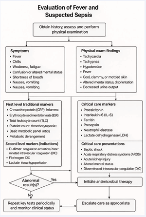
Figure 2: Algorithm for evaluation of fever and suspected sepsis.
This framework enhances diagnostic accuracy through judicious use of laboratory and imaging resources and strengthens antimicrobial stewardship by encouraging targeted therapy and reducing the risk of antimicrobial resistance.
Table 3: Diagnostic, prognostic, and predictive roles of routine hematological parameters, coagulation markers and critical care serological markers and, their kinetics (time to rise/fall)
|
Marker / Parameter |
Sample Type |
Diagnostic Role |
Prognostic / Predictive Role |
Kinetics (Time to Rise/ Peak/Fall) |
Reference Range |
Suggested Repeat Frequency |
Notes / Clinical Use |
|
ESR |
Whole blood (EDTA) |
Indicates chronic/ subacute inflammation |
Prolonged elevation suggests chronic disease |
Rises 24–48 h; peaks 3–5 d; slow fall (days–weeks) |
0–15 mm/h (M), 0–20 mm/h (F) |
Weekly or longer |
Useful in TB, autoimmune disease, vasculitis; affected by age/anemia |
|
CRP |
Whole blood (EDTA) |
Acute inflammation marker; differentiates bacterial vs viral patterns |
High CRP = severe disease; trend predicts response |
Rises 6–8 h; peaks ~48 h; falls 3–7 d |
<10 mg/L |
q24–48 h; daily in ICU |
Rapid decline with effective therapy; persistent elevation → reassess |
|
Total Leukocyte Count (TLC) |
Whole blood (EDTA) |
Infection/ inflammation screening |
Extreme values reflect risk and marrow suppression |
Rises 6–12 h |
4,000–10,000/mm³ |
Daily or clinically indicated |
Persistent leukopenia = worse prognosis |
|
Neutrophil-Lymphocyte Ratio (NLR) |
Whole blood (EDTA) |
Elevated in infection |
High NLR predicts mortality; guides triage |
Quick rise in acute inflammation |
0.78–3.58 |
Daily or alternate days |
Useful in resource-limited settings |
|
Platelet Count |
Whole blood (EDTA) |
Thrombocytopenia in viral sepsis; thrombocytosis in inflammation |
Low platelets predict severity |
Variable; depends on marrow activity |
1.5–4.5 lakh/mm³ |
2–3×/week or as needed |
Interpretation needs clinical context |
|
Plateletcrit |
Whole blood (EDTA) |
Reflects platelet mass |
Low PCT → poor prognosis, bleeding risk |
Tracks with platelet count |
0.22–0.24% |
2–3×/week |
Useful in dengue, sepsis, COVID-19 |
|
MPV (Mean Platelet Volume) |
Whole blood (EDTA) |
Platelet activation marker |
High MPV = vascular risk |
Slow kinetic changes |
7.2–11.8 fL |
Weekly or when platelets fluctuate |
Increased in sepsis/ reactive states |
|
Platelet Distribution Width (PDW) |
Whole blood (EDTA) |
Platelet size variability |
Indicates inflammatory burden |
Increases with heterogeneous platelet size |
9–17% |
2–3×/week |
Assesses sepsis complications risk |
|
Procalcitonin (PCT) |
Serum or plasma (SST) |
Bacterial infection marker |
Persistent elevation = complications |
Rises 4–6 h; peaks ~24 h; t½ ~24 h |
<0.1 ng/mL |
q24–48 h |
Guides antibiotic start/ stop decisions |
|
Critical care markers |
|||||||
|
Ferritin |
Serum or plasma (SST) |
Acute-phase reactant |
Very high ferritin = cytokine storm / HLH severity |
Rises in 24–48 h |
12–300 ng/mL |
q48–72 h |
>500–1000 ng/ mL suggests HLH/ hyperinflammation |
|
D-dimer |
Serum |
Coagulation activation, thrombosis |
High values = mortality risk; Predicts thrombotic events, anticoagulation needs |
Rises in 24 hours; declines with resolution |
< 0.50 mg/L FEU (i.e., < 500 ng/mL FEU) in adults; note age-adjusted cut-off in older adults is often used in practice |
2-3 times/week or per clinical need |
Used in COVID-19 and DIC management |
|
PT/INR – Prothrombin Time / International Normalized Ratio ; aPTT – Activated Partial Thromboplastin Time |
Plasma (citrate) |
Coagulation defects, DIC |
Derangement = risk of bleeding/ mortality |
Guides transfusion/ anticoagulant use; Changes with coagulation factor consumption |
PT ~11–13.5 s; INR 0.8–1.2 (method-dependent) aPTT ~25–37 s (method-dependent) |
Daily in ICU or bleeding/sepsis cases |
Essential in managing sepsis-induced coagulopathy |
|
Lactate |
Plasma (fluoride tube) |
Marker of hypoperfusion & sepsis severity |
High lactate predicts mortality; clearance improves survival |
Rises within hours; t½ 20–60 min |
0.5–2.2 mmol/L |
Every 4–6 h in shock |
Core component of sepsis bundles |
|
IL-6 (Interleukin-6) |
Serum or plasma (SST) |
Cytokine-storm marker |
High IL-6 = severe disease, ICU need |
Rapid rise within hours; falls over days |
<5 pg/mL |
q24–48 h |
Used in hyperinflammation (COVID-19, HLH) |
|
Lactate Dehydrogenase (LDH) |
Serum or plasma (SST) |
Tissue damage marker |
High levels predict MODS |
Rises 24 h; peaks 48–72 h |
140–280 U/L |
2–3×/week |
Elevated in lung/liver injury; non-specific |
|
Critical care markers (Research setting) |
|||||||
|
Presepsin |
Plasma (fluoride tube) |
Early bacterial sepsis marker |
Correlates with severity & mortality |
Rises 2–4 h; peaks early |
<320 pg/mL |
q24–48 h |
Emerging biomarker; more specific early |
|
Active Neutrophil Elastase (NE) |
Sputum |
Neutrophil activation marker |
High levels = tissue damage risk |
Rapid rise within hours |
56–136 ng/mL |
Daily (ICU) |
Useful in ARDS, sepsis, severe COVID-19 |
|
Fungal infection markers |
|||||||
|
Galactomannan (GM) |
Serum / BAL |
Marker for invasive aspergillosis |
Serial elevation = poor outcome |
Detectable early in disease |
1.92–7.3 mg/dL |
Twice weekly |
Better performance in neutropenic pts |
|
β-D-Glucan (BDG) |
Serum |
Broad fungal infection marker |
High BDG = fungal burden |
Rises 1–2 days after invasion |
<31 pg/mL |
Twice weekly |
False positives possible (IVIG, dialysis) |
|
Mannan / Anti-Mannan (Mn/A-Mn) |
Serum |
Candida infection marker |
Combined testing improves prognosis |
Variable; depends on immune status |
Assay-dependent |
Twice weekly |
Useful in immunocompromised patients |
EDTA: Ethylene diamine tetra acetic acid, SST: Serum separator tube, SFT: Sodium fluoride tube, BAL: Bronchoalveolar lavage fluid
Key Notes:
• Diagnostic: Identifies presence of infection/inflammation
• Prognostic: Correlates with severity/mortality
• Predictive: Guides therapeutic decisions or anticipates response
• Kinetics help guide when to test/retest.
• Frequency depends on clinical setting, disease trajectory, and resource availability: e.g., ICU = more frequent, OPD = less.
• Serial trends are more useful than isolated/single values.
Integrating these tests into clinical stewardship programs ensures targeted diagnostics, timely intervention, and rational antimicrobial use.
Table 3 provides a structured comparison of serological marker characteristics, outlining their diagnostic, prognostic, and predictive roles, associated kinetics, and recommended frequency of repeat testing. This framework supports standardized, evidence based decision making across varied clinical scenarios and is particularly valuable in critically ill patients, where appropriate marker selection and interpretation facilitate early diagnosis of severe infections, risk stratification, monitoring of therapeutic response, and informed escalation or de escalation of antimicrobial therapy. The tiered approach emphasized in Table?3 prioritizes initial clinical assessment supported by basic, widely available laboratory parameters such as CBC, CRP, and serum lactate for early identification and severity stratification of suspected infection and sepsis, with selective escalation to advanced biomarkers based on clinical deterioration, organ dysfunction, or inadequate response to therapy.
Table 4 complements Table 3 by mapping marker selection to distinct phases of critical illness, mirroring the stepwise care continuum from early evaluation and suspected sepsis through hyperinflammatory syndromes, suspected invasive fungal infection, and recovery. Together, these tables reinforce stewardship principles of diagnostic parsimony, context appropriate testing, and serial trend based interpretation.
Table 4: Clinical Stages of Critical Illness and Indications for Ordering Critical Care Serological Markers
| Clinical Stage | Key Serological Markers | Indications / Rationale |
| Initial Evaluation (Within 0-6 hours of ICU admission) | • Lactate | |
| • CRP | ||
| - PCT | • Rule out sepsis vs non-infectious causes of SIRS | |
| - TLC/NLR | • Assess severity and perfusion | |
| - IL-6 | • Establish inflammatory baseline | |
| • Presepsin (research setting) | ||
| Suspected Sepsis or Septic Shock | • Procalcitonin (PCT) | • Support early differentiation between bacterial and non-bacterial infection |
| • Lactate | • Guide antibiotic initiation/de-escalation | |
| • CRP | • Assess cytokine activation | |
| • IL-6 | ||
| • Presepsin (Research Setting) | ||
| HyperinflammatorySyndromes/CytokineStorm | - Ferritin | |
| - D-dimer | ||
| - CRP | Diagnose HLH, MAS, severe COVID-19, or secondary inflammation Guide immunomodulatory therapy (e.g., steroids, tocilizumab) | |
| - IL-6 | ||
| • LDH | ||
| Suspected Fungal Infections / Immunosuppression | • β-D-Glucan (BDG) - Galactomannan (GM) - Anti-Mannan / A-Mn IgG/IgM | Early detection of invasive fungal infections in neutropenic or immunocompromised patients |
| Targeted antifungal therapy | ||
| Multiorgan Dysfunction / ARDS / Worsening Organ Failure | • LDH | |
| • Ferritin | Reflect ongoing tissue damage and predict deterioration Indicate progression to ARDS or MODS | |
| • IL-6 | ||
| • Neutrophil Elastase | ||
| Coagulation Abnormalities / Suspected DIC | • D-dimer | Detect thrombotic complications or coagulopathy Guide anticoagulation, blood product support |
| • PT/INR, aPTT - Platelet indices (PCT, MPV, PDW) | ||
| Monitoring Response to Treatment | • PCT (every 24-48 hours) | Monitor improvement or need to escalate treatment Support decision to de-escalate antibiotics |
| • CRP (every 48-72 hours) | Track response to immunotherapy | |
| • Lactate (every 4-6 hours in shock) | ||
| • IL-6 / Ferritin / LDH | ||
| Weaning / Recovery Phase | • Lactate clearance | Ensure resolution of systemic inflammation Validate safety of de-escalation or ICU discharge |
| • CRP / PCT normalization Stabilization of inflammatory markers |
ICU – Intensive Care Unit; CRP – C-Reactive Protein; PCT – Procalcitonin; TLC – Total Leukocyte Count; NLR – Neutrophil-to-Lymphocyte Ratio; IL-6 – Interleukin-6; LDH – Lactate Dehydrogenase; HLH – Hemophagocytic Lymphohistiocytosis; MAS – Macrophage Activation Syndrome; COVID-19 – Coronavirus Disease 2019; β-D-Glucan (BDG) – Beta-D-Glucan; GM – Galactomannan; A-Mn IgG/IgM – Anti-Mannan Immunoglobulin G / Immunoglobulin M; ARDS – Acute Respiratory Distress Syndrome; MODS – Multiple Organ Dysfunction Syndrome; DIC – Disseminated Intravascular Coagulation; PT/INR – Prothrombin Time / International Normalized Ratio; aPTT – Activated Partial Thromboplastin Time; MPV – Mean Platelet Volume; PDW – Platelet Distribution Width
Key Stewardship Tips:
• Always combine serological data with clinical judgment and cultures/imaging.
• Trend values over time rather than relying on single readings/isolation.
Avoid unnecessary repetition unless there's clinical change or deterioration.
In evaluating fever, distinguishing infectious from non infectious inflammatory causes remains fundamental for accurate diagnosis and targeted management. Traditional haematological markers including CRP, ESR, total leukocyte count, and platelet indices, remain valuable first line investigations owing to their accessibility, cost effectiveness, and utility in identifying inflammatory responses and disease progression [85,86]. These markers aid clinicians in avoiding misdiagnosis and ensuring appropriate, aetiology driven therapy.
For critically ill patients or those with suspected sepsis, a stepwise, severity based approach to testing is essential. Advanced markers such as procalcitonin, lactate, interleukin 6, ferritin, and β D glucan significantly enhance diagnostic precision, facilitate early recognition of severe or invasive infections, and support stewardship through informed antimicrobial escalation or de escalation [54,85]. The Infectious Diseases Society of America (IDSA) specifically supports the judicious use of procalcitonin, particularly for guiding the duration and discontinuation of antibiotics rather than their initiation [87-89]. IDSA guidance consistently cautions that serological markers should not replace clinical judgment, cultures, or molecular diagnostics, and that their greatest value lies in serial trend analysis integrated into comprehensive clinical assessment and antimicrobial stewardship programs [87,88].
Integrating this tiered approach into hospital based antimicrobial stewardship and sepsis protocols promotes optimized resource utilization, enhances patient outcomes, and mitigates antimicrobial resistance. Ultimately, clinical stewardship ensures that serological and haematological markers are applied thoughtfully and effectively, strengthening both diagnostic accuracy and therapeutic decision making across the continuum of infection management.
REFERENCES
- Hotchkiss RS, Coopersmith CM, McDunn JE, Ferguson TA. The sepsis seesaw: tilting toward immunosuppression. Nat Med. 2009; 15: 496-497.
- Singh I, Smart D, Schreier J. Overdiagnosis and waste from inappropriate laboratory testing: findings from a nationwide analysis of 1 billion U.S. 2025; 30: A48-A49.
- National Heart, Lung, and Blood Institute (NHLBI). Routine lab tests are not a reliable way to diagnose long COVID. NHLBI News Release. 2024
- Suklan J, Cheaveau J, Hill S, Urwin SG, Green K, Winter A, et al. Utility of Routine Laboratory Biomarkers to Detect COVID-19: A Systematic Review and Meta-Analysis. Viruses. 2021; 13: 803.
- Karakonstantis S, Kalemaki D, Tzagkarakis E, Lydakis C. Pitfalls in studies of eosinopenia and neutrophil-to-lymphocyte count ratio. Infect Dis (Lond). 2018; 50: 163-174.
- Lass-Florl C, Alastruey-Izquierdo A, Gupta R, Chakroborti A. Interpretation, pitfalls of biomarkers in diagnosis of invasive fungal diseases. Indian J Med Microbiol. 2022; 40: 480-484.
- Theel ES, Doern CD. β-D-glucan testing is important for diagnosis of invasive fungal infections. J Clin Microbiol. 2013; 51: 3478-3483.
- Gabay C, Kushner I. Acute-phase proteins and other systemic responses to inflammation. N Engl J Med. 1999; 340: 448-54.
- Brigden ML. Clinical utility of the erythrocyte sedimentation rate. Am Fam Physician. 1999; 60: 1443-1450.
- Osei-Bimpong A, Meek JH, Lewis SM. ESR or CRP? A comparison of their clinical utility. Hematology. 2007; 12: 353-357.
- Marnell L, Mold C, Du Clos TW. C-reactive protein: ligands, receptors and role in inflammation. Clin Immunol. 2005; 117: 104-111.
- Cecconi M, Evans L, Levy M, Rhodes A. Sepsis and septic shock. Lancet. 2018; 392: 75-87.
- Vijayan AL, Vanimaya, Ravindran S, Saikant R, Lakshmi S, Kartik R, G M. Procalcitonin: a promising diagnostic marker for sepsis and antibiotic therapy. J Intensive Care. 2017; 5: 51.
- Singer M, Deutschman CS, Seymour CW, Shankar-Hari M, Annane D, Bauer M, et al. The Third International Consensus Definitions for Sepsis and Septic Shock (Sepsis-3). JAMA. 2016; 315: 801-810.
- Assal HH, Abdelrahman SM, Abdelbasset MA, Abdelaziz M, Sabry IM, Shaban MM. Presepsin as a Novel Biomarker in predicting In-hospital Mortality in Patients With COVID-19 Pneumonia. Int J Infect Dis. 2022; 118: 155-163.
- Chen L, Liu HG, Liu W, Liu J, Liu K, Shang J, et al. [Analysis of clinical features of 29 patients with 2019 novel coronavirus pneumonia]. Zhonghua Jie He He Hu Xi Za Zhi. 2020; 43: 203-208.
- Tanaka T, Narazaki M, Kishimoto T. IL-6 in inflammation, immunity, and disease. Cold Spring Harb Perspect Biol. 2014; 6: a016295.
- Nijman RG, Moll HA, Smit FJ, Gervaix A, Weerkamp F, Vergouwe Y, et al. C-reactive protein, procalcitonin and the lab-score for detecting serious bacterial infections in febrile children at the emergency department: a prospective observational study. Pediatr Infect Dis J. 2014; 33: e273-279.
- Pfeiffer CD, Fine JP, Safdar N. Diagnosis of invasive aspergillosis using a galactomannan assay: a meta-analysis. Clin Infect Dis. 2006; 42: 1417-1427.
- Levett PN. Leptospirosis. Clin Microbiol Rev. 2001; 14: 296-326.
- Largman-Chalamish M, Wasserman A, Silberman A, Levinson T, Ritter O, Berliner S, et al. Differentiating between bacterial and viral infections by estimated CRP velocity. PLoS One. 2022; 17: e0277401.
- Cherny SS, Brzezinski RY, Wasserman A, Adler A, Berliner S, Nevo D, Rosset S, Obolski U. Characterizing CRP dynamics during acute infections. Infection. 2025; 53: 1199-1203.
- Pepys MB, Hirschfield GM. C-reactive protein: a critical update. J Clin Invest. 2003; 111: 1805-1812.
- Póvoa P, Coelho L, Almeida E, Fernandes A, Mealha R, Moreira P, et al. Early identification of intensive care unit-acquired infections with daily monitoring of C-reactive protein: a prospective observational study. Crit Care. 2006; 10: R63.
- Ito A, Ishida T. Diagnostic markers for community-acquired pneumonia. Ann Transl Med. 2020; 8: 609.
- Kataja A, Tarvasmäki T, Lassus J, Sionis A, Mebazaa A, Pulkki K, et al. CardShock investigators. Kinetics of procalcitonin, C-reactive protein and interleukin-6 in cardiogenic shock - Insights from the Card Shock study. Int J Cardiol. 2021; 322: 191-196.
- Marshall H. NICE guidelines to family doctors on diagnosis of pneumonia. Lancet Respir Med. 2015; 3: 17.
- National Institute for Health and Care Excellence (NICE). Pneumonia in adults: diagnosis and management. Clinical guideline CG191. London: NICE; 2014.
- de Jager CP, Wever PC, Gemen EF, Kusters R, van Gageldonk-Lafeber AB, van der Poll T, et al. The neutrophil-lymphocyte count ratio in patients with community-acquired pneumonia. PLoS One. 2012; 7: e46561.
- Josse JM, Cleghorn MC, Ramji KM, Jiang H, Elnahas A, Jackson TD, et al. The neutrophil-to-lymphocyte ratio predicts major perioperative complications in patients undergoing colorectal surgery. Colorectal Dis. 2016; 18: 236-242.
- Buonacera A, Stancanelli B, Colaci M, Malatino L. Neutrophil to Lymphocyte Ratio: An Emerging Marker of the Relationships between the Immune System and Diseases. Int J Mol Sci. 2022; 23: 3636.
- Forget P, Khalifa C, Defour JP, Latinne D, Van Pel MC, De Kock M. What is the normal value of the neutrophil-to-lymphocyte ratio? BMC Res Notes. 2017; 10: 12.
- Lippi G, Franchini M. Platelets and immunity: the interplay of mean platelet volume in health and disease. Expert Rev Hematol. 2015; 8: 555-557.
- Budak YU, Polat M, Huysal K. The use of platelet indices, plateletcrit, mean platelet volume and platelet distribution width in emergency non-traumatic abdominal surgery: a systematic review. Biochem Med (Zagreb). 2016; 26: 178-193.
- Gasparyan AY, Ayvazyan L, Mikhailidis DP, Kitas GD. Mean platelet
volume: a link between thrombosis and inflammation? Curr Pharm Des. 2011; 17: 47-58.
- Nitin CB, Varma KV. Platelet Indices as a Predictive Marker in Type 2 Diabetes Complications: Micro Versus Macro Vascular. Int. J Med. Pharm. Res. 2024; 5: 221-228.
- Jeon K, Kim M, Lee J, Lee JS, Kim HS, Kang HJ, et al. Immature platelet fraction: A useful marker for identifying the cause of thrombocytopenia and predicting platelet recovery. Medicine (Baltimore). 2020; 99: e19096.
- Goyal S, Sharma CM, Kumar R, Mohan N. Platelet count and its indices as diagnostic markers of neonatal sepsis: a cross-sectional study. Int J Contemp Pediatr. 2024; 11: 951-956.
- Wang X, Li X, Shang Y, Wang J, Zhang X, Su D, et al. Ratios of neutrophil-to-lymphocyte and platelet-to-lymphocyte predict all-cause mortality in inpatients with coronavirus disease 2019 (COVID-19): a retrospective cohort study in a single medical centre. Epidemiol Infect. 2020; 148: e211.
- Noris P, Melazzini F, Balduini CL. New roles for mean platelet volume measurement in clinical practice? Platelets. 2016; 27: 607-612.
- Azim A. Presepsin: A Promising Biomarker for Sepsis. Ind J Crit Care Med. 2021; 25: 117-118.
- Turk V, Stoka V, Vasiljeva O, Renko M, Sun T, Turk B, Turk D. Cysteine cathepsins: from structure, function and regulation to new frontiers. Biochim Biophys Acta. 2012; 1824: 68-88.
- Zhao MM, Yang WL, Yang FY, Zhang L, Huang WJ, Hou W, et al. Cathepsin L plays a key role in SARS-CoV-2 infection in humans and humanized mice and is a promising target for new drug development. Signal Transduct Target Ther. 2021; 6: 134.
- Patel S, Homaei A, El-Seedi HR, Akhtar N. Cathepsins: Proteases that are vital for survival but can also be fatal. Biomed Pharmacother. 2018; 105: 526-532.
- Papayannopoulos V. Neutrophil extracellular traps in immunity and disease. Nat Rev Immunol. 2018; 18: 134-147.
- Lv H, Huang L, Yang X, Zhang C, Yu H, Shang X. The clinical effectiveness of sivelestat in treating sepsis patients with both acute respiratory distress syndrome and septic cardiomyopathy. J Cardiothorac Surg. 2024; 19: 399.
- Hashimoto S, Okayama Y, Shime N, Kimura A, Funakoshi Y, Kawabata K, et al. Neutrophil elastase activity in acute lung injury and respiratory distress syndrome. Respirology. 2008; 13: 581-584.
- Ito A, Ishida T. Diagnostic markers for community-acquired pneumonia. Ann Transl Med. 2020; 8: 609.
- Johansson N, Kalin M, Backman-Johansson C, Larsson A, Nilsson K, Hedlund J. Procalcitonin levels in community-acquired pneumonia - correlation with aetiology and severity. Scand J Infect Dis. 2014; 46: 787-791.
- Ivaska L, Elenius V, Mononen I, Ruuskanen O, Peltola V. Discrepancies between plasma procalcitonin and C-reactive protein levels are common in acute illness. Acta Paediatr. 2016; 105: 508-513.
- 51. Song JE, Kim MH, Jeong WY, Jung IY, Oh DH, Kim YC, et al. Mortality Risk Factors for Patients with Septic Shock after Implementation of the Surviving Sepsis Campaign Bundles. Infect Chemother. 2016; 48: 199-208.
- Kang HE, Park DW. Lactate as a Biomarker for Sepsis Prognosis? Infect Chemother. 2016; 48: 252-253.
- Gutiérrez HB, Concepción YA, Pérez JS, Lara YD, López FMR, Contreras PR. Prognostic Value of Serum Lactate Levels in Critically Ill Patients
in an Intensive Care Unit. J Crit Care Med (Targu Mures). 2020; 6: 59-64.
- Prescott HC, Antonelli M, Alhazzani W, Møller MH, Alshamsi F, Azevedo LCP, et al. Surviving Sepsis Campaign: International Guidelines for Management of Sepsis and Septic Shock 2026. Crit Care Med. 2026; 54: 725-812.
- Rosário C, Zandman-Goddard G, Meyron-Holtz EG, D’Cruz DP, Shoenfeld Y. The hyperferritinemic syndrome: macrophage activation syndrome, Still’s disease, septic shock and catastrophic antiphospholipid syndrome. BMC Med. 2013; 11: 185.
- Kernan KF, Carcillo JA. Hyperferritinemia and inflammation. Int Immunol. 2017; 29: 401-409.
- Carcillo JA, Simon DW, Podd BS. How We Manage Hyperferritinemic Sepsis-Related Multiple Organ Dysfunction Syndrome/ Macrophage Activation Syndrome/Secondary Hemophagocytic Lymphohistiocytosis Histiocytosis. Pediatr Crit Care Med. 2015; 16: 598-600.
- Zhang C, Wu Z, Li JW, Zhao H, Wang GQ. Cytokine release syndrome in severe COVID-19: interleukin-6 receptor antagonist tocilizumab may be the key to reduce mortality. Int J Antimicrob Agents. 2020; 55: 105954.
- Shekhawat J, Gauba K, Gupta S, Purohit P, Mitra P, Garg M, et al. Interleukin-6 Perpetrator of the COVID-19 Cytokine Storm. Indian J Clin Biochem. 2021; 36: 440-450.
- Kang S, Kishimoto T. Interplay between interleukin-6 signaling and the vascular endothelium in cytokine storms. Exp Mol Med. 2021; 53: 1116-1123.
- Zhang Y, Li X, Zhang X, Wang T, Zhang X. Progress in the study of pentraxin-3(PTX-3) as a biomarker for sepsis. Front Med (Lausanne). 2024; 11:1398024.
- Zein JG, Lee GL, Tawk M, Dabaja M, Kinasewitz GT. Prognostic Significance of Elevated Serum Lactate Dehydrogenase (LDH) in Patients with Severe Sepsis. CHEST. 2024; 126: 873.
- McPherson, Richard and Pincus, Matthew R., “Henry’s Clinical Diagnosis and Management by Laboratory Methods” (2017). Faculty Bookshelf. 81.
- Singer M, Deutschman CS, Seymour CW, Shankar-Hari M, Annane D, Bauer M, et al. The Third International Consensus Definitions for Sepsis and Septic Shock (Sepsis-3). JAMA. 2016; 315: 801-810.
- Lee SG, Song J, Park DW, Moon S, Cho HJ, Kim JY, et al. Prognostic value of lactate levels and lactate clearance in sepsis and septic shock with initial hyperlactatemia: A retrospective cohort study according to the Sepsis-3 definitions. Medicine (Baltimore). 2021; 100: e24835.
- Zhai C, Gu K, Zhai X, Wang J, Zhang J. Prognostic Value of Serum Lactate Dehydrogenase in Patients with Nasopharyngeal Carcinoma: a Meta-Analysis. Clin Lab. 2017; 63: 1777-1785.
- Levi M, van der Poll T. Coagulation and sepsis. Thromb Res. 2017; 149: 38-44.
- Iba T, Nisio MD, Levy JH, Kitamura N, Thachil J. New criteria for sepsis-induced coagulopathy (SIC) following the revised sepsis definition: a retrospective analysis of a nationwide survey. BMJ Open. 2017; 7: e017046.
- Gando S, Levi M, Toh CH. Disseminated intravascular coagulation. Nat Rev Dis Primers. 2016; 2: 16037.
- Fontaine T, Latgé JP. Galactomannan Produced by Aspergillus fumigatus: An Update on the Structure, Biosynthesis and Biological Functions of an Emblematic Fungal Biomarker. J Fungi (Basel). 2020; 6: 283
- Maertens J, Verhaegen J, Lagrou K, Van Eldere J, Boogaerts M. Screening for circulating galactomannan as a noninvasive diagnostic tool for invasive aspergillosis in prolonged neutropenic patients and stem cell transplantation recipients: a prospective validation. Blood. 2001; 97: 1604-1610.
- Chong GM, Maertens JA, Lagrou K, Driessen GJ, Cornelissen JJ, Rijnders BJ. Diagnostic Performance of Galactomannan Antigen Testing in Cerebrospinal Fluid. J Clin Microbiol. 2016; 54: 428-431.
- Reischies FM, Raggam RB, Prattes J, Krause R, Eigl S, List A, Quehenberger F, Strenger V, Wölfler A, Hoenigl M. Urine Galactomannan-to-Creatinine Ratio for Detection of Invasive Aspergillosis in Patients with Hematological Malignancies. J Clin Microbiol. 2016; 54: 771-774.
- White PL, Jones T, Whittle K, Watkins J, Barnes RA. Comparison of galactomannan enzyme immunoassay performance levels when testing serum and plasma samples. Clin Vaccine Immunol. 2013; 20: 636-638.
- Verweij PE, Weemaes CM, Curfs JH, Bretagne S, Meis JF. Failure to detect circulating Aspergillus markers in a patient with chronic granulomatous disease and invasive aspergillosis. J Clin Microbiol. 2003; 8: 3900-3901.
- Mercier T, Guldentops E, Lagrou K, Maertens J. Galactomannan, a Surrogate Marker for Outcome in Invasive Aspergillosis: Finally Coming of Age. Front Microbiol. 2018; 9: 661.
- Obayashi T. [The Plasma (1→3)-β-D-glucan Assay, a Japanese Contribution to the Diagnosis of Invasive Fungal Infection]. Med Mycol J. 2017; 58: J141-J147.
- Odabasi Z, Paetznick VL, Rodriguez JR, Chen E, McGinnis MR, Ostrosky-Zeichner L. Differences in beta-glucan levels in culture supernatants of a variety of fungi. Med Mycol. 2006; 44: 267-272.
- Sendid B, Dotan N, Nseir S, Savaux C, Vandewalle P, Standaert A, et al. Antibodies against glucan, chitin, and Saccharomyces cerevisiae mannan as new biomarkers of Candida albicans infection that complement tests based on C. albicans mannan. Clin Vaccine Immunol. 2008; 15: 1868-1877.
- Duettmann W, Koidl C, Krause R, Lackner G, Woelfler A, Hoenigl M. Specificity of mannan antigen and anti-mannan antibody screening in patients with haematological malignancies at risk for fungal infection. Mycoses. 2016; 59: 374-378.
- Mikulska M, Calandra T, Sanguinetti M, Poulain D, Viscoli C. Third European Conference on Infections in Leukemia Group. The use of mannan antigen and anti-mannan antibodies in the diagnosis of invasive candidiasis: recommendations from the Third European Conference on Infections in Leukemia. Crit Care. 2010; 14: R222.
- Christensson B, Sigmundsdottir G, Larsson L. D-arabinitol--a marker for invasive candidiasis. Med Mycol. 1999; 37: 391-396.
- 83. Vidal-García M, Redrado S, Domingo MP, Marquina P, Colmenarejo C, Meis JF, et al. Production of the Invasive Aspergillosis Biomarker Bis(methylthio)gliotoxin Within the Genus Aspergillus: In Vitro and in Vivo Metabolite Quantification and Genomic Analysis. Front Microbiol. 2018; 9: 1246.
- Li Q, Qu L, Miao Y, Li Q, Zhang J, Zhao Y, et al. A gene network database for the identification of key genes for diagnosis, prognosis, and treatment in sepsis. Sci Rep. 2023; 13: 21815.
- World Health Organization. Guidelines on the clinical management of sepsis. Geneva: WHO; 2024.
- Rhodes A, Evans LE, Alhazzani W, Levy MM, Antonelli M, Ferrer R, et al. Surviving Sepsis Campaign: International Guidelines for Management of Sepsis and Septic Shock: 2016. Intensive Care Med. 2017; 43: 304-377.
- Andre C. Kalil, Mark L. Metersky, Michael Klompas, John Muscedere, Daniel A, et al. Management of Adults With Hospital-acquired and Ventilator-associated Pneumonia: 2016 Clinical Practice Guidelines by the Infectious Diseases Society of America and the American Thoracic Society, Clinical Infectious Diseases. 2016; 63: e61–e111
- Metlay JP, Waterer GW, Long AC, Anzueto A, Brozek J, Crothers K, et al. Diagnosis and Treatment of Adults with Community-acquired Pneumonia. An Official Clinical Practice Guideline of the American Thoracic Society and Infectious Diseases Society of America. Am J Respir Crit Care Med. 2019; 200: e45-e67.
- Schuetz P, Wirz Y, Sager R, Christ-Crain M, Stolz D, Tamm M, et al. Procalcitonin to initiate or discontinue antibiotics in acute respiratory tract infections. Cochrane Database Syst Rev. 2017; 10: CD007498.
- Indian Council of Medical Research. Standard treatment workflows for management of sepsis and septic shock. New Delhi: ICMR; 2021