Clinical Value of Sonography and CT-Scan in the Diagnosis of Acute Cholecystitis: A Retrospective Analysis
- 1. Clinic for General and Visceral Surgery, Kepler University Clinic, Austria
- 2. Devision of Medical Controlling, Kepler University Clinic, Austria
- 3. Clinic for Gynecology, Kepler University Clinic, Austria
Abstract
Background: The diagnosis of acute cholecystitis is found on the basis of history, clinical assessment, a biochemical examination and sonography. Only in case of unclear clinical symptoms or discrepant findings a computed tomography is done. The aim of this retrospective study is to determine the accuracy of computed tomography and sonography in the diagnosis of acute cholecystitis.
Patients and methods: This is a retrospective study of patients in which a laparoscopic cholecystectomy was performed in case of acute cholecystitis between January 1st 2011 and December 31st 2015 at the Kepler University Clinic in Linz. The sensitivity and specifity of sonography and computed tomography were analyzed and compared to the intraoperative finding and histological results.
Results: A total of 475 patients underwent laparoscopic cholecystectomy for acute cholecystis. 404 patients underwent sonography and in 131 patients a computed tomography was done. Sonography showed a specificity of 95.56% and sensitivity of 74.65% in the diagnosis of acute cholecystitis. Computed tomography revealed a specificity of 85.71% and sensitivity of 90.32% in the diagnosis of acute cholecystitis.
Conclusion: Computed tomography had a high sensitivity and specificity in the diagnosis of acute cholecystitis, but Sonography is still the first choice .
Keywords
Acute cholecystitis; Computed tomography; Sonography; Laparoscopic cholecystectomy
Citation
Klugsberger B, Haslinger-Eisterer B, Oppelt P, Shamiyeh A (2017) Clinical Value of Sonography and CT-Scan in the Diagnosis of Acute Cholecystitis: A Retrospective Analysis. Ann Emerg Surg 2(4): 1021.
INTRODUCTION
Gallstones are a common finding, present in 10%-15% of the general population. Among patients with cholelithiasis, in 1%-4% of these will get symptoms per year and out of that 30% of these patients develop an acute cholecystitis [1,2]. Acute cholecystitis is one of the most common reasons for emergency admission to general surgical departments [3,4]. The diagnosis of acute cholecystitis is determined on the basis of typical anamnesis including recurrent or unrelenting right upper quadrant pain, fever, nausea and clinical examination findings of right upper quadrant tenderness, positive Murphy sign, elevated laboratory findings for acute inflammation and ultrasound [5,6]. Only in case of unclear clinical symptoms or discrepant findings a further radiological examination by computed tomography should be done.
In the current literature the accuracy of ultrasound diagnosis in the presence of acute cholecystitis is 81% [7]. In the literature, however, we have found only a few studies which analyzed the sensitivity and specificity of computed tomography in diagnosis of acute cholecystitis.
The aim of this retrospective study is to determine the accuracy of computed tomography and sonography in the diagnosis of acute cholecystitis.
PATIENTS AND METHODS
Approval from the regional ethic committee was obtained on February 17th 2016 (ref. no. K-95-16). This is a retrospective singlecenter cohort study of 475 patients who underwent laparoscopic cholecystectomy (LCHE) for acute cholecystitis between January 1st 2011, and December 31st 2015 at the department of general and visceral surgery, Kepler University Hospital in Linz, Austria. Demographic, clinical and pathologic data were retrieved from an electronic database. The diagnosis of acute cholecystitis was considered to be probable in patients with at least two of three diagnostic criteria described in the Tokyo guidelines: clinical parameter (fever), findings from blood chemistry (elevated white blood count) and positive ultrasound [8]. Only in case of unclear clinical symptoms or discrepant findings a further radiological examination is carried out by means of computed tomography (CT).
Sonographical findings and CT were compared with the histopathological analysis of the gallbladder. The sensitivity and specificity of sonography and CT for diagnosis of acute cholecystitis were analyzed.
Criteria for inclusion were laparoscopic cholecystectomy (LCHE) in case of acute choleystitis. Criteria for exclusion were konservative treatment of cholecystitis without cholecystectomy and elective laparoscopic cholecystectomy in case of gallbladder disease without cholecystitis. Patients with an open performed cholecystectomy were excluded from the analysis.
Sonography and CT was performed by radiologists on duty (senior residents or consultants).
LCHE was performed in a standardized procedure as usual in our clinic. The access was through the umbilical port (11mm), CO2 gas was established with a pressure at 12 mm mercury. After insertion of the optic (Storz, Germany) additional ports were placed under direct vision (one epigastric and one or two at the right upper quadrant of the abdomen.
Dissection of the Calot triangle following the “critical view of safety” technique [9], the cystic artery and the cystic duct was ligated, each by three titanium clips (two central and one peripheral) and divided. The electro cautery was used to dissect the gallbladder retrograde from the gallbladder bed. The specimen was removed through the umbilical incision. The fascia at the umbilical site was closed with non resorbable suture (Premilene 0 ® Braun, Tuttlingen, Germany). Skin closure was done by use of single knot suture 4/0. A consultant for pathology reviewed all surgical specimens.
Statistical analysis
We evaluated preoperative characteristics, including demographics (sex, mean age, and laboratory values (white blood cell count, C reactive protein, alkaline phosphatase, Gamma-GT, Bilirubin).
For specificity and sensitivity values two-sided 95% confidence intervals were calculated using the open-source R statistical software package, version 3.1.2 (The R Foundation for Statistical Computing, Vienna, Austria).
RESULTS
A total of 475 patients underwent LCHE for acute cholecystis from January 1st 2011 to December 31st 2015 at the department of general and visceral surgery, Kepler University Clinic in Linz. 404 patients underwent sonography (group A) and in 131 patients a computertomography (group B) was done. In 60 patients both an ultrasound and computertomography was performed (Table 1).
Table 1: number of performed sonography and CT in group A and group B.
|
sonography |
CT |
CT + sonography |
Total |
|
|
group A |
344 |
0 |
60 |
404 |
|
group B |
0 |
71 |
60 |
131 |
These patients were added to each group. Group A included 209 men and 195 women and in group B were 85 men and 46 women (Table 2).
Table 2: gender distribution in group A and group B.ca
| group A | n | % | group B | n | % | ||
| sex | male | 209 | 51.7 | sex | male | 85 | 64.9 |
| female | 195 | 48.3 | female | 46 | 35.1 | ||
| Total | 404 | 100 | Total | 131 | 100 |
The distribution of severity of cholecystitis in group A was in 234 cases (57, 9%) a mild form and in 170 cases (42, 1%) a moderate form. The distribution of severity of cholecystitis in group A was in 234 cases (57, 9%) a mild form and in 170 cases (42, 1%) a moderate form. The distribution of severity of acute cholecystitis in group B was in 42 cases (70%) a mild form and in 18 cases (30%) a moderate form. There were no cases of severe acute cholecystitis. If there was a severe acute cholecystitis, we performed an open cholecystectomy. Patients with an open performed cholecystectomy were excluded from the analysis.
The median age of all patients was 62.7 years (Q25 49.4, Q75 73.4). All patients showed an increase in cholestatic parameters: median alkine phosphatase (ALP) 79 U/l (Q25 62, Q75 111) and median Gamma-GT 50 U/l (Q25 25, Q75 119), the median Bilirubin value of all patients was 0.7 mg/dl (Q25 0.5, Q75 1.2). ). The inflammatory markers were elevated: the median white blood count of all patients was 11.5 G/l (Q25 8.3, Q75 15.1) and the median C-reactive protein (CRP) was 4.5 G/l (Q25 0.7, Q75 14.9).
Descriptive analysis group A
The median age in group A was 60.6 years (Q25 48.7, Q75 71.9). The median white blood count was 11.3 G/l (Q25 8.3, Q75 14.5) and the median C-reactive protein (CRP) was 3.6 G/l (Q25 0.6, Q75 11.9) (Figure 1,2).
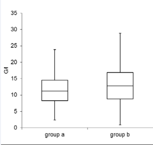
Figure 1: White blood count in group A and group B, G/l: per liter
The median alkaline phosphatase in group A was 78 U/l (Q25 61.5, Q75 107), the median Gamma-GT was 46 U/l (Q25 25, Q75 111) and median Bilirubin was 0.7 mg/ dl (Q25 0.5, Q75 1.2).
Descriptive analysis group B
The median age in group B was 69.8 years (Q25 57.8, Q75 78.2). The median white blood count was 12.8 G/l (Q25 8.8, Q75 16.8) and the median C-reactive protein (CRP) was 12.0 G/l (Q25 3.6, Q75 20.4) (Figure 1,2).
Figure 2: C - reactive protein in group A and group B; milligrams per deciliter.
The median alkaline phosphatase in group B was 82.5 U/l (Q25 62, Q75 123), the median Gamma-GT was 57.5 U/l (Q25 30.5, Q75 121) and median Bilirubin was 0.9 mg/dl (Q25 0.5, Q75 1.5).
Sonography (group A) showed a specificity of 95.56% and sensitivity of 74.65% with an overall accuracy of 77.0% in the diagnosis of acute cholecystitis. Computed tomography (group B) revealed a specificity of 85.71% and sensitivity of 90.32% with an overall accuracy of 90.1% in the diagnosis of acute cholecystitis (Figure 3).
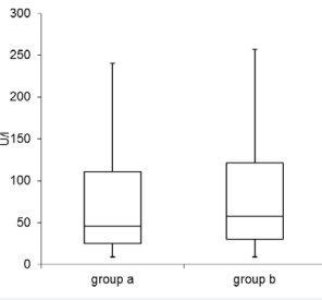
Figure 3: Gamma-GT in group A and group B; U/l: units per liter
DISCUSSION
Sonography was considered the first-line imaging technique for the evaluation of patients who are clinically suspected for acute cholecystitis [7,10]. In our Clinic, CT was performed if ultrasound findings are equivocal or when clinical findings are nonspecific.
Our study showed in group B (CT) a low false-negative rate in diagnosis of acute cholecystitis with 5.3%. In contrast, group A showed a false-negative rate in diagnosis of acute cholecystitis of 11.1%. A comparison of both groups is difficult, because only the patients with discrepant findings underwent CT. Group B (CT) had higher elevated inflammatory markers, despite this, the diagnosis of acute cholecystitis was considered with CT.
We showed that computed tomography yielded a specificity of 85.7% and a sensitivity of 90.3% with an overall accuracy of 90.1%. Sonography had a specificity of 95.6% and a sensitivity of 74.7% with an overall accuracy of 77.0% in our retrospective analysis. Regarding to the analysis of the laboratory values, we found that leukocytosis and elevation of C-reactive protein were preoperative predictors of acute cholecystitis (Figure 4-6).
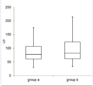
Figure 4: Alkaline phosphatasis in group A and group B; U/l: units per liter.
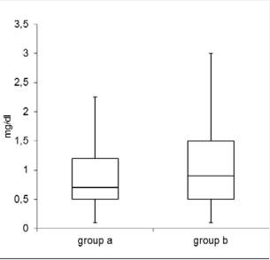
Figure 5: Bilirubine in group A and group B, mg/dl: milligrams per decilitre.
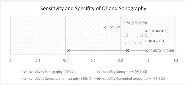
Figure 6: Sensitivity and specifity of sonography (group A) and computed tomography (group B) in the diagnosis of acute cholecystitis.
These figures were in keeping with those reported by other studies carried out for identifying preoperative prognostic parameters for acute cholecystitis [10-13]. Our study showed that computed tomography had a high sensitivity and specificity in case of acute cholecystitis. Nevertheless, sonography had the main advantages: high sensitivity for cholelithiasis, lack of ionizing radiation or contrast injection, fast availability and relatively low cost [14].
There was a limitation in our study: We analysed only patients treated with laparoscopic cholecystectomy. Patients treated with antibiotics were excluded.
CONCLUSION
We recommend ultrasound as a first choice for acute right upper abdominal pain with suspected history of gall stone disease. If the findings are unclear, computed tomography provides a useful supplement radiological examination with a high overall accurance in diagnosis of acute cholecystitis.
ACKNOWLEDGEMENTS
We thank Dr. Wolfgang Schimetta for statistical support and the Medical Society of Upper Austria (Medizinische Gesellschaft für Oberösterreich).
REFERENCES
- Halldestam I, Enell EL, Kullman E, Borch K. Development of symptoms and complications in individuals with asymptomatic gallstones. Br J Surg. 2004; 91: 734738.
- Bennett GL, Balthazar EJ. Ultrasound and CT evaluation of emergent gallbladder pathology. Radiol Clin North Am. 2003; 41: 12031216.
- Stoker J, van Randen A, Laméris W, Boermeester MA. Imaging patients with acute abdominal pain. Radiology. 2009; 253: 3146.
- Bingener J, Schwesinger WH, Chopra S, Richards ML, Sirinek KR. Does the correlation of acute cholecystitis on ultrasound and at surgery reflect a mirror image? Am J Surg. 2004; 188: 7037.
- Trowbridge RL, Rutkowski NK, Shojania KG. Does this patient have acute cholecystitis? JAMA. 2003; 289: 80-86.
- Hirota M, Takada T, Kawarada Y, Nimura Y, Miura F, Hirata K, et al. Diagnostic criteria and severity assessment of acute cholecystitis: Tokyo guidelines. J Hepatobiliary Pancreat Surg. 2007; 14: 78-82.
- Kiewiet JJ, Leeuwenburgh MM, Bipat S, Bossuyt PM, Stoker J, Boermeester MA. A systematic review and meta-analysis of diagnostic performance of imaging in acute cholecystitis. Radiology. 2012; 264: 708-720.
- Yamashita Y, Takada T, Kawarada Y, Nimura Y, Hirota M, Miura F, et al. Surgical treatment of patients with acute cholecystitis: Tokyo Guidelines. J Hepatobiliary Pancreat Surg. 2007; 14: 91-97.
- Strasberg SM, Brunt LM. Rationale and use of the critical view of safety in laparoscopic cholecystectomy. J Am Coll Surg. 2010; 211: 132-138.
- Nguyen L, Fagan SP, Lee TC, Aoki N, Itani KM, Berger DH, et al. Use of a predictiveequation for diagnosis of acute gangrenous cholecystitis. Am J Surg. 2004; 188: 463-466.
- Nikfarjam M, Niumsawatt V, Sethu A, Fink MA, Muralidharan V, Starkey G, et al. Outcomes of contemporarymanagement of gangrenous and non-gangrenous acute cholecystitis. HPB (Oxford). 2011; 13: 551-558.
- Wu B, Buddensick TJ, Ferdosi H, Narducci DM, Sautter A, Setiawan L, et al. Predicting gangrenous cholecystitis. HPB (Oxford). 2014; 16: 801-806.
- Aydin C, Altaca G, Berber I, Tekin K, Kara M, Titiz I. Prognostic parameters for the prediction of acute gangrenous cholecystitis. J Hepatobiliary Pancreat Surg. 2006; 13: 155-159.
- Fagenholz PJ, Fuentes E, Kaafarani H, Cropano C, King D, de Moya M, et al. Computed Tomography is more sensitive than Ultrasound for the diagnosis of acute cholecystitis. Surg Infect (Larchmt). 2015; 16: 509-512.











































































































































