Epstein–Barr Virus: Molecular Biology, Pathogenesis, Oncogenic Potential, and Clinical Implications
- 1. Department of Water Pollution Research, Environment and Climate Change Research Institute, National Research Center, Giza, 12622, Egypt
Abstract
Epstein–Barr virus (EBV), also known as Human herpesvirus 4 (HHV-4), infects more than 90% of the global population and establishes lifelong latency in host B lymphocytes. Although primary infection is often asymptomatic, EBV is etiologically linked to infectious mononucleosis, multiple lymphoid and epithelial malignancies, and emerging autoimmune disorders. EBV is carcinogenic due to its established role in cancers such as Burkitt lymphoma, Hodgkin lymphoma, nasopharyngeal carcinoma, and EBV-associated gastric carcinoma. Recent advances in molecular virology, immunology, and epidemiology have refined our understanding of EBV latency programs, immune evasion strategies, and oncogenic mechanisms. Moreover, longitudinal studies have strengthened the causal association between EBV and multiple sclerosis. This review synthesizes updated evidence regarding EBV molecular biology, viral persistence, tumorigenesis, and clinical implications, highlighting current challenges and future directions in prevention and therapeutic development.
Keywords
• Epstein–Barr Virus
• EBV Latency
• Oncogenic Virus
• Nasopharyngeal Carcinoma
• Burkitt Lymphoma
• Immune Evasion
• Multiple Sclerosis
Citation
Ahmed NI (2026) Epstein–Barr Virus: Molecular Biology, Pathogenesis, Oncogenic Potential, and Clinical Implications. Ann Virol Res 10(1): 1042.
INTRODUCTION
The Epstein-Barr Virus (EBV) is known as human herpesvirus 4, which is an enveloped, double-stranded DNA virus and belongs to the Herpesviridae family. EBV particles are enveloped viruses approximately 120–180 nm in diameter, as shown in Figure (a).
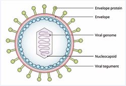
Figure (a) Structure of EBV [6].
The envelope of the virus has glycoproteins, which are important for attachment and entry into the host cells; it mostly infects B lymphocytes and epithelial cells and can cause lots of health problems [1].
The virus encodes several proteins, including latent membrane proteins (LMPs) and nuclear antigens (EBNAs), which play crucial roles in cell transformation and immune evasion [2].
EBV could be primarily transmitted by oral transfusion with saliva through direct contact with an infected person, sharing drinks or food, or sharing personal items [3]. Sexual Contact: EBV DNA has been detected in semen and genital secretions, suggesting that sexual contact may serve as a transmission route beyond salivary exchange [4]. EBV can be transferred via blood transfusions. However, salivary transmission remains the dominant mode [5].
MOLECULAR BIOLOGY OF EBV
Epstein–Barr virus (EBV) primarily infects B lymphocytes, which are essential components of the adaptive immune system. Viral entry into B cells occurs through the interaction between the viral envelope glycoprotein gp350 and the cellular receptor CD21 (complement receptor 2, CR2) on the B-cell surface [7,8]. This interaction facilitates viral attachment and internalization, enabling the viral capsid to enter the host cell and deliver the viral genome to the nucleus [9]. In addition to B lymphocytes, EBV can also infect epithelial cells, particularly those in the oropharyngeal mucosa, which facilitates viral replication and shedding in saliva [4].
A fundamental characteristic of Epstein–Barr virus (EBV) is its capacity to transition between two principal phases: the lytic and latent states. In the lytic phase, the virus actively replicates, generating new infectious particles that enable transmission between hosts [10]. Conversely, after the initial infection, EBV commonly establishes a latent state in which viral replication is downregulated to evade immune detection [11]. During latency, the virus expresses a limited set of molecules, including Epstein–Barr nuclear antigens (EBNAs), latent membrane proteins (LMPs), and EBV-encoded small RNAs (EBERs), which are involved in modulating host cellular signaling and ensuring the persistence of the viral genome as a circular episome within dividing cells [2-12].
EBV latency is categorized into distinct gene expression patterns known as latency programs. Latency I (EBNA1 only) is seen in Burkitt lymphoma; latency II (EBNA1, LMP1, and LMP2) is associated with Hodgkin lymphoma and nasopharyngeal carcinoma; and latency III (full complement of EBNAs and LMPs) is common in immunocompromised individuals [4-8]. These programs reflect the virus’s journey through B-cell differentiation, starting with the growth-promoting latency III in naïve B cells and eventually reaching a quiescent state in memory B cells for lifelong persistence [11-13].
Key latency genes drive these processes: LMP1 acts as a viral oncogene by mimicking a constitutively active CD40 receptor, triggering the NF-κB pathway to promote cell survival and proliferation [14]. EBNA2 serves as a master transcriptional activator, while EBNA1 is essential for tethering and maintaining the viral episome during host cell division [10-15] as shown in Figure (b).
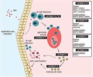
Figure (b) Latency types across the EBV life cycle. Following the entry and infection of host B-cells, EBV undergoes latency III expression, which eventually degrades into latency II expression of its latent genome. As infected cells enter the germinal center and undergo maturation, the EBV genome is suppressed into latency II expression of EBERs, LMPs, and EBNA1, which further devolves into latency I expression as mature B-cells exit the germinal center. Fully mature memory B-cells exhibit a near-quiescent EBV latent genome, where only EBERs are expressed in minimal quantities. Lytic reactivation of the virus enables viral capsid replication and further infection of cells, both lymphocytic and epithelial. [16]
PATHOGENESIS AND ONCOGENIC POTENTIAL
Epstein–Barr virus (EBV) is linked to multiple lymphoproliferative disorders, including Burkitt lymphoma and Hodgkin lymphoma, in which it promotes oncogenesis through mechanisms such as chromosomal abnormalities and impaired immune regulation [17,18]. Furthermore, EBV can undergo reactivation in immunocompromised individuals, contributing to the development of disorders such as post-transplant lymphoproliferative disease (PTLD) [19].
EBV contributes to approximately 200,000 new cancer cases annually worldwide [20]. EBV is linked to several malignancies, including Burkitt lymphoma, Hodgkin lymphoma, nasopharyngeal carcinoma, and gastric carcinoma. The virus is also implicated in autoimmune diseases and neurodegenerative disorders, highlighting its broad impact on human health [2].
Gene sequences of the Epstein–Barr virus have been detected in benign breast tissues 1 to 11 years prior to the development of EBV-positive breast cancer [21], thus providing a substantial causal assessment.
Studies indicate that EBV may increase breast cancer risk through mechanisms such as immune stimulation and the activation of oncogenic signaling pathways. A case control study in Yemen found that 94.7% of breast cancer patients tested positive for EBV IgG, compared to 64.0% of healthy controls. This analysis shows a strong link [22]. A further study indicated that 27.9% of breast cancer cases had reported EBV infections, markedly higher than the 8.02% observed in control tissues [23]. The role of Epstein Barr virus (EBV) in breast cancer remains controversial, as current evidence is inconsistent and does not establish a clear causal relationship. While some studies report the presence of EBV in breast tumors, the wide variation in results suggests that differences in detected methods and populations may influence these findings rather than a true biological effect. In particular, PCR-based techniques may detect viral DNA from surrounding immune cells rather than tumor cells, leading to possible misinterpretation. Therefore, EBV should be considered a potential but unconfirmed factor in breast cancer, and further well designed studies are needed to clarify its role.
CLINICAL IMPLICATIONS
The Epstein-Barr Virus (EBV) has significant clinical implications, affecting a wide range of health conditions from infectious mononucleosis to various malignancies and autoimmune diseases. Understanding these implications is crucial for effective diagnosis and management, particularly in immunocompromised patients. The clinical outcome of EBV infection often depends on the host immune response, age at primary infection, and environmental or genetic factors [8-24].
INFECTIOUS MONONUCLEOSIS AND OTHER CLINICAL MANIFESTATIONS
Infectious mononucleosis is one of the most common clinical outcomes of Epstein–Barr virus (EBV) infection, particularly when primary exposure occurs during adolescence or early adulthood. The condition typically presents with symptoms such as fever, pharyngitis, lymphadenopathy, fatigue, and splenomegaly [25]. In most immunocompetent individuals, it follows a self-limiting course and resolves within a few weeks without the need for specific antiviral therapy. However, some patients may experience prolonged fatigue or develop complications, including hepatitis, neurological manifestations, or splenic rupture, although such events are relatively uncommon. Notably, the clinical manifestations are primarily driven by the host immune response to EBV-infected B lymphocytes rather than by direct viral cytotoxicity.
Beyond systemic manifestations, EBV infection may also contribute to several oral health conditions, particularly in immunocompromised individuals. These include oral hairy leukoplakia and certain periodontal diseases, which are considered important oral indicators of EBV reactivation or immune suppression [26]. In addition, EBV is strongly associated with the development of several malignancies, including Burkitt lymphoma and nasopharyngeal carcinoma, highlighting the virus’s significant oncogenic potential [25-27].
PREVENTION OF EPSTEIN–BARR VIRUS INFECTION
Prevention of Epstein–Barr virus (EBV) infection primarily focuses on minimizing exposure to infected saliva and maintaining practices that support immune health. Since EBV is commonly transmitted through saliva, good personal hygiene such as avoiding the sharing of drinking utensils, toothbrushes, and other personal items, as well as regular hand washing can help reduce the risk of transmission. Limiting close contact with infected individuals, including avoiding kissing or sharing food with those showing symptoms of infectious mononucleosis, is also recommended. Maintaining a healthy immune system through adequate rest, proper hydration, and a balanced diet may further help the body respond effectively to viral exposure. Although EBV transmission occurs mainly through saliva, practicing safe sexual behaviors may also reduce potential exposure. Currently, no licensed vaccine exists for EBV; however, ongoing research aims to develop effective vaccines to reduce the global burden of EBV associated diseases [28].
CONCLUSION
Epstein-Barr virus (EBV) is a highly prevalent human herpesvirus that establishes lifelong latency and can lead to a range of clinical manifestations, from mild infectious mononucleosis to serious malignancies. Early diagnosis, supportive treatment, and preventive strategies such as good hygiene practices and future vaccine development are crucial in managing EBV-related diseases.
REFERENCES
- Houen G, Trier NH. Epstein-Barr Virus and Systemic Autoimmune Diseases. Front Immunol. 2021; 11: 587380.
- Murata T. Epstein-Barr virus: the molecular virology and theassociated diseases. Fujita Med J. 2023; 9: 65-72.
- Castillo A, Giraldo S, Guzmán N, Bravo LE. Factors associated with the presence of the Epstein-Barr virus in the oral cavity of high school students from the city of Cali (Colombia). Enferm Infecc Microbiol Clin (Engl Ed). 2022; 40: 113-120.
- Yu H, Robertson ES. Epstein-Barr Virus History and Pathogenesis. Viruses. 2023; 15: 714.
- Centers for Disease Control and Prevention (CDC): About Epstein- Barr virus (EBV). 2024.
- Cohen JI. Epstein-Barr virus. In D. M. Knipe and P. M. Howley (Eds.), Fields Virology (6th ed., pp. 1898–1959). Lippincott Williams and Wilkins. 2023.
- Smith NA, Coleman CB, Gewurz BE, Rochford R. CD21 (Complement Receptor 2) Is the Receptor for Epstein-Barr Virus Entry into T Cells. J Virol. 2020; 94: e00428-e00520.
- Young LS, Rickinson AB. Epstein-Barr virus: 40 years on. Nat Rev Cancer. 2004; 4: 757-768.
- Wang J, Wang H, Ding Y, Cao N, Nan F, Wu F, et al. Gp350-targeted CAR-T therapy in EBV-positive Burkitt lymphoma: preclinical development of gp350 CAR-T. J Transl Med. 2025; 23: 171.
- Murata T, Sugimoto A, Inagaki T, Yanagi Y, Watanabe T, Sato Y, et al. Molecular Basis of Epstein-Barr Virus Latency Establishment and Lytic Reactivation. Viruses. 2021; 13: 2344.
- Thorley-Lawson DA, Gross A. Persistence of the Epstein-Barr virus and the origins of associated lymphomas. N Engl J Med. 2004; 350: 1328-1337.
- Wang M, Hutt-Fletcher LM, Zeng Y. Epstein–Barr Virusq. Reference Module in Biomedical Sciences. 2018.
- Coleman CB, Lang J, Sweet LA, Smith NA, Freed BM, Pan Z, et al. Epstein- Barr Virus Type 2 Infects T Cells and Induces B Cell Lymphomagenesis in Humanized Mice. J Virol. 2018; 92: e00813-e00818.
- Huang J, Zhang X, Nie X, Zhang X, Wang Y, Huang L, et al. Assembly and activation of EBV latent membrane protein 1. Cell. 2024; 187: 4996-5009.e14.
- Tempera I, Lieberman PM. Epigenetic regulation of EBV persistence and oncogenesis. Semin Cancer Biol. 2014; 26: 22-29.
- Di Pietro A. Epstein-Barr Virus Promotes B Cell Lymphomas by Manipulating the Host Epigenetic Machinery. Cancers (Basel). 2020; 12: 3037.
- Kimura H, Murata T. Understanding the Role of EBV Infection in Lymphomagenesis. 2024; 233-245.
- Bronson AS, Zhu Y, Lilley CM, Crane GM, Mirza KM. Clinicopathologic Insights and Molecular Oncogenesis: Understanding Epstein-Barr Virus-Induced B-cell Lymphoproliferations. Int J Surg Pathol. 2025; 33: 502-515.
- Ali AS, Al-Shraim M, Al-Hakami AM, Jones IM. Epstein- Barr Virus: Clinical and Epidemiological Revisits and Genetic Basis of Oncogenesis. Open Virol J. 2015; 9: 7-28.
- Sung H, Ferlay J, Siegel RL, Laversanne M, Soerjomataram I, Jemal A, etal. Global Cancer Statistics 2020: GLOBOCAN Estimates of Incidence and Mortality Worldwide for 36 Cancers in 185 Countries. CA Cancer J Clin. 2021; 71: 209-249.
- Lawson JS, Glenn WK. Multiple oncogenic viruses are present in human breast tissues before development of virus associated breast cancer. Infect Agent Cancer. 2017; 12: 55.
- Al-Shiabani RK, Al-Jaufy A, Al-Moyed KA, Al-Shamahy HA. Correlation of epstein-barr virus with breast cancer: a case control study. Universal J Pharmaceutical Res. 2024; 9: 75-81.
- Agolli A, Ishak A, Viswanathan M, Co EL, Shivakumar J, Agolli O. Epstein-Barr Viral Infection and the Risk for Breast Cancer: A Systematic Review. Int J Hematol Oncol Stem Cell Res. 2023; 17: 114- 124.
- Cohen JI. Epstein-Barr virus infection. N Engl J Med. 2000; 343: 481-492.
- Jenson HB. Epstein-Barr virus. Pediatr Rev. 2011; 32: 375-83.
- Sriram S, Jayakanth M, Buch SA, Alam T, Hasan S. Epstein–Barr Virus and Oral Health: Clinical Manifestations, Diagnostic Insights, and Implications for Dental Practice. 2025051645. 2025.
- Gärtner BC. Epstein-Barr Virus. Manual of Clinical Microbiol. 2023; 1-22.
- Shannon-Lowe C, Rickinson AB, Bell AI. Epstein-Barr virus-associated lymphomas. Philos Trans R Soc Lond B Biol Sci. 2017; 372: 20160271.