Erythema Nodosum Associated with Idiopathic Granulomatous Mastitis in a Pregnant Woman: A Case Report
- 1. Department of Dermatology-venerology, Ibn Rochd University Hospital, School of Medicine and Pharmacy, Aïn Chok University, Morocco
- 2. Department of Obstetrics and Gynaecology C, Ibn Rochd University Hospital, School of Medicine and Pharmacy, Aïn Chok University, Morocco
Abstract
A 35-year-old woman, gravida 5 para 4, at 20 weeks gestation, presented with fever, painful breast mass, painful nodular lesions on both lower limbs, and inflammatory arthralgias evolving for 2 months. No history of tuberculosis, sarcoidosis, breast trauma, or family breast disease was reported. Examination revealed multiple tender erythematous subcutaneous nodules on the legs and a large, irregular, infiltrated left breast mass without nipple discharge or retraction. Laboratory tests showed elevated C-reactive protein, normal blood count and negative autoimmune markers. Breast ultrasonography indicated mastitis (BIRADS 3), and core needle biopsy demonstrated non-caseating epithelioid and giant-cell granulomas with inflammatory infiltrate, without infection or malignancy. GeneXpert for Mycobacterium tuberculosis was negative. The diagnosis of idiopathic granulomatous mastitis associated with erythema nodosum and arthralgias was established. Cutaneous lesions improved rapidly after short-course oral corticosteroids, and a clinical follow-up strategy for the breast mass was adopted.
Keywords
• Corticosteroids
• Erythema nodosum
• Idiopathic granulomatous mastitis
• Pregnancy
• Pseudotumoral
• lesion
Citation
Essekakri K, Baghad B, Bensouda M, Chiheb S (2026) Erythema Nodosum Associated with Idiopathic Granulomatous Mastitis in a Pregnant Woman: A Case Report. J Dermatolog Clin Res 14(1): 1171.
INTRODUCTION
Idiopathic granulomatous mastitis (IGM) is a rare inflammatory breast disorder, representing 0.5 to 1.8% of Breast disorders [1,2]. It predominantly affects young women of reproductive age and often presents with misleading clinical and radiologic features, mimicking deep infection or neoplastic processes [3,4]. The diagnosis, which is one of exclusion, relies on a comprehensive work up and histopathological examination, demonstrating epithelioid and multinucleated giant-cell granulomas without any identifiable infectious agent [5].
Although its pathogenesis remains incompletely understood, several findings suggest the involvement of autoimmune or autoinflammatory mechanisms [2,3]. Supporting this theory, various systemic manifestations have been reported, including inflammatory arthritis, episcleritis, hidradenitis suppurativa, and, more rarely, erythema nodosum [6-9]. The association between IGM and erythema nodosum remains exceptional, with fewer than thirty cases documented in the literature [6-9].
IGM occurring in pregnant patients remains exceptionally rare, with only a few isolated cases reported in the literature. The development of IGM during pregnancy adds significant diagnostic and therapeutic complexity [10-12].
We report here the case of a pregnant woman presenting with IGM associated with erythema nodosum and inflammatory arthralgias.
CASE PRESENTATION
A 35-year-old woman, gravida 5 para 4, at 20 weeks gestation, under regular obstetric follow-up, no history of tuberculosis, sarcoidosis, breast trauma, or family breast disease was reported. She was referred by her obstetrician to the dermatology department For a non pruritic painful breast mass associated with a febrile cutaneous eruption and arthralgias evolving for 2 months. On physical examination, the patient was febrile at 38.8°C and presented with painful inflammatory subcutaneous nodules on both lower limbs, consistent with erythema nodosum (Figure 1).
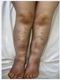
Figure 1: Inflammatory subcutaneous nodules on lower limbs
The rest of the examination revealed a large irregular mass located in the upper outer quadrant of the left breast, with inflammatory skin overlying the lesion without areolar involvement or skin retraction, measuring approximately 10 cm (Figure 2).
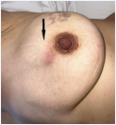
Figure 2: Left mastitic mass
On musculoskeletal examination, the patient exhibited arthralgias, consistent with an inflammatory pattern. No lymphadenopathy was noted.
An initial radiologic work-up of the breast mass was performed. Breast ultrasonography demonstrated features consistent with left-sided mastitis, classified as BIRADS 3 according to the American College of Radiology. Core needle biopsy revealed breast tissue disrupted by polymorphous granulation tissue consisting of lymphocytes and plasma cells, with sheets of foamy histiocytes and non-caseating epithelioid granulomas containing giant cells. Lymphoid follicles and small foci of suppuration were also noted. No pathogenic organism or tumor proliferation was identified. GeneXpert testing for Mycobacterium tuberculosis DNA was negative.
A work-up for erythema nodosum was performed, showing elevated C-reactive protein. Other investigations including complete blood count, serum electrolytes with calcium levels, liver and renal function tests, antistreptolysin O were normal. Immunological investigations such as antinuclear antibodies, anti-double stranded DNA antibodies, and angiotensin-converting enzyme levels, were within normal limits.
A skin biopsy was performed on the lower-limb lesions, revealing inflammatory changes predominantly involving the septal component of the sub cutis. The infiltrate was composed of lymphocytes and neutrophils, with some eosinophils and occasional giant cells, without evidence of tuberculoid or sarcoid-type granulomas. No coagulative or fibrinoid necrosis was identified, nor any features of vasculitis. The adipose lobules were infiltrated peripherally, with preservation of the lobular architecture.
The diagnosis of idiopathic granulomatous mastitis associated with erythema nodosum and inflammatory arthralgias was established. The erythema nodosum lesions improved rapidly following initiation of short course oral corticosteroid therapy at 1 mg/kg/day, alongside with vitamine C and strict bed rest. Regarding IGM, A slight improvement was noted after the short course of systemic corticosteroids, after which the patient was initiated on topical corticosteroids with close clinical follow-up.
DISCUSSION
Our case illustrates a rare association between Idiopathic Granulomatous Mastitis (IGM) and erythema nodosum during pregnancy, with fewer than thirty cases reported since the first description by Adams in 1987 [1]. IGM, already considered a rare cause of nonpuerperal inflammatory mastopathy [2], typically affects young women of childbearing age, including pregnant patients [3]. Pregnancy, due to the profound hormonal and immunologic changes, may represent a physiopathologic context conducive to the emergence or exacerbation of this condition [4].
Erythema Nodosum (EN) is a panniculitis which may have various causes, such as sarcoidosis, tuberculosis, infections or drugs, or it can be idiopathic [5]. It is a rare systemic manifestation of IGM [6,7].
IGM, clinically, most often presents as a painful pseudotumoral mass, sometimes suppurative, mimicking breast carcinoma and necessitating histopathologic evaluation [8]. Histology typically reveals non-caseating epithelioid and giant-cell granulomatous inflammation centered on the lobules, without caseous necrosis or detectable microorganisms [9]. The diagnosis remains a diagnosis of exclusion, requiring ruling out infectious, neoplastic or systemic causes of granulomatous mastitis, such as mycobacterial infection, sarcoidosis, or vasculitis [10], obtaining core specimens by true-cut biopsy for pathological study is paramount.
Extramammary manifestations are uncommon but support a possible autoimmune or autoinflammatory mechanism [11]. Erythema nodosum, observed in our pregnant patient, is considered one of these unusual extramammary manifestations. The pathogenesis of this uncommon lesion remains obscure. Several hypotheses have been put forward, such as pregnancy, breastfeeding, contraceptive pills, smoking, α1-antitrypsin deficiency and hyperprolactinemia [12-18].
No consensus therapeutic guidelines currently exist for IGM. Systemic corticosteroids remain the first-line treatment, although refractory forms have been reported [19]. Surgery most should be approached cautiously due to the risk of cosmetic sequelae and recurrence [20]. Among alternative treatments for resistant cases, TNF-α inhibitors have shown promising results, though evidence remains limited.
CONCLUSIONS
In conclusion, our case underscores that the occurrence of erythema nodosum in a pregnant woman should raise suspicion for IGM among other possible etiologies. This rare but meaningful association should alert clinicians, as it may guide the diagnostic process toward a challenging condition requiring histologic confirmation and tailored management.
REFERENCES
- Jacquin-Porretaz C, Devalland C, Delapparent T, Nardin C, Dupond AS. Mastite granulomateuse idiopathique associée à un érythème noueux [Idiopathic granulomatous mastitis associated with erythema nodosum]. Ann Dermatol Venereol. 2019; 146: 571-576.
- Kessler E, Wolloch Y. Granulomatous mastitis: a lesion clinically simulating carcinoma. Am J Clin Pathol. 1972; 58: 642-646.
- Baslaim MM, Khayat HA, Al-Amoudi SA. Idiopathic granulomatous mastitis: a heterogeneous disease with variable clinical presentation. World J Surg. 2007; 31: 1677-1681.
- Azmat S, Heller DS. Reproductive hormonal influences in granulomatous mastitis. Breast J. 2017; 23: 606-607.
- Koufakis T, Gabranis I. Erythema nodosum leads to the diagnosis of pulmonary tuberculosis. Pan Afr Med J. 2014; 18: 291.
- Adams DH, Hubscher SG, Scott DG. Granulomatous mastitis--a rare cause of erythema nodosum. Postgrad Med J. 1987; 63: 581-582.
- Nakamura T, Yoshioka K, Miyashita T, Ikeda K, Ogawa Y, Inoue T, et al. Granulomatous mastitis complicated by arthralgia and erythema nodosum successfully treated with prednisolone and methotrexate. Intern Med. 2012; 51: 2957-2960.
- Krawczyk N, Kühn T, Ditsch N, et al. Idiopathic granulomatous mastitis: a review of current literature.1. Arch Gynecol Obstet. 2016; 293: 1-7.
- Yin Y, Liu X, Meng Q, Han X, Zhang H, Lv Y. Idiopathic Granulomatous Mastitis: Etiology, Clinical Manifestation, Diagnosis and Treatment. J Invest Surg. 2022; 35: 709-720.
- Bani-Hani KE, Yaghan RJ, Matalka II, Shatnawi NJ. Idiopathic granulomatous mastitis: time to avoid unnecessary mastectomies. Breast J. 2004; 10: 318-322.
- Hamsho S, Alaswad M, Alsmodi H, Sleiay M, Hoha G, Alcheikh S. Idiopathic granulomatous mastitis with extramammary manifestations: a case report. Ann Med Surg (Lond). 2023; 85: 6192-6195.
- Altintoprak F, Kivilcim T, Ozkan OV. Aetiology of idiopathic granulomatous mastitis. World J Clin Cases. 2014; 2: 852-858.
- Benson JR, Dumitru D. Idiopathic granulomatous mastitis: presentation, investigation and management. Future Oncol. 2016; 12: 1381-1394.
- Altintoprak F, Karakece E, Kivilcim T, Dikicier E, Cakmak G, Celebi F, et al. Idiopathic granulomatous mastitis: an autoimmune disease? Scientific World J. 2013; 2013: 148727.
- Sheybani F, Naderi HR, Gharib M, Sarvghad M, Mirfeizi Z. Idiopathic granulomatous mastitis: Long-discussed but yet-to-be-known. Autoimmunity. 2016; 49: 236-239.
- Ozel L, Unal A, Unal E, Kara M, Erdo?du E, Krand O, et al. Granulomatous mastitis: is it an autoimmune disease? Diagnostic and therapeutic dilemmas. Surg Today. 2012; 42: 729-733.
- Lin CH, Hsu CW, Tsao TY, Chou J. Idiopathic granulomatous mastitis associated with risperidone-induced hyperprolactinemia. Diagn Pathol. 2012; 7: 2.
- Jeon J, Lee K, Kim Y, Chun YS, Park HK. Retrospective analysis of idiopathic granulomatous mastitis: its diagnosis and treatment. J Breast Dis. 2017; 5: 82-88.
- Schmajuk G, Genovese MC. Treatment of granulomatous mastitis with corticosteroids. Semin Arthritis Rheum. 2009; 39: 146-150.
- Seo HR, Na KY, Yim HE. Surgical outcomes in granulomatous mastitis. Breast. 2012; 21: 365-369.