Thoracic Endovascular Aortic Repair after Amplatzer Failure in a Patient with an Aberrant Right Subclavian Artery: A Case Report
- 1. Department of Neurosurgery, Harvard Medical School, USA
- 2. Virgin Research Institute, México
- 3. Universidad Autonoma de Guadalajara, Mexico
Abstract
Aberrant right subclavian artery (arteria lusoria) is an uncommon aortic arch anomaly that may cause dysphagia due to extrinsic esophageal compression. Management can be especially challenging in pediatric patients, in whom no well-established standard of care exists. We report the case of a 13-year old boy with a 6-month history of progressive dysphagia and weight loss. Computed tomography angiography demonstrated an aberrant right subclavian artery associated with a 9.61 mm Kommerell diverticulum. A staged hybrid approach was undertaken. The first stage consisted of a right common carotid subclavian bypass through a supraclavicular approach using a ringed expanded polytetrafluoroethylene graft. Endovascular exclusion was then attempted with an Amplatzer AVP II device; however, proximal deployment failed because of inadequate anchoring related to the Kommerell diverticulum and worsening esophageal compression. The device was repositioned distally, and thoracic endovascular aortic repair was subsequently performed to achieve definitive exclusion while avoiding sternotomy. The postoperative course was uncomplicated, with complete resolution of dysphagia within the first postoperative week. At one, three, six, and 12 months, the patient remained asymptomatic, with weight gain, no neurologic or vascular complications, and imaging confirmation of complete exclusion, graft patency, and absence of endoleak. This case supports the technical feasibility of a staged hybrid repair with TEVAR after unsuccessful plug exclusion in a selected pediatric patient.
Keywords
• Aberrant Right Subclavian Artery
• Kommerell Diverticulum
• Dysphagia Lusoria
• Amplatzer
• TEVAR
Citation
Hernandez DA, Zuloaga A, Avila R, Lopez JA, Siordia RE, et al. (2026) Thoracic Endovascular Aortic Repair after Amplatzer Failure in a Patient with an Aberrant Right Subclavian Artery: A Case Report. Ann Cardiovasc Dis 10(2): 1051.
INTRODUCTION
Aortic arch anomalies result from embryologic defects of the branchial arch system and are identified in up to 19% of the population [1]. In normal anatomy, the right subclavian artery arises from the brachiocephalic trunk; however, in patients with an aberrant right subclavian artery (ARSA), the subclavian artery originates as the final branch of the aortic arch [2]. ARSA can be associated with a Kommerell diverticulum, defined as a diverticular or aneurysmal dilation at the origin of the aberrant vessel, which has been reported in up to 60% of affected cases [3]. Although most patients are asymptomatic, some may develop dysphagia secondary to extrinsic esophageal compression, referred to as dysphagia lusoria [4]. Symptomatic arteria lusoria is uncommon, but when present, treatment is necessary [5]. Its management can be challenging and often requires a multidisciplinary approach. In the absence of a well-established standard of care, particularly in children, our main objective is to report our experience with a distinctive two-stage therapeutic approach.
CASE REPORT
A 13-year-old male presented to our clinic with a 6-month history of progressive, previously undiagnosed dysphagia and weight loss. The dysphagia initially affected solids and later gradually progressed to liquids. He did not report chest pain or dyspnea. He had no significant past medical history and was not taking any medications. His family history was unremarkable. His psychosocial history was notable for anxiety and stress related to his inability to eat and drink properly. He had not undergone any prior interventions before receiving care from our team. The patient failed the bedside swallow test, and further gastrointestinal workup was performed. Our main clinical suspicion was the presence of either an intrinsic or extrinsic esophageal obstruction. Gastrointestinal studies were unremarkable. Computed tomography angiography (CTA) demonstrated an aberrant right subclavian artery associated with a 9.61 mm Kommerell diverticulum (Figures 1 and 2).
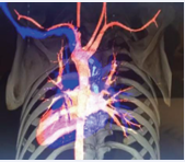
Figure 1: CT angiography showing an aberrant right subclavian artery at the distal portion of the aortic arch.
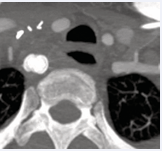
Figure 2: CT scan showing dysphagia lusoria secondary to esophageal compression.
Based on the CTA findings, a diagnosis of arteria lusoria was made, and a two-stage therapeutic intervention was proposed. The first stage involved surgical intervention, followed by endovascular treatment. A supraclavicular approach was performed through a 4 cm supraclavicular incision. Upon surgical exploration and deep neck dissection, the right subclavian artery was identified and appeared significantly dilated and tortuous. The right carotid artery appeared normal. A lateral-to-lateral bypass was created between the right common carotid artery and the right subclavian artery using a ringed expanded polytetrafluoroethylene (ePTFE) graft measuring 8 mm in diameter and 4 cm in length (W. L. Gore) in order to preserve perfusion to the right upper extremity and the subclavian artery territory (Figure 3).
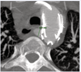
Figure 3: CT scan showing dysphagia lusoria secondary to esophageal compression.
Ligation of the vessel at its distal end was considered; however, the vessel was left open with the intention of introducing an Amplatzer AVP II device through the orifice. In addition, transection of the vessel was not feasible from this surgical approach. In order to avoid sternotomy, it was decided to attempt endovascular closure using an 18 mm Amplatzer AVP II device (Abbott Cardiovascular). Proximal deployment of the Amplatzer device was unsuccessful because of the presence of the Kommerell diverticulum (Figure 4).
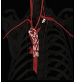
Figure 4: Arteriography showing a carotid-subclavian graft, Amplatzer device, and aortic stent graft.
Due to insufficient anchoring of the device and further exacerbation of the esophageal compression by the device itself, it was decided to place the device more distally, just proximal to the bypass, in order to close the distal orifice of the vessel. Subsequently, thoracic endovascular repair had to be performed through surgical exposure of the right femoral artery. An aortic endograft (Zenith Alpha 26 × 105 mm, 16 Fr) was advanced and deployed just distal to the left subclavian artery (Figure 5). The risk of endoleak was carefully considered before the procedure, and the decision to proceed was made based on the anticipated overall benefit to the patient and the avoidance of sternotomy. No endoleak was detected after deployment of the aortic endograft. After deployment, we confirmed the absence of leakage from the right subclavian artery and patency of the left subclavian artery.
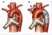
Figure 5: Schematic representation of the anatomy and staged hybrid repair. (A) Aberrant right subclavian artery arising from a Kommerell diverticulum and coursing posterior to the esophagus. (B) Postoperative anatomy after right common carotid—right subclavian bypass, distal occlusion of the aberrant right subclavian artery, and TEVAR, with exclusion of the aberrant artery origin and preservation of right upper-extremity perfusion. By, right common carotid-right subclavian bypass; O, occlusion site; T, thoracic endograft.
Follow-up and Outcomes
The immediate postoperative course was uncomplicated. The patient was admitted to the ICU for close hemodynamic monitoring. Complete resolution of dysphagia was achieved within the first postoperative week. At one, three, six, and 12-month follow-up, the patient remained asymptomatic, without neurological deficits or vascular compromise. Weight gain and return to normal daily activities were documented. CTA performed at three and 12 months confirmed complete exclusion of the aberrant right subclavian artery and continued patency of the carotid-subclavian bypass graft, absence of endoleak, and resolution of the previously observed esophageal compression.
DISCUSSION
Strengths and Weaknesses
In this case report, we described a severe presentation of dysphagia lusoria. The uniqueness of this case resided in the multidisciplinary treatment strategy, in which surgical and endovascular techniques were used not as competing alternatives but as complementary approaches for the patient’s benefit. A major strength of this report was the emphasis on maintaining a broad differential diagnosis when evaluating a pediatric patient with dysphagia and associated weight loss [6]. In addition, this case underscored the clinical relevance of Kommerell diverticulum in patients with arteria lusoria, particularly its potential to interfere with Amplatzer device anchoring, and highlighted the role of TEVAR, an approach increasingly utilized in pediatric thoracic aortic pathology [7]. Moreover, because an aberrant right subclavian artery rarely requires intervention in pediatric patients, this case is notable in that it documents surgical treatment in a child. The principal limitation, as with all case reports, was the limited level of evidence inherent to this study design, which precludes formal treatment recommendations. Rather, this report should be interpreted as a hypothesis generating contribution to the literature.
Comparison with Current Literature
Current literature describes open, endovascular, and hybrid approaches for symptomatic ARSA with Kommerell diverticulum, with treatment selection determined by patient characteristics and anatomy [8]. In our patient, the combination of surgical revascularization and endovascular exclusion allowed preservation of perfusion while avoiding sternotomy; however, TEVAR in this pediatric setting should be regarded as an individualized rescue strategy rather than an established standard of care.
The application of TEVAR in pediatric patients raises unique concerns regarding long-term endograft durability in the setting of continued somatic and aortic growth. Available pediatric experience is limited and derived mostly from adolescents with blunt thoracic aortic injury (BTAI) [9,10]. In the abscence of aortic coarctation or BTAI , the utility of TEVAR is poorly described in literature with evidence of its utility coming mainly from case reports similar at the one described aboved. Byrne et al. reported the first percutaneous covered-stent exclusion of a descending thoracic aortic aneurysm in a child with tuberous sclerosis [11]. No previously published pediatric case report exists with the use of TEVAR for the management of an aberrant right subclavian artery. Reported experience with this condition has predominantly involved open resection with subclavian transposition [8].
Furthermore, the long-term impact of continued aortic growth on a fixed-diameter endograft remains incompletely characterized. Somatic growth-induced endograft migration has not been clearly described in the pediatric literature, only current available evidence demonstrates no stent migration over 8–15 years follow up [9,10]. Nevertheless progressive aorta-graft mismatch manifests over time as relative stenosis, pseudocoarctation, loss of seal, or endoleak, but the incidence and clinical significance of these complications during continued aortic growth remain unknown. Accordingly, the absence of endoleak or graft-related complications at 12 months in our 13-year old patient represents a favorable early clinical and imaging outcome but cannot establish lifelong durability. Given his remaining somatic growth and the paucity of pediatric experience with this anatomy, long-term imaging surveillance into adulthood will be performedl to monitor aortic growth, endograft position and seal, and any future need for reintervention.
Take-Away Lessons
The management of an aberrant right subclavian artery with Kommerell diverticulum requires individualized planning based on anatomy, symptoms, and patient age. A Kommerell diverticulum may prevent stable plug fixation, as demonstrated in this case, and alternative exclusion strategies should be anticipated when endovascular occlusion is planned. TEVAR may provide a rescue strategy in selected patients when conventional exclusion is unsuccessful; however, its long-term durability in children with remaining aortic growth remains unknown and requires prolonged surveillance.
CONCLUSION
This case demonstrates the technical feasibility of a staged hybrid approach for symptomatic arteria lusoria with Kommerell diverticulum after unsuccessful plug exclusion. The presence of a Kommerell diverticulum may compromise Amplatzer device deployment, and TEVAR may serve as a rescue strategy to achieve definitive closure while avoiding open thoracic reconstruction. However, long-term endograft durability in children with remaining aortic growth is unknown, underscoring the need for long term surveillance.
Informed Consent
Yes — written informed consent for publication of this case and accompanying images was obtained.
ACKNOWLEDGEMENTS
The authors wish to thank the nurses involved in the care of the patient and the department of radiology of Hospital Mexico Americano for providing the diagnostic images. We also acknowledge our medical school faculty for providing resources that supported the literature review and preparation of this manuscript.
Declaration of Conflicts of Interest
The authors declare that they do not have conflicts of interest.
Funding: No funding was received for this work.
Ethical Approval
Ethical approval was not required for this case report in accordance with the policies of Virgin Research Institute
Informed Consent
Written informed consent was obtained by the parents of the patient.
Data Accessibility Statement
Data sharing is not applicable to this case report, since the only available data for this manuscript is the medical record, which is confidential.
REFERENCES
- Popieluszko P, Henry BM, Sanna B, Hsieh WC, Saganiak K, P?kala PA, et al. A systematic review and meta-analysis of variations in branching patterns of the adult aortic arch. J Vasc Surg. 2018; 68: 298-306.e10.
- Giuliani L, Di Toro A, Urtis M, Narula N, Grasso M, Pelenghi S, et al. Prevalence and Complications of Aberrant Subclavian Artery in Patients With Heritable and Nonheritable Arteriopathies. J Am Coll Cardiol. 2023; 81: 979-991.
- Isselbacher EM, Preventza O, Hamilton Black J 3rd, Augoustides JG, Beck AW, Bolen MA, et al. 2022 ACC/AHA Guideline for the Diagnosis and Management of Aortic Disease: A Report of the American Heart Association/American College of Cardiology Joint Committee on Clinical Practice Guidelines. Circulation. 2022; 146: e334-e482.
- Janssen M, Baggen MG, Veen HF, Smout AJ, Bekkers JA, Jonkman JG, et al. Dysphagia lusoria: clinical aspects, manometric findings, diagnosis, and therapy. Am J Gastroenterol. 2000; 95: 1411-1416.
- Dieffenbach BV, Sharma G, Shah SK, Menard MT, Belkin M. Aberrant subclavian artery division and revascularization by a supraclavicular approach for definitive or staged treatment of dysphagia lusoria. J Vasc Surg. 2020; 72: 219-225.
- Hamersley ERS, Baldassari C. Pediatric esophageal dysphagia. Otolaryngol Clin North Am 2024; 57: 581–587.
- Grubbs H, Snyder KB, Stewart K, Cross A, Landmann A, Johnson J, et al. Pediatric blunt thoracic aortic injuries: Understanding the role of patient size and utilization of thoracic endovascular aortic repair. J Trauma Acute Care Surg. 2025; 99: 412-417.
- Bath J, D’Oria M, Rogers RT, Colglazier JJ, Braet DJ, Coleman DM, Scali ST, et al. Contemporary outcomes after treatment of aberrant subclavian artery and Kommerell’s diverticulum. J Vasc Surg. 2023; 77: 1339-1348.e6.
- Aimanan K, Ahmad A, Arvind M, Pian PM, Pillay KV, Hussein H. Long-Term Durability of TEVAR for Blunt Traumatic Aortic Injury in Young Patients. Ann Vasc Surg. 2026; 131: 35-43.
- Gennai S, Leone N, Mezzetto L, Veraldi GF, Santi D, Spaggiari G, et al. Systematic review and meta-analysis of long-term reintervention following thoracic endovascular repair for blunt traumatic aortic injury. J Vasc Surg. 2023; 78: 540-547.e4.
- Byrne RD, Lahiri S, Bansal M, Jacob B, Stapleton G. Endovascular Repair of a Descending Thoracic Aortic Aneurysm in a Pediatric Patient with Tuberous Sclerosis: A Case Report and Review of the Literature. Pediatr Cardiol. 2022; 43: 238-243.