A Novel GATA3 Exon 2 Deletion in a Pediatric Patient with Hypoparathyroidism: Expanding the Genotypic Spectrum of Barakat Syndrome
- 1. The University of Arizona College of Medicine-Phoenix, USA
- 2. Phoenix Children’s Hospital, USA
Abstract
Objective: Barakat syndrome, also known as HDR syndrome, is a rare disorder caused by pathogenic variants in GATA3 characterized by hypoparathyroidism, sensorineural deafness, and renal anomalies. This case highlights a previously unreported GATA3 variant in a pediatric patient and underscores the diagnostic value of repeat genetic testing when clinical suspicion remains high.
Case Presentation: An 11-year-old African American male presented with seizures due to severe hypocalcemia from hypoparathyroidism. Initial genetic testing was non-diagnostic. The patient was treated with calcium and calcitriol and discharged with outpatient endocrine follow-up. After a two-year lapse in care, he re-presented with recurrent hypocalcemia. Repeat testing using an expanded next-generation sequencing panel identified a novel pathogenic deletion encompassing exon 2 of the GATA3 gene, consistent with Barakat syndrome. Subsequent workup revealed mild bilateral sensorineural hearing loss and a simple renal cyst but normal renal function. The patient was re-established with endocrinology, nephrology, and audiology for long-term multidisciplinary management.
Conclusion: This case adds to the expanding genotypic spectrum of GATA3-related HDR syndrome and illustrates the importance of updated genetic testing in cases with a suggestive clinical phenotype but initially negative results. Timely diagnosis facilitated targeted subspecialty care and avoided unnecessary investigations, emphasizing the need for ongoing evaluation in evolving genetic landscapes.
Keywords
- Barakat syndrome
- HDR syndrome
- GATA3
- Hypoparathyroidism
- Hypocalcemia
- Genetic testing
Citation
Varda N, Grebe T, Chawla R (2025) A Novel GATA3 Exon 2 Deletion in a Pediatric Patient with Hypoparathyroidism: Expanding the Geno typic Spectrum of Barakat Syndrome. Pediatr Child Health 13(3): 1359.
INTRODUCTION
Barakat syndrome, also known as HDR syndrome for its triad of classic symptoms: hypoparathyroidism, deafness, and renal disease, is a rare disorder first described in 1977 by Barakat et al. Two male siblings, both with nephrotic syndrome, sensorineural deafness, and hypoparathyroidism passed away before the age of 10 from renal failure. On autopsy, the parathyroid glands were either absent or hypoplastic [1]. While the exact prevalence is unknown, Barakat is a rare syndrome with an estimated 200 reported cases in literature worldwide. Patient presentation can vary considerably. Deafness is the most consistently reported symptom with 96% of patients experiencing early-onset hearing loss that can range from moderate to severe and is typically bilateral. Hypoparathyroidism has been reported to occur in 93% of patients. Hypocalcemia, tetany, afebrile seizures, and neuromuscular irritability are all sequala of hypoparathyroidism. Renal disease occurs in 72% of reported cases; manifestations include dysplastic kidneys, vesicoureteral reflux, hematuria, nephrotic syndrome, chronic kidney disease, and pelvicalyceal deformity. The likelihood of symptom occurrence increases with age, and while it is estimated that only about 54% [2], of patients present with the triad of symptoms, most patients will have developed the three pathognomonic features into adulthood. Additional features include electrolyte abnormalities, congenital heart disease, growth failure, cognitive disability, and facial and ocular abnormalities [3,4].Barakat Syndrome is an autosomal dominant disorder caused by pathogenic variants in GATA3 gene on chromosome 10p14, or deletions in chromosome 10p14 that encompass GATA3. GATA3 encodes a protein belongs to a family of dual zinc-finger transcription factors which serves an important regulator of T-cell development and endothelial cell biology. Notably it is involved in the embryologic development of the parathyroid glands,auditory system, kidneys, thymus, and central nervous system. Heterozygous GATA3 pathogenic variants lead to an absent or reduced DNA-binding affinity, causing developmental abnormalities [5].
CASE PRESENTATION
An 11-year-old African American male presented to the emergency department in November 2020 following an episode of seizure-like activity, which was accompanied by urinary incontinence, altered mental status, and ocular deviations. Initial laboratory tests in the emergency department revealed severe hypocalcemia, which was determined to be secondary to hypoparathyroidism. His calcium level was critically low at 5.1 mg/dL (8.9-10.4), with an intact parathyroid hormone (PTH) of 14 pg/mL (15-65), normal magnesium levels, and an elevated phosphorus level of 8.8 mg/dL (3.4-5.5). Additionally, he was found to be vitamin D deficient, with a 25-hydroxyvitamin D level of 11.2 ng/mL (30-80). He was born full term with no prior history of hypocalcemia. He experienced a febrile seizure at age 2. The patient denied any history of developmental delay, opportunistic infections, or hearing loss. Growth chart showed height was at 31st percentile, weight was at 71st percentile. Family history was negative for any known calcium or adrenal disorders, though the father was adopted and reported some subjective hearing loss in mother.On physical examination, no dysmorphic features were noted apart from slight hypertelorism. Exam was notable for a negative Chvostek sign. He was given an IV calcium gluconate bolus and was then admitted to the pediatric intensive care unit (PICU) where treatment with an intravenous calcium gluconate drip was initiated. Further workup revealed that the patient was euthyroid with a TSH of 1.74 uIU/mL (0.67–4.16) and a free T4 of 1.24 ng/ dL (0.86 – 1.40). Adrenal function was intact with a robust AM cortisol of 20.4 ug/dL (>10 is considered sufficent), and an ACTH of 24.3 pg/mL (7.2-63.3). Additionally, 21-hydroxylase antibodies were negative. Physical examination findings were consistent with Tanner stage 3 pubertal development. During his hospitalization he was started on enteral calcium and calcitriol and weaned of his IV calcium drip. The patient was discharged on day 5 of hospitalization with a calcium level of 9.2 mg/dL (8.9-10.4) and an ionized calcium level of 5.15 mg/dL (4.6-5.17). He was discharged home on calcium carbonate 2000mg every 6 hours=75mg/kg/day of elemental calcium, calcitriol 1 mcg BID, and Vit D 50,000 IU/weekly x 6 weeks. During his hospitalization, a chromosomal microarray (CMA) was sent that was normal, ruling out 22q11.2 Deletion Syndrome (historically referred to as DiGeorge Syndrome) as a cause of hypocalcemia. Over the next several months, he was regularly followed by endocrinology as an outpatient for calcium monitoring and adjustments to his home medications. Goal was to keep calcium in the low normal range and minimize hypercalciuria and development of nephrocalcinosis. Further genetic testing was completed as an outpatient. Invitae hypoparathyroidism panel was completed and was negative for pathogenic variants in AP251, CASR, CDC73, CDKN1B, GNA11, MEN1, and RET.The patient was lost to follow-up between April 2021 and April 2023, when he returned with a calcium level of 6.6 mg/dL (8.9-10.4). His medications were reinitiated, and after counseling on the importance of adherence, his calcium levels increased to 10.4 mg/dL (8.9-10.4) Invitae hypoparathyroidism panel was repeated as it now included testing for AIRE, CDH7, CYP24A1, FAM111A, GATA3, GCM2, GNAS, HADHA, HADHB, PDE4D, PRKAR1A, PTH, STX16, TBCE, TBX1 (Table 1),for specific disease associations. The more extensive genetic panel revealed a pathogenic GATA3 variant, specifically a deletion of exon 2, consistent with Barakat syndrome-a previously unreported finding. Consequently, the patient was referred to nephrology and audiology for further evaluation, as Barakat syndrome is associated with renal dysfunction and sensorineural hearing loss. Renal ultrasound showed a simple, benign cyst in the upper pole of the right kidney measuring 1.5 x 1.6 x 1.6 cm, with no evidence of septa, internal debris, or calcifications. The right kidney measured 9.4 cm, and the left kidney was 8.2 cm, both with normal anatomy and no congenital abnormalities in the kidneys or urinary tract. The patient had no hematuria, urinalysis was unremarkable for nephrotic syndrome, and creatinine levels were normal. A follow-up renal ultrasound in January 2024 confirmed that the cyst has remained stable. Audiological testing revealed mild sloping to moderate sensorineural hearing loss in the right ear, with slight to mild loss in the left ear. The patient continues to be actively,
Table 1: Genes sequenced in the Invitae hypoparathyroidism panel and conditions associated with aberrant calcium metabolism.
|
Gene |
Disorder(s) |
|
CASR |
Familial Hypocalciuric Hypercalcemia (FHH), Neonatal Severe Hyperparathyroidism |
|
GATA3 |
Barakat (HDR) Syndrome |
|
GCM2 |
Primary Hyperparathyroidism |
|
MEN1 |
Multiple Endocrine Neoplasia Type 1 |
|
RET |
Multiple Endocrine Neoplasia Type 2 |
|
AIRE (APS1) |
Autoimmune Polyendocrinopathy |
|
TBCE |
Hypoparathyroidism–Retardation–Dysmorphism (HRD), Kenny–Caffey Syndrome |
Caption: Additional genes that were investigated as part of the diagnostic panel include AP251, CDC73, CDKN1B, FAM111A, GNA11, GNAS, HADHA, HADHB, PDE4D, PRKAR1A, PTH, STX16, and TBX1. These genes are less frequently involved in parathyroid disorders but may contribute to their pathophysiology in certain cases.
followed by endocrinology, audiology, and nephrology. His calcium levels have remained stable with ongoing treatment, including calcitriol (0.5 mcg twice a day), calcium carbonate (2000 mg three times a day) = 40mg/ kg/day of elemental calcium, and cholecalciferol (1000 IU daily).
DISCUSSION
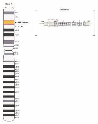
Barakat, or HDR syndrome, is a rare congenital disorder caused by haploinsufficiency of zinc-finger transcription factor GATA3 located on chromosome 10p14. GATA3 is a 29,886 nucleotide long gene [5], belonging to a family of transcription factors that bind to a GATA motif (A/TGATAA/G) through a highly conserved zinc finger domain. The six members of this family all show distinct tissue-specific expression, playing a critical role in vertebrate development; GATA3 expression can be detected as early as the fourth gestational week, and its transcripts are found in various developing tissue, including but not limited to the developing kidney, parathyroids, and the inner ear. Pathogenic variants in the GATA3 gene lead to the absence of DNA binding, resulting in haploinsufficiency that produces the phenotypic triad characteristic of Barakat [6]. The effect of mutant protein was discovered when scientists were studying 22q11.2 Deletion Syndrome. Some patients who exhibited clinical features similar to 22q11.2 Deletion Syndrome were found to have a partial deletion of chromosome 10p. Molecular analyses revealed two distinct, non-overlapping regions on chromosome 10: terminal 10p deletions (10p14), were linked to hypoparathyroidism, deafness, and renal malformations, while interstitial deletions (10p13-14), were associated with immune deficiencies and heart malformations. The GATA3 gene is located within the HDR critical region, positioned between these two non overlapping regions on chromosome 10p. Additionally, the region on chromosome 10p13, when deleted, results in a phenotypic presentation similar to 22q11.2 Deletion Syndrome and is referred to as DiGeorge critical region 2 (DGCR2) [7]. The two aforementioned microdeletion syndromes and their respective locations on chromosome 10 are illustrated in Figure 1.
Figure 1: Chromosome 10p microdeletion syndromes: Genomic Loci of HDR and DGCR2 and an illustration of the novel whole exon 2 deletion described our patient Caption: Schematic representation of human chromosome 10, including the HDR (at 10p14) and DGCR2 (at 10p13). This diagram also includes an illustration of the 6 codons of the GATA3 gene located on 10p14. Exon 2, which houses the initiator codon has been outlined in dashed red box to demonstrate the whole exon deletion being described in this case report. The hatch marked lines, flanked by the start (ATG) and stop (TAG) codons, represent the area of the wild type GATA3 gene that is normally transcribed when not mutated.
Loss of function variants of GATA3 are pathogenic, but a wide array of genetic variability of disease-causing GATA3 pathogenic variants exists, exemplified by this case of Barkat syndrome. Ali et al., categorized GATA3 variants into three groups based on their functional impact on DNA binding. The first group, which makes up over 90% of all variants, results in truncated proteins that are unable to bind DNA due to the loss of the carboxyl-terminal zinc finger. The second group decreases DNA binding affinity.The third group is linked to conformational changes but does not affect DNA binding or affinity [8,9]. According to a 2020 review of genetic causes of HDR syndrome by Lemos and Thakkar, the spectrum of pathogenic variants in GATA3 consisted of 40% frameshift deletions or insertions, 23% missense, 14% nonsense, 6% splice site, 1% in-frame deletions or insertions, 15% whole-gene deletions, and 1% whole-gene duplications [10]. Refer to Table 2 for a more detailed review of the known genetic variants registered with the NIH. For conciseness, only the mutations classified as a pathogenic and likely pathogenic for HDR syndrome have been included [10].
Table 2: Summary of Pathogenic Variations of HDR Syndrome according to NIH’s Clinical Genome Resource [10].
|
Variant Type |
Count |
Functional Impact |
Large Deletions (Whole Exons) |
Size |
|
Nonsense |
6 |
Premature Stop |
— |
— |
|
Frameshift |
17 |
Altered Reading Frame |
— |
— |
|
Splice Site |
8 |
Splicing Alteration |
— |
— |
|
Missense |
10 |
Amino acid Substitution |
— |
— |
|
Large Deletions (Whole Exons) |
3 |
Large Genomic Deletion |
10p14: g.4,689,760_1,912,088del; 10p15 (250 Kb deletion); 10p15 (900 Kb deletion) |
14.43 Mb, 250 Kb, 900 Kb |
|
In-frame Deletion |
1 |
Amino acid Deletion without Frameshift |
— |
— |
Caption: Table 2 summarizes the spectrum of genomic variants identified as pathogenic for HDR Syndrome, including variant class, count, functional impact, genomic coordinates, and size of large deletions encompassing whole exons.
individuals with GATA3-related disorders. The deletion was confined solely to exon 2. The structural organization of the GATA3 gene and the identified exon deletion are depicted in Figure 1. Both exon 1 of GATA3 and the upstream TAF3 gene were found to be intact based on CMA results, suggesting that the deletion is localized, and the likelihood of additional gene involvement is minimal. The GATA3 gene comprises six exons spanning approximately 17 kilobases (kb) of DNA [11]. CMA is validated to detect copy number variants (CNVs) of 50 kb or larger with approximately 95% accuracy. Given that the identified microdeletion falls below this detection threshold, it is likely that this size discrepancy accounts for the initial false-negative result on genomic testing. While whole exome sequencing was not performed in this case given its limited clinical utility in the context of both a positive genetic finding and a consistent clinical presentation, it would be required to definitively exclude the involvement of neighboring genes.
Our patient case demonstrates the value of follow up genetic evaluation after negative results for updated testing. Genome discovery is progressing at a rapid rate and improved genomic testing methods are being developed which facilitate the identification of previously undetected pathogenic variants. Therefore, when testing is initially negative/inconclusive for a patient whose presentation strongly suggests a genetic disorder, repeat testing is warranted. Additionally in a child presenting with hypocalcemia, APS-1 (autoimmune polyglandular syndrome type 1) remains on the differential until proven otherwise, and should prompt frequent screening for other co-morbid autoimmune conditions. After genetic confirmation of Barakat syndrome, unnecessary screening and testing was avoided. Ultimately, diagnosis of Barakat syndrome allowed our patient to establish care early on with nephrology as renal disease is a significant source of morbidity in patients with Barakat syndrome and the prognosis is ultimately dependent on the severity of renal disease.
LEARNING POINTS
We report a novel pathogenic variant in GATA3 (deletion of exon 2) as a cause of Barakat/HDR Syndrome in a pediatric patient-Because of continued improvements in available genetic testing, updated testing is warranted when clinical suspicion remains high despite initially negative results. -Pathogenic variants in GATA3 should be considered in all forms of pediatric hypoparathyroidism in order to promptly identify and treat comorbid renal disease and auditory abnormalities.
REFERENCES
- Barakat AY, D’Albora JB, Martin MM, Jose PA. Familial nephrosis, nerve deafness, and hypoparathyroidism. J Pediatr. 1977; 91: 61-64.
- Lemos MC, Thakker RV. Hypoparathyroidism, deafness, and renal dysplasia syndrome: 20 years after the identification of the first GATA3 mutations. Hum Mutat. 2020; 41: 1341-1350.
- National Organization for Rare Disorders. Barakat syndrome – Symptoms, causes, treatment. 2023.
- Berkešová BA, Borbély Z. Barakat syndrome. Vnit?ní Léka?ství. 2023; 69: 16-19.
- National Center for Biotechnology Information. GATA3 GATA binding protein 3 [Homo sapiens (human)]. National Institutes of Health. 2025.
- Muroya K, Kato M, Okamoto N. GATA3 abnormalities and the phenotypic spectrum of HDR syndrome. J Med Genet. 2001; 38: 374-380.
- Rare Chromosome Disorder Support Group. (n.d.). 10p deletions. 2025.
- Spennato U, Siegwart J, Hartmann B, Fischer EJ, Bracco C, Capraro J, et al. Barakat syndrome diagnosed decades after initial presentation. Endocrinol Diabetes Metab Case Rep. 2023: 23-0018.
- Ali A, Christie PT, Grigorieva IV, Harding B. A novel loss-of- function mutation of GATA3 (p.R299Q) in a Japanese family with Hypoparathyroidism, Deafness, and Renal Dysplasia (HDR) syndrome. Human Molecular Genet. 2007; 16: 265-275.
- National Center for Biotechnology Information. (n.d.). ClinVar. National Institutes of Health. 2025.
- Labastie MC, Bories D, Chabret C, Grégoire JM, Chrétien S, Roméo PH. Structure and expression of the human GATA3 gene. Genomics. 1994; 21: 1-6.


