Transoral resection of a lingual thyroglossal duct cyst: a case report
- 1. Pediatrics Residency Program, Maringá Regional University Hospital, State University of Maringá, Brazil
- 2. Department of Medicine, State University of Maringá, Brazil
Abstract
Background: Lingual thyroglossal duct cysts are rare, representing only 0.5% to 3% of all thyroglossal duct cysts cases (TDC). They can present in early infancy with severe respiratory and feeding symptoms, often posing a diagnostic challenge. The aim of this report is to describe a rare case of a lingual TDC in an infant and the successful transoral surgical approach used.
Case presentation: A 4-month-old female patient presented with respiratory stridor, wheezing, and difficulty sleeping and breastfeeding since her first week of life. Flexible laryngoscopy identified a nodular lesion at the base of the tongue, while computed tomography confirmed a 1.3 cm hypodense nodular lesion at the foramen cecum, with a normally positioned thyroid gland. The patient underwent transoral resection with drainage of mucous content. Healing occurred by secondary intention, resulting in immediate respiratory improvement and a favorable outcome, with no recurrence over a 2-year follow-up period.
Conclusion: This case underscores the importance of early diagnosis using laryngoscopy and imaging to differentiate lingual TGDC from more common pediatric conditions, such as laryngomalacia. The transoral surgical approach proved to be a safe and effective treatment, ensuring long-term resolution of symptoms.
Keywords
• Infant
• Surgical Procedures
• Thyroglossal Duct Cyst
• Tongue
Citation
Francischini RN, Cardoso de Campos R, Galindo AMR, Beltrame MHA, Egger PA (2026) Transoral resection of a lingual thyroglossal duct cyst: a case report. Pediatr Child Health 14(1): 1363.
ABBREVIATIONS
TDCs: Thyroglossal duct cysts; TDC: Thyroglossal duct cyst; CT: Computed Tomography
INTRODUCTION
Thyroglossal duct cysts (TDCs) result from failure of obliteration of the thyroglossal duct, a transient embryonic structure connecting the foramen cecum at the base of the tongue to the definitive position of the thyroid gland along the cervical midline. This type of failure represents the most common congenital anomaly of the cervical region, accounting for approximately 70% of cervical masses in children [1]. The thyroglossal duct typically regresses completely by the tenth week of gestation [2]. Although most TDCs occur in the infrahyoid region (65% to 80% of cases) [3], in about 0.5% to 3% of patients, the cyst is located in a lingual position [4,5], producing specific clinical features. Lingual TDCs often present early, combined with stridor, apnea, and feeding difficulties, particularly in the neonatal period, with potentially severe implications [6]. This form of presentation may mimic other conditions that are more common in infants, such as laryngomalacia, gastroesophageal reflux, hemangiomas, or vascular malformations [7,8]. The diagnosis of TDCs requires laryngoscopic evaluation of the base of the tongue, in addition to imaging studies, such as computed tomography (CT) or magnetic resonance imaging, which are essential for lesion characterization and for confirming the presence of orthotopic thyroid tissue [9,10]. Regarding treatment, the Sistrunk procedure remains the mainstay, involving resection of the cyst, the ductal tract, and the central portion of the hyoid bone, thereby significantly reducing the risk of recurrence [11]. However, in purely lingual lesions, this approach may be excessive.
Transoral excision has been established as an effective alternative for lingual presentations, resulting in reduced morbidity and more favorable outcomes, as evidenced by recent research [6]. Given the rarity of the lingual localization of TDCs and the lack of specific guidelines for their management, the present case report aims to contribute to the clinical recognition, diagnosis, and appropriate surgical management of this entity, underscoring the importance of prolonged postoperative follow-up and individualized therapeutic approaches.
CASE PRESENTATION
A 4-month-old female infant was brought to a university hospital in northwestern Paraná State, Brazil, with a clinical presentation of dyspnea. According to the mother, the child had exhibited wheezing and inspiratory stridor since the first week of life, with progressive worsening of symptoms, including rhonchi and difficulty sleeping and breastfeeding. The pregnancy was uneventful, with a gestational age of 39 weeks and 5 days and vaginal delivery. The birth weight was 3240 g, and the Apgar score 8/10. The infant was exclusively breastfed and had previously experienced only one episode of upper respiratory tract infection at 3 months of age, treated with symptomatic medicines alone.
On physical examination, the patient was in fair general condition, well hydrated, and well perfused. Pulmonary auscultation revealed bilateral vesicular breath sounds, with diffuse rhonchi and few wheezes, along with signs of respiratory distress (subcostal and suprasternal retractions). No palpable cervical lymphadenomegaly was noted. Otolaryngological examination revealed cerumen in the auditory canal, and oroscopy showed no apparent lesions. Flexible laryngoscopy was performed and showed a nodular lesion at the base of the tongue (Figure 1 (A)). Subsequently, CT of the cervical region was requested, revealing a hypodense nodular lesion with regular borders, measuring 1.3 cm in diameter, located at the foramen cecum (Figure 1(B)). The thyroid gland was present in its normal anatomical location, with no structural abnormalities.
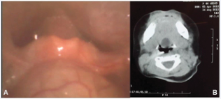
Figure 1 (A) – Laryngoscopic examination showing a sublingual thyroglossal duct cyst. (B) – Axial computed tomography scan showing a hypodense nodular lesion with regular borders in the region of the foramen cecum.
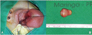
Figure 2 (A) – Postoperative view showing the incision made for cyst dissection. (B) – Surgical specimen submitted to anatomopathological analysis.
In view of these findings, the diagnostic hypothesis of a lingual TDC was established. Surgical resection of the lesion via a transoral approach was indicated, given the lingual location and the absence of cervical involvement. The surgical procedure was performed under general anesthesia with orotracheal intubation. After adequate exposure, an incision was made around the lesion, followed by complete excision. Careful dissection of the cystic capsule was then performed, respecting anatomical planes and preserving adjacent structures. Resection was complete and uneventful, with layered suturing of the tongue and closure of the surgical wound (Figure 2(A)). Anatomopathological examination of the surgical specimen revealed a cystic structure lined by cuboidal epithelium with squamous metaplasia, surrounded by connective tissue containing seromucous glands (Figure 2 (B)). No atypia or signs of malignancy were observed, thereby confirming the diagnosis of a TDC. In the immediate postoperative period, the patient showed significant respiratory improvement. She was discharged on the fifth postoperative day without complications. After 40 days, she was completely asymptomatic. No signs of lesion recurrence were observed during 2 years of outpatient follow-up.
DISCUSSION
In the present report, the persistence of stridor since the neonatal period and the identification of a submucosal mass on flexible laryngoscopy were crucial in guiding the diagnostic strategy. Ultrasonography is the ideal initial imaging modality in the evaluation of cervical masses, as it allows distinguishing between solid and cystic lesions in most cases and provides accurate information on the location of the mass in relation to adjacent anatomical structures. However, when the lesion is located at the base of the tongue, as in the case described herein, ultrasonography may hinder its visualization [12], making CT necessary for accurate assessment. Accordingly, cervical CT not only confirmed the cystic nature of the lesion but also was essential to evaluate the position of the thyroid gland and to rule out ectopia [13]. The examination revealed a well-defined lesion adjacent to the foramen cecum, without cervical extension or thyroid abnormalities, allowing selection of the transoral approach.
The Sistrunk procedure remains the gold standard for cervical TDCs, with recurrence rates ranging from 2.6% to 5% [10]. However, for exclusively lingual lesions, transoral resection has gained acceptance as a less invasive alternative. Recent studies have demonstrated the success of this approach, evidenced by the low morbidity and absence of recurrence during medium-term follow-up [6], as also observed in the present case. The decision to pursue a transoral approach was based on the lesion’s confinement to the base of the tongue and the absence of extension to the hyoid bone, as previously proposed [11]. Prolonged follow-up (2 years) was essential to confirm cure, since recurrence of lingual TDCs may occur in up to 10% of cases, particularly when ductal remnants persist [3,9]. The patient’s favorable outcome supports previous findings [4], which highlighted immediate resolution of respiratory symptoms following complete resection.
The reported case is notable because of its anatomical rarity (0.5% to 3% of TDCs) and its relevant clinical implications. Comparison with the literature showed that the presentation of progressive stridor and feeding difficulties followed the pattern described in other reports [4,5], reinforcing the importance of early flexible laryngoscopy for differential diagnosis from conditions such as laryngomalacia, which occurs in approximately 40% of cases [7]. Suturing of the surgical wound provided adequate lingual reconstruction, representing a valuable approach with excellent functional outcomes and no recurrence after 2 years of follow-up.
Findings from this case, supported by the existing literature, reinforce the feasibility of a minimally invasive approach for isolated lesions and point toward a new paradigm in the management of these congenital anomalies, integrating emerging technologies with conventional surgical techniques. The scarcity of specific guidelines for lingual TDCs underscores the relevance of this report, which contributes to both current clinical practice and the development of future management strategies.
REFERENCES
- Allard RH. The thyroglossal cyst. Head Neck Surg. 1982; 5: 134-146.
- Moore KL, Persaud TVN. The Developing Human: Clinically Oriented Embryology. 8th ed. Saunders; 2008.
- Mondin V. Lingual thyroglossal duct cyst: a unique surgical approach. Head Neck. 2005; 27: 897-901.
- Wong KK. Lingual thyroglossal duct cysts: a 10-year experience. Laryngoscope. 2019; 129: 1001-1006.
- Lin Y. Neonatal lingual thyroglossal duct cyst: a case series. Int J Pediatr Otorhinolaryngol. 2017; 92: 154-157.
- Shin JJ. Transoral excision versus Sistrunk procedure for lingual thyroglossal duct cysts. J Pediatr Surg. 2016; 51: 2037-2040.
- Rahbar R, Nicollas R, Roger G, Triglia JM, Healy GB, McGill TJ, et al. The biology and management of subglottic hemangioma: past, present, future. Laryngoscope. 2005; 115: 1880-1891.
- Perkins JA. Emerging concepts in pediatric head and neck vascular anomalies. Int J Pediatr Otorhinolaryngol. 2010; 74: 267-272.
- Geller KA. Risk factors for thyroglossal duct remnant recurrence. JAMA Otolaryngol Head Neck Surg. 2019; 145: 48-53.
- Maddalozzo J. High rate of recurrence after Sistrunk procedure for thyroglossal duct remnant surgery. Otolaryngol Head Neck Surg. 2012; 147: 78-82.
- Brousseau VJ. Thyroglossal duct cysts: a 30-year experience with transoral laser excision. Arch Otolaryngol Head Neck Surg. 2003; 129: 537-539.
- Wong KT, Lee YY, Rei AD, Ahuja AT. Imagem de massas císticas ou semelhantes a cistos. Clin Radiol. 2008; 63: 613-622.
- Huang T. Imaging of thyroglossal duct cysts: variations and complications. AJNR Am J Neuroradiol. 2013; 34: 1738-1743.