Ectodermal Dysplasia: Series of 8 Cases
- 1. Department of Dermatology’s Diseases, CHU Ibn Rochd Casablanca, Morocco
Abstract
Introduction: Hypohidrotic ectodermal dysplasia is a genetic disease of ectoderm development characterized by malformations of ectodermal structures such as the skin, hair, teeth and sweat glands.
Material and methods: We report the cases of 8 children.
Results: The mode of diagnosis, the clinical signs and the therapeutic option are detailed.
Discussion: Hypohidrotic ectodermal dysplasia is characterized by the triad of manifestations including sparse hair (atrichosis / hypotrichosis), missing teeth (anodontia / hypodontia) or abnormal (for example conical) and reduced or absent sweating due to the absence of sweat glands (anhidrosis / hypohidrosis) leading to heat intolerance. The treatment is based on the eviction of uncontrolled exposure to heat with an early dental treatment which aims to restore dental function and improve the appearance of teeth as well as orthodontic treatment. Most patients have normal life expectancy with early diagnosis and appropriate management.
Keywords
- Ectodermal dysplasia
- Anodontia
- Hypohidrosis
- Hypotrichosis
CITATION
: KHEDIM N, HALI F, Chiheb S (2022) Ectodermal Dysplasia: Series of 8 Cases. Arch Paediatr Dev Pathol 5(1): 1024
INTRODUCTION
The Hypohidrotic Ectodermal Dysplasia (HED) is a genetic disease of the development of the ectoderm characterized by malformations of the skin, hair, and teeth essentially. Skin and dental damage are in the foreground. They are an integral part of the treatment, both from an aesthetic point of view and in order to promote self-esteem and social integration of the patient. The diagnosis is usually relatively late; it is most often made in connection with delayed tooth eruption. Due to the high prevalence of oral anomalies related to HED. Like any other genodermatosis, genetic counseling for the affected person and their family members is essential. In order to inform about the risk of occurrence or recurrence of the disease.
The care of patients requires the collaboration of dermatologists, dentists, ENT specialists, ophthalmologists, pneumologists and psychologists.
We report a series of 8 cases of HED.
MATERIAL AND METHODS
It’s a retrospective study that included all the cases of hypohidrotic ectodermal dysplasia diagnosed in pediatric consultations from 2018 and 2021, at the dermatology department of the Ibn Rochd University Hospital of Casablanca.
RESULTS
Eight cases were identified including 6 boys and 2 girls, the sex ratio was 3. The average age was 5 years old with extremes ranging from 3 months old to 14 years old. 1 case of consanguinity was reported and one similar case among the siblings in a family. All our patients had a good psychomotor development.
our patients had a good psychomotor development. The clinical exam of our patients revealed all the typical clinical signs (hypohidrosis, hypotrichosis, hypodontia) with a called senile face. The hair was light colored, thin, brittle, and a receding hairline. The eyebrows were none existent. The skin was dry and the palpebral hyperpigmentation was responsible of very marked dark circles (figure 1). One of our patients had been hospitalized for fever without localizing signs (Table 1).
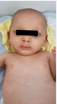
Figure 1 Senile face with dark circles and light colored hair with thin eyebrows.
Dental involvement was predominant and affected all of our patients and was present as an abnormal shape (conoid tooth) (figure 2) or number “hypodontia”, delayed eruption or the absence of teeth in the mandible.
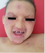
Figure 2 Senile face with dark circles and light colored hair with thin eyebrows , conoidal aspect of the teeth and eczema lesions.
Our patients were put on emollient with a preparation based on dermocorticoids and fusidic acid for the eczema lesions with good improvement of the skin lesions, and an odontostomatology consultation for a prosthetic treatment allowing a better functionality and the correction of the aesthetic damage.
DISCUSSION
HED is characterized by isolated anomalies of the epidermis and its appendages due to a defect in the embryonic development of the ectodermal layer, the nature of which is until now unknown[1], or associated with damage to another organ in the syndromic form (eye, ear…).
Table 1: Different clinical manifestations of our patients.
| Patient 1 | Patient 2 | Patient 3 | Patient 4 | Patient 5 | Patient 6 | Patient 7 | Patient 8 | |
| Consanguinity | Yes | No | No | No | No | No | No | No |
| Similar case among the siblings in a family | No | Yes | No | No | No | No | No | No |
| Hypohidrosis, | Yes | Yes | Yes | Yes | Yes | Yes | Yes | Yes |
| Hypotrichosis, | Yes | Yes | Yes | Yes | Yes | Yes | Yes | Yes |
| Hypodontia | Yes | Yes | Yes | Yes | Yes | Yes | Yes | Yes |
| Hyperthermal access | No | No | Yes | No | No | No | No | No |
More than 180 ectodermal dysplasias have been identified. Hypohidrotic / anhidrotic ectodermal dysplasia, MIM 305100 represents the most frequent form with an estimated worldwide incidence of 1 in 100,000, without ethnic predominance [2].
Four genes are identified in over 90% of cases: EDAR, EDARADD, WNT10A and EDA1 which is found in over 50% of cases which explains the male predominance [3].
The diagnosis is essentially clinical with a characteristic senile facies corresponding to a hypoplasia of the middle level of the face characterized by the prominence of the forehead with a periorbital depigmentation and a rarefaction of the eyelashes and eyebrows as well as a possibility of mongoloid obliquity of the palpebral fissure with weakening of the corneal epithelium responsible for photophobia associated with the triad of anhidrosis (or hypohidrosis), hypotrichosis, and anodontia (or oligodontia) [4].
The scarcity of sweat glands is the cause of hypohidrosis, which is responsible for thermoregulation disorders within tolerance to temperatures above 23-24 °C leading to frequent hyperthermic attacks in young children involving the vital prognosis during the first years mainly. Cutaneous xerosis with an amber aspect, hyperkeratotic appearance is reported with regularly fissured palmoplantar keratoderma at the end of the second decade [5]. A mid-facial, periocular and perioral pigmentation is observed with healing difficulties in some patients, that requires a close attention to these patients.
Skin biopsy of an area rich in sweat glands (palms, soles of the feet, axillary regions) shows normal epidermis in the majority of cases, sometimes atrophied or hyperkeratotic with a decrease or absence of sweat glands and pilo sebaceous follicles.
The hair growth is almost constant and concerns the hair giving a sparse, fine, dry, rough, brittle appearance, often white or depigmented, the eyelashes, the eyebrows and the hairs of the axillary and pubic region with a regular respect of the beard. Trichogramma analysis can characterize the hairs, their size and growth rate [6], helping to adapt the management strategy.
Hypodontiare mains more common than anodontia and requires early treatment. It is more severe in the mandible than in the maxilla [7]. The teeth take on a conical appearance called “pig’s teeth or wolf teeth or teeth in bottles” with weakening of the enamel leading to the appearance of early caries. The panoramic X-ray of the patient subject strongly guides the diagnosis and specifies the extent of dental anomalies.
Other impairments are reported, particularly ophthalmological, ENT area which requires a multidisciplinary approach involving the dermatologist, dentist, ENT specialist, ophthalmologist, pulmonologist and psychologist.
A genetic counseling consultation is desirable to inform the patient and his family about the risk of the onset or recurrence of the disease.
To date, the management of patients remains symptomatic, aiming to treat the various clinical signs and prevent complications, with no tendency to cure the disease. Frequent moisturizing of the skin, feet and palms of the hands and daily nasal hygiene are recommended.
Dentures can be placed as early as 2.5 years of age and should be changed regularly to accommodate growth. Implants can be placed as early as 6 years of age in case of anodontia. Final dental rehabilitation will only take place at the end of growth and in most cases requires the use of bone grafts. Air conditioning is recommended at home, at work and possibly at school when possible. It is important to monitor young children because of hyperthermia, which can be life-threatening.
No gene therapy for humans has been reported for HED. However, there are some reports in animal models showing evidence for a potential short-term treatment with a recombinant ectodysplasin protein that can correct some of the developmental genetic defects observed in HED patients [8].
CONCLUSION
The diagnosis of hypohidrotic ectodermal dysplasia is easy in the major forms where the clinical triad is present. Most patients have a normal life expectancy with early diagnosis and appropriate management.
REFERENCES
5. Lambert D, Nivelon-Chevalier A, Nivelon JL, Chapuis JL. Dysplasie ectodermique anhydrotique. Ann DermVenereol, 1977;104: 298-303.


