Core Regulators of Epithelial Mesenchymal Transition in Keloid Identified Through Progressive Bioinformatics Analysis and Experimental Validation
- 1. Linyi People’s Hospital, Shandong Second Medical University, China.
- 2. First Peoples Hospital of Ningyang, China
- †. These authors contributed equally to this work
Abstract
Objective: Keloids are benign fibrotic conditions with increased proliferation, migration, and invasion, resembling tumor metastasis. Epithelial-mesenchymal transition (EMT) regulates tumor metastasis; thus, identifying EMT-related biomarkers may clarify keloid pathogenesis.
Methods: Transcriptomic datasets (GSE83286, GSE212954, GSE90051) were analyzed using WGCNA, differential expression, and machine learning (LASSO, SVM-RFE, RF, XGBoost) to identify key EMT-related genes. Immune infiltration and single-cell analyses were performed, followed by experimental validation in human keloid tissues.
Results: Twenty-nine EMT-related genes were identified. FN1 and RUNX2 emerged as key genes, with high diagnostic accuracy. Immune infiltration revealed reduced plasma cells, B cells, and dendritic cells, alongside increased resting mast cells and M2 macrophages in keloids. Single-cell and experimental validation confirmed FN1 and RUNX2 upregulation in keloid tissues.
Conclusion: FN1 and RUNX2 are key EMT-related genes in keloids, offering potential diagnostic markers and therapeutic targets.
Keywords
• Keloid
• Epithelial-mesenchymal transition
• FN1 • RUNX2
• Immune infiltration
Citation
Wu J, Wu YK, Zhang Y, Zhang Y, Zhang J (2026) Core Regulators of Epithelial-Mesenchymal Transition in Keloid Identified Through Progres sive Bioinformatics Analysis and Experimental Validation. JSM Burns Trauma 9(1): 1054.
ABBREVIATIONS
EMT: Epithelial-Mesenchymal Transition; WGCNA: Weighted Gene Co-expression Network Analysis; DEGs: Differentially Expressed Genes; FN1: Fibronectin 1; RUNX2: Runt-related transcription factor 2
INTRODUCTION
Keloids are pathological scars characterized by excessive fibroblast proliferation and collagen deposition, leading to firm, raised lesions that extend beyond the original wound boundary [1,2]. They cause significant physical and psychological distress, yet effective treatments remain limited [3]. Epithelial-mesenchymal transition (EMT), a process where epithelial cells acquire mesenchymal traits, enhances migration, invasion, and apoptosis resistance [4,5]. Emerging evidence links EMT to keloid progression, with keratinocytes exhibiting an EMT-like state regulated by TGF-β1 [6]. However, systematic identification of EMT-related key genes in keloids using integrated bioinformatics and machine learning approaches remains scarce. This study aims to identify core EMT regulators in keloids through comprehensive transcriptomic analysis and experimental validation.
MATERIALS AND METHODS
Transcriptomic datasets GSE83286, GSE212954 (merged as training set), and GSE90051 (validation set) were obtained from GEO. EMT-related genes were sourced from dbEMT 2.0. WGCNA identified keloid-associated gene modules Figure 1. DEGs were analyzed with thresholds P < 0.05 and |logFC| > 1. Intersection of WGCNA genes, DEGs, and EMT genes yielded EMT-related genes (EMTRGs). GO/KEGG enrichment and PPI network analyses were performed. Four machine learning algorithms—LASSO, SVM-RFE, Random Forest, and XGBoost—were applied to identify key genes. A diagnostic nomogram was constructed and validated. Immune infiltration was assessed via CIBERSORT. Single-cell analysis used GSE163973. Finally, qRT-PCR and Western blot validated FN1 and RUNX2 expression in three keloid and three normal skin samples.
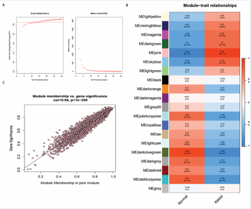
Figure 1: WGCNA identifies keloid-associated core genes. (A) Soft-thresholding power selection plot; (B) Module-trait correlation heatmap; (C) Distribution of gene connectivity within the pink module.
RESULTS AND DISCUSSION
Type or copy/paste your text here WGCNA identified the pink module as most correlated with keloids (cor = 0.95, P = 8×10??), yielding 1,140 genes. Intersection with DEGs (n = 740) and EMT genes (n = 1,185) produced 29 EMTRGs. GO/KEGG analyses revealed enrichment in mesenchymal differentiation, epithelial proliferation, and cancer-related pathways. PPI network highlighted FN1, POSTN, TGFB1, RUNX2, and MMP14 as top hub genes. Machine learning intersection identified FN1 and RUNX2 as key EMT-related genes. Nomogram models based on FN1 and RUNX2 showed excellent diagnostic performance (AUC > 0.9 in both training and validation sets).
Immune infiltration analysis revealed decreased plasma cells, B cells, and dendritic cells, and increased resting mast cells and M2 macrophages in keloids. FN1 and RUNX2 expression positively correlated with resting mast cells and negatively with plasma cells, suggesting immune dysregulation may promote EMT and keloid progression.
Single-cell analysis confirmed significantly higher FN1 and RUNX2 AddModuleScore in keloid samples. Experimental validation in human tissues showed consistent upregulation of FN1 and RUNX2 at both mRNA and protein levels in keloids compared to normal skin.
RUNX2 is known to activate PI3K-AKT signaling in keloids, enhancing cell proliferation. FN1, though less studied in keloids, promotes tumor cell migration and invasion via integrin signaling and ECM remodeling. Our findings suggest FN1 may similarly drive keloid fibrosis. The observed immune landscape—reduced plasma cells and increased mast cells—may further facilitate EMT through TGF-β and inflammatory mediator release
CONCLUSION
This study identifies FN1 and RUNX2 as core EMT-related genes in keloids, with high diagnostic value. Their upregulation correlates with immune infiltration changes, implicating them in keloid pathogenesis. These findings provide potential diagnostic biomarkers and therapeutic targets, warranting further mechanistic and translational studies.
ACKNOWLEDGEMENTS
The authors thank all colleagues who supported this research.
REFERENCES
- Bogari A, Jomah M, Alshehri N, Alrabiah HF, Binghaith A, AlOsaimi K, et al. 5-Fluorouracil in Facial Plastic Surgery: Indications, Efficacy, and Safety - A Systematic Review and Narrative Synthesis. Facial Plast Surg. 2026; 42: 28-38.
- Atefi N, Jafari MA, Roohaninasab M, Dehghani A, Jafarzadeh A, Peighambari SB, et al. Evaluating the effectiveness and safety of pulsed dye laser alone, the combination of pulsed dye laser and botulinum toxin type A, and the combination of pulsed dye laser and triamcinolone injection in the treatment of hypertrophic and keloid scars: a three-arm randomized controlled clinical trial. Lasers Med Sci. 2025; 40: 92.
- Kong J, Zhou C, Qin H, Li C, Wu Z, Zhang L. Identifying key genes, miRNAs, and pathways in keloid formation: A bioinformatics and experimental study. J Plast Reconstr Aesthet Surg. 2025; 102: 313- 322.
- Hahn JM, McFarland KL, Combs KA, Supp DM. Partial epithelial- mesenchymal transition in keloid scars: regulation of keloid keratinocyte gene expression by transforming growth factor-β1. Burns Trauma. 2016; 4: 30.
- Zhang M, Liu S, Guan E, Liu H, Dong X, Hao Y, et al. Hyperbaric oxygen therapy can ameliorate the EMT phenomenon in keloid tissue. Medicine (Baltimore). 2018; 97: e11529.
- Lv W, Wu M, Ren Y, Luo X, Hu W, Zhang Q, et al. Treatment of keloids through Runx2 siRNA-induced inhibition of the PI3K/AKT signaling pathway. Mol Med Rep. 2021; 23: 55.












































































































































