Cholecystokinin – More Than Just a Gallbladder Emptier
- 1. Department of Clinical Biochemistry, Rigshospitalet, University of Copenhagen, Denmark
Abstract
The existence of cholecystokinin (CCK) as a gut hormone was suggested in 1928 based on hormonal regulation of gallbladder emptying, later also on stimulation of pancreatic enzyme secretion, and modification of gastrointestinal motor activity. In 1968, a CCK peptide was purified from gut extracts and identified as a 33 amino acid peptide (CCK-33). Based on that structure, research has since expanded the insight into CCK biology remarkably: Thus, CCK was the first discovered intestinal satiety signal from gut to brain. Moreover, the CCK gene is now known to be expressed in different peptide forms not only in the gut, but much so in central and peripheral neurons, in addition to extra-intestinal endocrine cells, immune cells, cardiomyocytes, spermatogenic cells, and certain fat cells. Accordingly, CCK peptides function not only as hormones. They are also neurotransmitters, paracrine growth and satiations factors, anti-inflammatory cytokines, incretins, adipokins, myokins, potential fertility factors, and tumor markers. Thus, CCK research has now opened windows for insights into the pathophysiology of not only gallbladder disease, but also metabolic disorders, neuropsychiatric diseases, cardiac diseases, as well as the diagnosis of neuroendocrine and pediatric tumors.
Keywords
• Cholecystokinin;
• Cytokine
• Gut hormone
• Neurotransmitter
• Satiation factor
Citation
Rehfeld JF (2026) Cholecystokinin – More Than Just a Gallbladder Emptier. JSM Gastroenterol Hepatol 13(2): 1141.
INTRODUCTION
In modern endocrinology, gastrointestinal hormones have recently surfaced as a strikingly dynamic area with considerable clinical impact [1]. Particularly well known examples are glucagon-like peptide-1 (GLP-1) and glucose dependent insulinotropic polypeptide (GIP) from which drugs have already been derived and used in the treatment of diabetes and obesity, with additional candidates in the pipelines for therapy of cardiovascular, renal and perhaps cerebral diseases [2-4].
Other gastrointestinal hormones might also be of interest as drug targets. Among these is not least cholecystokinin (CCK), whose role as an intestinal regulator of gallbladder emptying actually may turn out to be secondary compared to some of the extraintestinal activities implicated in the complex biology of CCK [5,6].
Following a summary of the CCK history, this article will briefly overview the new CCK biology including phylogeny, biogenesis, cellular expression with cell-specific peptide patterns, receptors, and subsequently derived functions.
A SHORT HISTORY
More than a century ago, European physiologists observed that administration of acid into the duodenum stimulated bile flow from the liver and gallbladder contraction [7-10,11]. In 1928, Ivy and Oldberg provided evidence that the gallbladder effect was due to a new gut hormone, which they named CCK [12]. However, not until Mutt and Jorpes established a large plant for purification from vast amounts of porcine jejunal mucosa did structure identification become possible. The structure was published in 1968 [13] as a peptide sequence of 33 amino acid residues, a C-terminal sequence homologous to that of gastrin, and besides it was O-sulfated (Figure 1).
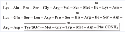
Figure 1: The structure of the 33 amino acid residues of cholecystokinin as published in 1968 (see ref. [12]). Note that the tyrosyl residue in position 27 is O-sulfated and that the phenylalanyl residue in position 33 is carboxyamidated. The C-terminal boxed sequence is the bioactive, receptor-bound epitope of CCK.
Identification of the structure became a decisive milestone. With synthetic fragments of CCK-33, material became available for physiological studies, for production of antibodies as well as for identification of the CCK gene and new molecular forms of CCK peptides. These tools together with other methods paved the way for a number of paradigmatic shifts that have unfolded a multifaceted biology. Consequently, CCK is now to be seen as a ubiquitous peptide messenger system in the body [5,6].
THE NEW BIOLOGY
Phylogenesis
CCK is member of a family that includes the antral hormone gastrin [14,15]; the amphibian skin peptides, the caeruleins [16,17]; and in protochordates, the neuropeptide cionin [18]. Insect neuropeptides, the sulfokinins [19,20] are also structurally related (Figure 2). Consequently, evolutionary studies indicate that CCK peptides originated almost 600 million years ago [21-24].
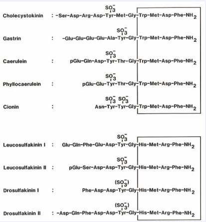
Figure 2: The decisive homologous structure in members of the cholecystokinin peptide family in mammals, amphibians, protochordates (Ciona Intestinalis), and insects (a cockroach (Leucophaea Madera) and a fruitfly (drosophilia melanogaster)). Note that the boxed C-terminal tetrapeptide sequences are all carboxyamidated and that this tetrapeptide amide as such is the core structure necessary for receptor binding. Note also O-sulfation of the more N-terminal tyrosyl residues. The tyrosyl-sulfation is a derivatization that modifies the affinity and hence the specificity of receptor bindings
Biogenesis and Molecular heterogeneity of peptides
There is one CCK gene [25,26] which is transcribed to a single mRNA of 750 bases [25-27]. The mRNA molecule is then translated to a precursor protein (preproCCK) with a sequence of 115 amino acid residues [27]. The N-terminal signal or pre-sequence is probably removed during translation, leaving a proCCK of 95 amino acids, which subsequently undergoes posttranslational modifications by a multitude of processing enzymes acting along cellular secretory or synaptic pathways [28-37]. The modifications comprise cleavages at mono- and dibasic sites, amino acid derivatizations and N- or C-terminal trimmings. As a result of the posttranslational processing, a number of different bioactive CCK peptides are accumulated in the secretory granules or presynaptic vesicles: CCK-83, -58, -33, -22, -8, and -5. The first five occur in both O-sulfated and nonsulfated forms, whereas CCK-5 is unsulfated. The peptide patterns, however, are cell-specific [6-37].
The Tissue and cell expression
In accordance with the role as gut hormone, specific endocrine cells in the intestinal mucosa, I-cells, have been identified in the duodenum and jejunum as the major source of intestinal CCK [38-40]. A majority of CCK in plasma originates from these cells. The I-cells, however, are present also in the ileum, and sporadically in the proximal colon [41-43].
Most CCK is, however, not synthesized in the gut, but in neurons, central as well as peripheral [30-48]. The CCK neurons also express “classic” transmitters, such as GABA in interneurons and glutamate in pyramidal and thalamic neurons [48,49].
Although cerebral neurons and intestinal I-cells are major expression sites, specific expression has in addition been detected in other cells (Table 1). They include pituitary corticotrophs [50,51], thyroid C-cells [52,53], adrenal medullary cells [54], spermatogenic cells [55,56], cardiac myocytes [57], and circulating monocytes [58-60].
Table 1: Tissue concentrations of bioactive cholecystokinin (CCK) peptides in mammals.
|
Tissue |
Tissue contenta (pmol/g) |
|
Intestinal Tract: |
|
|
Duodenal mucosa |
200 |
|
Jejunal mucosa |
150 |
|
Ileal mucosa |
20 |
|
Colonic mucosa |
5 |
|
|
|
|
Central Nervous System: |
|
|
Cerebral cortex |
400 |
|
Hippocampus |
350 |
|
Hypothalamus |
200 |
|
Cerebellum |
2 |
|
Spinal cord |
40 |
|
|
|
|
Peripheral Nervous System: |
|
|
Vagal nerve |
25 |
|
Sciatic nerve |
15 |
|
Nerveplexes in colonic wall |
5 |
|
|
|
|
Extraintestinal Endocrine Glands: |
|
|
Adenohypophysis |
25 |
|
Neurohypophysis |
20 |
|
Thyroid gland |
2 |
|
Adrenal medulla |
1 |
|
|
|
|
Urogenital Tract: |
|
|
Testicles |
5 |
|
Spermatozoas |
1 |
|
|
|
|
Cardiovascular System: |
|
|
Atrial myocytes |
10 |
|
Ventricular myocytes |
2 |
|
|
|
|
Mononuclear Immune Cellsb: |
++ |
aOrders of magnitude based on measurement of tissue extracts from several mammalian species.
bExpression determined by immunocytochemistry of monocytes.
Cell-specificity of CCK peptide expression
There are at least three different cell patterns of CCK. First, in central and peripheral neurons, O-sulfated CCK 8 predominates, but also unsulfated CCK-5 is present in significant amounts. Second, gut endocrine I-cells express a considerably broader pattern that includes larger molecular forms (CCK-58, -33, and -22) as well as short forms (CCK-8 and -5) [61]. The four longest CCK peptides in I-cells all occur also in nonsulfated variants [35]. Finally, specific patterns are found in lower concentrations in pituitary corticotrophs, thyroid C-cells, spermatogenic cells, cardiac myocytes, and circulating monocytes [50-59].
CCK Receptors
The distribution and distinction between “alimentary” (CCKA ) and “brain” (CCKB ) receptors were studied already in the 1970s and 1980s [64,65]. In 1992, however, the structures of the CCKA and the gastrin/CCKB receptors were deduced after cDNA cloning [66,67]. The receptors are now named CCK1 and CCK2 . Later, informative reviews of receptor distribution, physiology, and pharmacology have been published [68,69]. The binding specificity of the CCK1 and CCK2 receptors differs. Thus, all carboxyamidated CCK and gastrin peptides irrespective of sulfation are bound with similar affinities to the gastrin/CCK2 receptor, whereas the CCK1 receptor is more selective and binds only tyrosyl O-sulfated CCK peptides. A novel CCK receptor was recently detected in brain tissue [70]. Further characterization is awaited.
CCK functions
An overview of the functions of CCK requires recognition of the new biology in its entirety: On one hand, there are the different cellular origins and release mechanisms for CCK peptides. On the other hand, there are the targets in terms of CCK receptors and the routes by which the peptides travel to receptors expressed on target cells (endocrine, neurocrine, paracrine, autocrine, spermiocrine ways and synaptic transmissions). The CCK receptors are expressed in a majority of organs and tissues in the body: central and peripheral neurons as well as glial cells; the entire gastrointestinal tract; myocytes of the gallbladder and the sphincter Oddi muscles; exocrine and endocrine pancreatic cells; the cardiovascular system; the urogenital system; and the immune system [68,69]. Consequently, CCK peptides have many labels and may act independently as neurotransmitters, hormones, growth factors, satiation factors, cytokines, myokines, and perhaps adipokines and fertility factors.
On top of this picture, it is of fundamental significance to realize that the target cells for CCK peptides also express receptors for a multitude of other bioactive peptide systems. Therefore, different peptide systems interact and cross talk along intracellular signal-transduction pathways. Well-known examples are the mutual potentiation of CCK peptides with secretin in their stimulation of exocrine pancreatic enzyme and bicarbonate secretion [71,72]; the CCK potentiation of GLP-1 in stimulation of pancreatic beta cell growth [73]; the interaction of CCK with GLP-1 and PYY in signaling to the brain via afferent vagal fibers to regulate food intake [74]; and, for instance, the balance between sulfated and nonsulfated CCK peptides from CCKomas in the inhibition or stimulation of gastric acid secretion [75]. Probably, studies of the interactive physiology between CCK and other bioactive peptide systems as well as the interaction with non-peptidergic extracellular messenger molecules will be an essential part of the future physiology and pharmacology of CCK [76].
CCK and Gallbladder diseases
The association between CCK and gallbladder function is per definition the basis for the century-old biology of CCK as reflected also in the name of the hormone [11]. Today it is an established fact that CCK peptides from I-cells in the proximal small intestine in a concentration related manner control the intestinal phase of gallbladder emptying via CCK1 receptors expressed on gallbladder muscles [77]. The mechanistic details and the relationship to gastrointestinal disorders, pancreatic insufficiency, and gallstone/gallbladder diseases have systematically been studied not least by Masclee and Lamers et al., [77-83].
The results are summarized in Table 2, which shows that the occurrence of gallstones frequently accompanies the examined disorders. Although gallstone-attacks are painful and annoying, surgical gallbladder removal is today fast and uncomplicated, and life without a gallbladder is almost normal. Remember that the successful rodent, the rat, does not have a gallbladder. Thus, perhaps the pancreozymic effect of CCK should be considered more essential for digestion than the gallbladder-emptying effect, as demonstrated also in normal rats.
Table 2: Cholecystokinin secretion and gallbladder motility in some gastrointestinal disorders.a
|
Disorder |
Stimulus |
CCK secretion |
Gallbladder motility |
Gallstone prevalence |
Mechanism |
|
Partial gastrectomy |
Oral meal |
Increased |
Increased |
Increased |
Rapid gastric emptying |
|
Sleeve gastrectomy |
Oral meal |
Increased |
Normal |
- |
Rapid gastric emptying |
|
Roux-en-Y bypass |
Oral meal |
Increased |
Decreased |
Increased |
Rapid gastric emptying with bypass of prox. gut |
|
Celiac disease |
Oral meal |
Decreased |
Decreased |
Increased |
- |
|
Exocrine pancreatic insufficiency |
Oral meal |
Decreased |
Decreased |
Increased |
Delayed intestinal fat digestion |
|
Whipple operation |
Oral meal |
Decreased |
Gallbladder removed |
- |
Reduced pancreatic enzyme secretion |
|
Colectomy |
Oral meal |
Increased |
Normal |
Increased |
Changes in bile composition |
|
Parenteral nutrition |
No enteral feeding |
Decreased |
Stasis |
Increased |
No enteral stimulus |
aSee also ref. (77) for further details
ACKNOWLEDGEMENT
The skillful and patient linguistic as well as secretarial assistance of Connie Bundgaard (MA) is gratefully acknowledged.
Data availability statement
Data sharing is not relevant for the present perspective article.
Funding statement
The writing of this article received no funding. Earlier cholecystokinin studies in the laboratory of the author, however, received support from the Danish Medical Research Council, the Danish Cancer Union, and the Novo Nordic Foundation.
Conflicts of interest
The author declares no conflicts of interests.
REFERENCES
- Rehfeld JF, Goetze JP. Gastrointestinal hormones: History, biology, and measurement. Adv Clin Chem. 2024; 118: 111-154.
- Madsbad S, Holst JJ. The promise of glucagon-like peptide 1 receptor agonists (GLP-1RA) for the treatment of obesity: a look at phase 2 and 3 pipelines. Expert Opin Investig Drugs. 2025; 34: 197-215.
- Marso SP, Daniels GH, Brown-Frandsen K, Kristensen P, Mann JF, Nauck MA, et al. Liraglutide and cardiovascular outcomes in type 2 diabetes. N Engl J Med. 2016; 375: 311-322.
- Jastreboff AM, Aronne LJ, Ahmad NN, Wharton S, Connery L, Alves B, et al. Tirzepatide once weekly for the treatment of obesity. N Engl J Med. 2022; 387: 205-216.
- Rehfeld JF. Cholecystokinin – from local gut hormone to ubiquitous messenger. Front Endocrinol.2017; 8: art.47.
- Rehfeld JF. Cholecystokinin – portrayal of an unfolding peptide messenger system. Peptides. 2025: 186: 171369.
- Bernard C. Mémoire sur le pancreas. Compte Rend Acad Sci.1856; 55: 379-563.
- Wertheimer E. De l’action des acides et du choral sur la secretion biliaire. Compte Rend Soc Biol. 1903; 55: 286-287.
- Fleig C. Réflex de l’acide sur la secretion biliaire. Compte Rend Soc Biol. 1903; 55: 353-355.
- Okada S. On the secretion of bile. J Physiol. 1914; 49: 457-482.
- Rehfeld JF, Feinle-Bisset C. Milestones in the history of CCK. In: C. Feinle-Bisset, J.F. Rehfeld (Eds.) Cholecystokinin – from gallbladder to cognition and beyond. Elsevier/Academic Press, 2025; pp. 3-19.
- Ivy AC, Oldberg E. Hormone mechanism for gallbladder contraction and evacuation. Am J Physiol. 1928; 86: 599-613.
- Mutt V, Jorpes JE, Structure of porcine cholecystokinin-pancreozymin. I. Cleavage with thrombin and with trypsin. Eur J Biochem. 1968; 6:156-162.
- Gregory R, Tracy H. The constitution and properties of two gastrins extracted from hog antral mucosa. Gut. 1964; 5: 103-117.
- Gregory H, Hardy PM, Jones DS, Kenner GW, Sheppard RC. The antral hormone gastrin. Structure of gastrin. Nature. 1964; 204: 931-933.
- Anastasi A, Erspamer V, Endean R. Isolation and amino acid sequence of caerulein, the active decapeptide of the skin of hyla caerulea. Arch Biochem Biophys. 1968; 125: 57-68.
- Anastasi A, Bertaccini G, Cei JM, De Caro G, Erspamer V, Impicciatore M. Structure and pharmacological actions of phyllocaerulein, a caerulein-like nonapeptide: its occurrence in extracts of the skin of Phyllomedusa sauvagei and related Phyllomedusa species. Br J Pharmacol. 1969; 37:198-206.
- Johnsen AH, Rehfeld JF. Cionin: a disulfotyrosyl hybrid of cholecystokinin and gastrin from the neural ganglion of the protochordate Ciona intestinalis. J Biol Chem. 1990; 265: 3054-3058.
- Nachman RJ, Holman GM, Haddon WF, Ling N. Leucosulfakinin, a sulfated insect neuropeptide with homology to gastrin and cholecystokinin. Science. 1986; 234: 71-73.
- Nichols R, Schneuwly SA, Dixon JE. Identification and characterization of a drosophila homologue to the vertebrate neuropeptide cholecystokinin. J Biol Chem. 1988; 263: 12167-12170.
- Johnsen AH. Phylogeny of the cholecystokinin/gastrin family. Front Neuroendocrinol.1998; 19:73-99.
- Baldwin GS, Patel O, Shulkes A. Evolution of gastrointestinal hormones: the cholecystokinin/gastrin family. Curr Opin Endocrinol Diabetes Obes. 2010; 17: 77-88.
- Dupré D, Tostivint H. Evolution of the gastrin-cholecystokinin gene family revealed by synteny analysis. Gen Comp Endocrinol.2014; 195: 164-173.
- Nässel DR, Wu SF. Evolutionary conserved roles of cholecystokinin signaling. In: C. Feinle-Bisset, J.F. Rehfeld (Eds.) Cholecystokinin – from gallbladder to cognition and beyond. Elsevier/Academic Press, 2025; 21-69.
- Deschenes RJ, Haun RS, Funckes CL, Dixon JE. A gene encoding rat cholecystokinin. Isolation, nucleotide sequence, and promoter activity. J Biol Chem. 1985; 260: 1280-1286.
- Takahashi Y, Kato K, Hayashizaki Y, Wakabayashi T, Ohtsuka E, Matsuki S, et al. Molecular cloning of the human cholecystokinin gene by use of a synthetic probe containing deoxyinosine. Proc Natl Acad Sci USA. 1985; 82: 1931-1935.
- Gubler U, Chua AO, Hoffman BJ, Collier KJ, Eng J. Cloned cDNA to cholecystokinin mRNA predicts an identical preprocholecystokinin in pig brain and gut. Proc Natl Acad Sci USA. 1984; 81: 4307-4310.
- Rehfeld JF, Bundgaard JR. Cell-specific precursor processing. In: J.F. Rehfeld, J.R. Bundgaard (Eds.) Cellular peptide hormone synthesis and secretory pathways, Springer Verlag. Berlin, Heidelberg. 2010; 45-62.
- Dockray GJ. Immunochemical evidence of cholecystokinin-like peptides in brain. Nature. 1976; 264: 568-570.
- Rehfeld JF. Immunochemical studies on cholecystokinin. II. Distribution and molecular heterogeneity in the central nervous system and small intestine of man and hog. J Biol Chem. 1978; 253: 4022-4030.
- Goltermann NR, Rehfeld JF, Roigaard-Petersen H. In vivo biosynthesis of cholecystokinin in rat cerebral cortex. J Biol Chem. 1980; 255: 6181-6185.
- Rehfeld JF, Hansen HF. Characterization of preprocholecystokinin products in the porcine cerebral cortex. Evidence of different processing pathways. J Biol Chem. 1986; 261: 5832-5840.
- Reeve JR, Eysselein V, Walsh JH, Ben-Avram CM, Shively JE. New molecular forms of cholecystokinin. Microsequence analysis of forms previously characterized by chromatographic methods. J Biol Chem. 1986; 261:16392-16397.
- Shively J, Reeve JR, Eysselein VE, Ben-Avram C, Vigna SR, Walsh JH. CCK-5: sequence analysis of a small cholecystokinin from canine brain and intestine. Am J Physiol. 1987; 252: G272-G75.
- Agersnap M, Rehfeld JF. Nonsulfated cholecystokinins in the small intestine of pigs and rats. Peptides. 2015; 71: 121-127.
- Rehfeld JF. The endoproteolytic maturation of progastrin and procholecystokinin. J Mol Med. 2006; 84: 544-550.
- Rehfeld JF, Feinle-Bisset C. The biogenesis and cell-specific expression of cholecystokinin peptides. In: C. Feinle-Bisset, J.F. Rehfeld (Eds.), Cholecystokinin – from gallbladder to cognition and beyond. Elsevier/Academic Press, 2025; 71-85.
- Polak JM, Pearse AGE, Bloom SR, Buchan AMJ, Rayford PL, Thompson JC. Identification of cholecystokinin and secretin cells. Lancet. 1975; 2: 1016-1017.
- Buffa R, Solcia E, Go VLW. Immunohistochemical identification of the cholecystokinin cell in the intestinal mucosa. Gastroenterology. 1976; 70: 528-532.
- Buchan AMJ, Polak JM, Solcia E, Capella C, Hudston D, Pearse AGE. Electron immunocytochemical evidence for the human intestinal I cell as the source of CCK. Gut. 1978; 19: 403-447.
- Larsson LI, Rehfeld JF. Distribution of gastrin and CCK cells in the rat gastrointestinal tract. Histochemistry. 1978; 58: 23-31.
- Fakhry J, Wang J, Martins P, Fothergill LJ, Hunne B, Prieur P, et al. Distribution and characterization of CCK enteroendocrine cells in the mouse small and large intestine. Cell Tissue Res. 2017; 369: 245-253.
- Gilliam-Vigh H, Jorsal T, Rehfeld JF, Pedersen J, Poulsen SS, Vilsbøll T, et al. Expression of cholecystokinin and its receptors in the intestinal tract of type 2 diabetes patients and healthy controls. J Clin Endocrinol Metab. 2021; 106: 2164-2170.
- Larsson LI, Rehfeld JF. Localization and molecular heterogeneity of cholecystokinin in the central and peripheral nervous system. Brain Res. 1979; 165: 201-218.
- Emson PC, Rehfeld JF, Rossor MN. Distribution of cholecystokinin-like peptides in the human brain. J Neurochem. 1982; 38: 1177-1179.
- Marley PD, Rehfeld JF, Emson PC. Distribution and chromatographic characterisation of gastrin and cholecystokinin in the rat central nervous system. J Neurochem. 1984; 42: 1523-1535.
- Crawley JN. Comparative distribution of cholecystokinin and other neuropeptides. Why is this peptide different from all other peptides? Ann N Y Acad Sci. 1985; 448: 1-8.
- Hökfelt T, Barde S, Zhong W, He J. Cholecystokinin in the central and peripheral nervous system. In: C. Feinle-Bisset, J.F. Rehfeld (Eds.), Cholecystokinin – from gallbladder to cognition and beyond. Elsevier/Academic Press, 2025; pp. 101-160.
- Schultzberg M, Hökfelt T, Nilsson G, Terenius L, Rehfeld JF, Brown M, et al. Distribution of peptide- and catecholamine-containing neurons in the gastro-intestinal tract of rat and guinea-pig: immunohistochemical studies with antisera to substance P, vasoactive intestinal polypeptide, enkephalins, somatostatin, gastrin/ cholecystokinin, neurotensin and dopamine beta-hydroxylase. Neuroscience. 1980; 5: 689-744.
- Rehfeld JF. Accumulation of nonamidated preprogastrin and preprocholecystokinin products in porcine pituitary corticotrophs. Evidence of post-translational control of cell differentiation. J Biol Chem. 1986; 261: 5841-5847.
- Rehfeld JF. Preprocholecystokinin processing in the normal human anterior pituitary. Proc Natl Acad Sci USA. 1987; 84: 3019-3023.
- Laurberg P, Rehfeld JF. Cholecystokinin peptides as local modulators of thyroidal calcitonin secretion in the dog? J Endocrinol. 1987; 115:77-82.
- Rehfeld JF, Johnsen AH, Ødum L, Bardram L, Schifter S, Scopsi L. Non-sulphated cholecystokinin in human medullary thyroid carcinomas. J Endocrinol. 1990; 124: 501-506.
- Bardram L, Hilsted L, Rehfeld JF. Cholecystokinin, gastrin and their precursors in pheochromocytomas. Acta Endocrinol. 1989; 120: 479-484.
- Persson H, Ericsson A, Schalling M, Rehfeld JF, Hökfelt T. Detection of cholecystokinin in spermatogenic cells. Acta Physiol Scand. 1988; 134: 565-566.
- Persson H, Rehfeld JF, Ericsson A, Schalling M, Pelto-Huikko M, Hökfelt T. Transient expression of the cholecystokinin gene in male germ cells and accumulation of the peptide in the acrosomal granule: possible role of cholecystokinin in fertilization. Proc Natl Acad Sci USA. 1989; 86: 6166-6170.
- Goetze JP, Johnsen AH, Kistorp C, Gustafsson F, Johnbeck CB, Rehfeld JF. Cardiomyocyte expression and cell-specific processing of procholecystokinin. J Biol Chem. 2015; 290: 6837-6843.
- Okahata H, Nishi Y, Muraki K, Sumii K, Miyachi Y, Usui T. Gastrin/ cholecystokinin-like immunoreactivity in human blood cells. Life Sci. 1985; 36: 369-373.
- Sacerdote P, Breda M, Barcellini W, Meroni PL, Panerai AE. Age-related changes of beta-endorphin and cholecystokinin in human and rat mononuclear cells. Peptides. 1991; 12: 1353-1356.
- Panza G, Monzani E, Sacerdote P, Panerai AE. Beta-endorphin, vasoactive intestinal peptide and cholecystokinin in peripheral blood mononuclear cells from healthy subjects and from drug-free and haloperidol-treated schizophrenic patients. Acta Psychiatr Scand. 1992; 85: 207-210.
- Rehfeld JF, Bundgaard JR, Hannibal J, Zhu X, Norrbom C, Steiner DF, et al. The cell-specific pattern of cholecystokinin peptides in endocrine cells versus neurons is governed by the expression of prohormone convertases 1/3, 2, and 5/6. Endocrinology. 2008; 149: 1600-1608.
- Rehfeld JF, Sun G, Christensen T, Hillingsø JG. The predominant cholecystokinin in human plasma and intestine is cholecystokinin-33. J Clin Endocrinol Metab. 2001; 86: 251-258.
- Eysselein VE, Eberlein GA, Hesse WH, Singer MV, Goebell H, Reeve JR Jr. Cholecystokinin-58 is the major circulating form of cholecystokinin in canine blood. J Biol Chem. 1987; 262: 214-217.
- Miller LJ. Biochemical characterization of receptors for the cholecystokinin family of hormones. In: J.C. Thompson (Ed.) Gastrointestinal endocrinology: receptors and post-receptor mechanisms. Academic Press, New York, London, Tokyo 1990. 81-93.
- Jensen RT, Huang SC, von Schrenk T, Wank SA, Gardner JD. Cholecystokinin receptor antagonists: ability to distinguish various classes of cholecystokinin receptors. In: J.C. Thompson (Ed.) Gastrointestinal endocrinology: receptors and post-receptor mechanisms. Academic Press, New York, London, Tokyo 1990. 95-113.
- Wank SA, Harkins R, Jensen RT, Shapira H, de Weerth A, Slattery T. Purification, molecular cloning, and functional expression of the cholecystokinin receptor from rat pancreas. Proc Natl Acad Sci USA. 1992; 89: 3125-3129.
- Kopin AS, Lee YM, McBride EW, Miller LJ, Lu M, Lin HY, et al. Expression cloning and characterization of the canine parietal cell gastrin receptor. Proc Natl Acad Sci USA. 1992; 89: 3605-3009.
- Wank SA. Cholecystokinin receptors. Am J Physiol. 1995; 269: G628-G646.
- Dufresne M, Seva C, Fourmy D. Cholecystokinin and gastrin receptors. Physiol Rev. 2006; 86: 805-847.
- He L, Shi H, Zhang G, Peng Y, Ghosh A, Zhang M, et al. A novel CCK receptor GPR173 mediates potentiation of GABAergic inhibition. J Neurosci. 2023; 43: 2305-2325.
- Henriksen FW, Worning H. The interaction of secretin and pancreozymin on the exocrine pancreatic secretion in dogs. Acta Physiol Scand. 1967; 70: 241-249.
- Wormsley KG. A comparison of the response to secretin, pancreozymin and a combination of these hormones, in man. Scand J Gastroenterol.1969; 4: 411-417.
- Suarez-Pinzon WL, Power RF, Yan Y, Wasserfall C, Atkinson M, Rabinovitch A. Combination therapy with glucagon-like peptide-1 and gastrin restores normoglycemia in diabetic NOD mice. Diabetes. 2008; 57: 3281-3288.
- Vana V, Lærke MK, Rehfeld JF, Arnold M, Dmytrieva O, Langhans W, et al. Vagal afferent cholecystokinin receptor activation is required for glucagon-like peptide-1-induced satiation. Diabetes Obes Metab. 2022; 24:268-80.
- Rehfeld JF. The CCKoma syndrome and its relation to the Zollinger-Ellison syndrome: a diagnostic challenge. Scand J Gastroenterol. 2024; 59: 533-542.
- Rehfeld JF, Feinle-Bisset C. Reflections on cholecystokinin research: past, present and future. In: C. Feinle-Bisset, J.F. Rehfeld (Eds.), Cholecystokinin – from gallbladder to cognition and beyond. Elsevier/Academic Press, 2025; pp. 597-600.
- Masclee AAM, Masclee GMC. Cholecystokinin and the gallbladder. In: C. Feinle-Bisset, J.F. Rehfeld (Eds.). Cholecystokinin – from gallbladder to cognition and beyond. Elsevier/Academic Press, 2025, pp. 253-79.
- Hopman WP, Kerstens PJ, Jansen JB, Rosenbusch G, Lamers CB. Effect of graded physiologic doses of cholecystokinin on gallbladder contraction measured by ultrasonography. Determination of threshold, dose-response relationships and comparison with intraduodenal bilirubin output. Gastroenterology. 1985; 89: 1242-1247.
- Masclee AA, Jansen JB, Corstens FH, Lamers CB. Reversible gall bladder dysfunction in severe pancreatic insufficiency. Gut. 1989; 30: 866-872.
- Masclee AA, Jansen JB, Driessen WM, Geuskens LM, Lamers CB. Delayed plasma cholecystokinin and gallbladder responses to intestinal fat in patients with Billroth I and II gastrectomy. Surgery. 1989; 106: 502-508.
- Masclee AA, Jansen JB, Driessen WM, Geuskens LM, Lamers CB. Effect of truncal vagotomy on cholecystokinin release, gallbladder contraction, and gallbladder sensitivity to cholecystokinin in humans. Gastroenterology. 1990; 98: 1338-1344.
- de Boer SY, Masclee AA, Jebbink MC, Schipper J, Lemkes HH, Jansen JB, et al. Effect of acute hyperglycaemia on gall bladder contraction induced by cholecystokinin in humans. Gut. 1993; 34:1128-1132.
- Masclee AAM, Geuskens LM, Driessen WMM, Jansen JBM, Lamers CBHW. Effect of aging on plasma cholecystokinin secretion and gallbladder emptying. AGE. 1988; 11:136-140.