Adult abdominal cystic lymphangioma revealed by intra peritoneal hemorrhage: A case report
- 1. Department of radiology, Mahmoud El Matri Hospital, Ariana, Tunisia
Abstract
Adult abdominal cystic lymphangioma was a very rare none yet elucidated pathology. In our case, it was revealed by intraperitoneal hemorrhage. Complete surgical excision allowed complication treatment and relapse avoiding.
Keywords
• Case report
• Cystic lymphangioma
• Intra peritoneal hemorrhage
Citation
Landolsi S, Youssfi R, Aouida A, Omri A, Ridene I, et al. (2023) Adult abdominal cystic lymphangioma revealed by intra peritoneal hemorrhage: A case report. JSM Gen Surg Cases Images 5(1): 1048.
INTRODUCTION
Adult abdominal cystic lymphangioma was a very rare none yet elucidated pathology. Clinical presentation was variable. Complications were rare but potentially fatal [1].
Herein we present a case of adult abdominal cystic lymphangioma revealed by abdominal pain and anemia secondary to intra peritoneal hemorrhage. The work has been reported in line with SCARE criteria [2].
PATIENT AND OBSERVATION
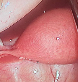
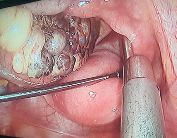
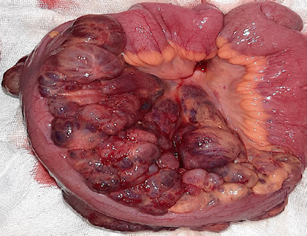
A 24-years old woman with a medical history of chronic anemia thought to be secondary to metrorrhagia, complained about pain in the lower half of the abdomen lasting for one day without other signs. Physical examination revealed a temperature of 38°C with pale conjunctiva, and tenderness in the lower half of the abdomen. Gynecologic exam was normal. Biological exams confirmed an iron deficiency anemia. BetahCG levels were within normal range. After a brief resuscitation, the patient had an exploratory laparoscopy for suspected complicated appendicitis. Peroperative exploration revealed a hemoperitoneum in the Pouch of Douglas (Figure 1) with a 10 cm multicystic lesion arising from small bowel mesentery (Figure 2). No other lesions were associated. Because of the size of the lesion, laparotomy was performed. This lesion corresponded to surface scattered multiple cysts well-encapsulated with thin walls (Figure 3). Total excision was performed with is peristaltic latero-lateral ileo-ileal anastomosis. The postoperative course was uneventful. Histopathological exam revealed a multilocular intra-abdominal cystic lymphangioma with various-sized cystic spaces lined by attenuated endothelial cells arranged in a single layer. No recurrence was noticed after a regular follow up of 12 months.
DISCUSSION
Our case illustrated a complicated extremely rare pathology in adults revealed by abdominal pain associated to misdiagnosed chronic anemia. In fact, adult abdominal cystic lymphangioma accounted for less than 5% of all cystic lymphangiomas [3]. Its etiology is still unknown even though primary lymphatic cysts failure to converge with the main lymphatic system was suggested [4,5]. As in our case, abdominal pain constituted the most revealing symptom [6,7]. Unlike our case, imaging features allowed positive diagnosis with conservative treatment in asymptomatic cases [1]. It presented as a well-defined lesion with anechoic content and fine fibrous separating septa in abdominal ultrasound and as a homogenous hypo dense lesion with nonenhanced contrast walls [8]. Surgical removal was indicated in front of complication as in our case [1]. Laparotomy was preferred for huge lesions [1]. Total resection as performed for our patient reduced relapse in comparison with partial excision [1,9].
CONCLUSION
Adult abdominal cystic lymphangioma still a not fully understood pathology with various presentations. It has to be suspected in presence of evocator aspects. Complications implicate complete excision in order to treat the complication and to avoid recurrence.
PATIENT CONSENT
A writing informed consent was obtained from the patient to public this report in accordance with the journal’s patient consent policy.
REFERENCES
- Xiao J, Shao Y, Zhu S, He X. Characteristics of adult abdominal cystic Lymphangioma: a single-center Chinese cohort of 12 cases. BMC Gastroenterology. 2020; 20: 244.
- Agha RA, Borrelli MR, Farwana R, Koshy K, Fowler A, Orgill DP, SCARE Group. The SCARE 2018 Statement: Updating Consensus Surgical Case Report (SCARE) Guidelines. Int J Surg. 2018; 60: 132-136.
- Kosir MA, Sonnino RE, Gauderer MW. Pediatric abdominal lymphangiomas: a plea for early recognition. J Pediatr Surg. 1991; 26: 1309-1313.
- Weeda VB, Booij KA, Aronson DC. Mesenteric cystic lymphangioma: a congenital and an acquired anomaly? Two cases and a review of the literature. J Pediatr Surg. 2008; 43: 1206-1208.
- Goh BK, Tan YM, Ong HS, Chan WH, Yap CK, Wong WK. Endoscopic ultrasound diagnosis and laparoscopic excision of an omental lymphangioma. J Laparoendosc Adv Surg Tech A. 2005; 15: 630-633.
- Makni A, Chebbi F, Fetirich F, Ksantini R, Bedioui H, Jouini M, et al. Surgical management of intra-abdominal cystic lymphangioma. Report of 20 cases. World J Surg. 2012; 36: 1037-1043.
- Tarcoveanu E, Moldovanu R, Bradea C, Vlad N, Ciobanu D, Vasilescu A, et al. Laparoscopic treatment of Intraabdominal cystic Lymphangioma. Chirurgia (Bucur). 2016; 111: 236-241.
- Verdin V, Seydel B, Detry O, Daele DV, Meunier P, Honore P, et al. [Clinical case of the month. Cystic lymphangioma of the mesentery]. Rev Med Liege. 2010; 65: 615-618.
- Kim SH, Kim HY, Lee C, Min HS, Jung SE. Clinical features of mesenteric lymphatic malformation in children. J Pediatr Surg. 2016; 51: 582– 587.


